![Western Blot: Bad Antibody [NB100-56080]](https://resources.rndsystems.com/images/products/Bad-Antibody-Western-Blot-NB100-56080-img0002.jpg "Western Blot: Bad Antibody [NB100-56080]")
Loading...
Key Product Details
Validated by
Biological Validation
Species Reactivity
Human
Applications
Immunohistochemistry, Immunohistochemistry-Paraffin, Western Blot, Immunocytochemistry/ Immunofluorescence, Immunoprecipitation
Label
Unconjugated
Antibody Source
Polyclonal Rabbit IgG
Format
BSA Free
Loading...
Product Specifications
Immunogen
A full length recombinant protein corresponding to human Bad was used as immunogen.
Clonality
Polyclonal
Host
Rabbit
Isotype
IgG
Scientific Data Images for Bad Antibody - BSA Free
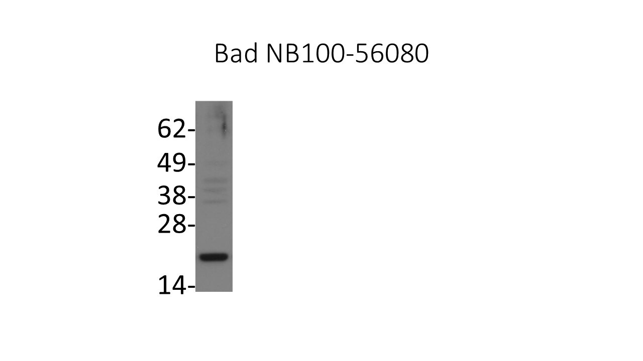
Western Blot: Bad Antibody [NB100-56080]
Western Blot: Bad Antibody [NB100-56080] - Analysis of Bad in Daoy whole cell lysate using anti-Bad antibody. Image from verified customer review.![Immunocytochemistry/ Immunofluorescence: Bad Antibody [NB100-56080]](https://resources.rndsystems.com/images/products/Bad-Antibody-Immunocytochemistry-Immunofluorescence-NB100-56080-img0003.jpg "Immunocytochemistry/ Immunofluorescence: Bad Antibody [NB100-56080]")
Immunocytochemistry/ Immunofluorescence: Bad Antibody [NB100-56080]
Immunocytochemistry/Immunofluorescence: Bad Antibody [NB100-56080] - Immunofluorescence microscopy of BAD using NB100-56080 at 1:2000. Du145 human prostate carcinoma cells were cultured without (A) or with (B) and (C) 1 uM of the triphosphatase inhibitor thapsigargin (THG) for 12 hr. A and B, staining with BAD antibody, followed by a FITC-conjugated secondary antibody. C, staining with a mitochondrial marker (antibody to mitochondrial Hsp60), followed by a rhodamine-conjugated secondary antibody. THG induces Ca2+ release from internal stores which can promote apoptosis. BAD staining was located diffusely throughout the cytoplasm of untreated cells (A), and localized to the mitochondria in treated cells (B).![Immunohistochemistry-Paraffin: Bad Antibody [NB100-56080]](https://resources.rndsystems.com/images/products/Bad-Antibody-Immunohistochemistry-Paraffin-NB100-56080-img0004.jpg "Immunohistochemistry-Paraffin: Bad Antibody [NB100-56080]")
Immunohistochemistry-Paraffin: Bad Antibody [NB100-56080]
Immunohistochemistry-Paraffin: Bad Antibody [NB100-56080] - IHC analysis of formalin fixed paraffin-embedded (FFPE) human liver using 1:2000 conc. of Bad antibody on a Bond Rx autostainer (Leica Biosystems). The assay involved 20 minutes of heat induced antigen retrieval (HIER) using 10mM sodium citrate buffer (pH 6.0) and endogenous peroxidase quenching with peroxide block. The sections were incubated with primary antibody for 30 minutes and Bond Polymer Refine Detection (Leica Biosystems) with DAB was used for signal development followed by counterstaining with hematoxylin. Whole slide scanning and capturing of representative images was performed using Aperio AT2 (Leica Biosystems). Cytoplasmic staining of Bad was observed. Staining was performed by Histowiz.Applications for Bad Antibody - BSA Free
Application
Recommended Usage
Immunocytochemistry/ Immunofluorescence
1:500-1:2000
Immunohistochemistry
1:2000
Immunohistochemistry-Paraffin
1:2000
Immunoprecipitation
1:50-1:200
Western Blot
1:1000-1:2000
Reviewed Applications
Read 1 review rated 5 using NB100-56080 in the following applications:
Formulation, Preparation, and Storage
Purification
Unpurified
Formulation
Whole antisera
Format
BSA Free
Preservative
0.02% Sodium Azide
Concentration
This product is unpurified. The exact concentration of antibody is not quantifiable.
Shipping
The product is shipped with polar packs. Upon receipt, store it immediately at the temperature recommended below.
Stability & Storage
Store at 4C short term. Aliquot and store at -20C long term. Avoid freeze-thaw cycles.
Background: Bad
Long Name
Bcl-xL/Bcl-2 Associated Death Promoter
Alternate Names
BBC6, BCL2L8
Entrez Gene IDs
572 (Human)
Gene Symbol
BAD
Additional Bad Products
Product Documents for Bad Antibody - BSA Free
Certificate of Analysis
To download a Certificate of Analysis, please enter a lot or batch number in the search box below.
Product Specific Notices for Bad Antibody - BSA Free
This product is for research use only and is not approved for use in humans or in clinical diagnosis. Primary Antibodies are guaranteed for 1 year from date of receipt.
Related Research Areas
Customer Reviews for Bad Antibody - BSA Free (1)
5 out of 5
1 Customer Rating
Have you used Bad Antibody - BSA Free?
Submit a review and receive an Amazon gift card!
$25/€18/£15/$25CAN/¥2500 Yen for a review with an image
$10/€7/£6/$10CAN/¥1110 Yen for a review without an image
Submit a review
Customer Images
Showing
1
-
1 的
1 review
Showing All
Filter By:
-
Application: Western BlotSample Tested: Whole cell lysate from Daoy cellsSpecies: HumanVerified Customer | Posted 07/14/2016Western blot analysis of Bad in Daoy cell lysate (25ug)

There are no reviews that match your criteria.
Protocols
Find general support by application which include: protocols, troubleshooting, illustrated assays, videos and webinars.
- Antigen Retrieval Protocol (PIER)
- Antigen Retrieval for Frozen Sections Protocol
- Appropriate Fixation of IHC/ICC Samples
- Cellular Response to Hypoxia Protocols
- Chromogenic IHC Staining of Formalin-Fixed Paraffin-Embedded (FFPE) Tissue Protocol
- Chromogenic Immunohistochemistry Staining of Frozen Tissue
- ClariTSA™ Fluorophore Kits
- Detection & Visualization of Antibody Binding
- Fluorescent IHC Staining of Frozen Tissue Protocol
- Graphic Protocol for Heat-induced Epitope Retrieval
- Graphic Protocol for the Preparation and Fluorescent IHC Staining of Frozen Tissue Sections
- Graphic Protocol for the Preparation and Fluorescent IHC Staining of Paraffin-embedded Tissue Sections
- Graphic Protocol for the Preparation of Gelatin-coated Slides for Histological Tissue Sections
- ICC Cell Smear Protocol for Suspension Cells
- ICC Immunocytochemistry Protocol Videos
- ICC for Adherent Cells
- IHC Sample Preparation (Frozen sections vs Paraffin)
- Immunocytochemistry (ICC) Protocol
- Immunocytochemistry Troubleshooting
- Immunofluorescence of Organoids Embedded in Cultrex Basement Membrane Extract
- Immunofluorescent IHC Staining of Formalin-Fixed Paraffin-Embedded (FFPE) Tissue Protocol
- Immunohistochemistry (IHC) and Immunocytochemistry (ICC) Protocols
- Immunohistochemistry Frozen Troubleshooting
- Immunohistochemistry Paraffin Troubleshooting
- Immunoprecipitation Protocol
- Preparing Samples for IHC/ICC Experiments
- Preventing Non-Specific Staining (Non-Specific Binding)
- Primary Antibody Selection & Optimization
- Protocol for Heat-Induced Epitope Retrieval (HIER)
- Protocol for Making a 4% Formaldehyde Solution in PBS
- Protocol for VisUCyte™ HRP Polymer Detection Reagent
- Protocol for the Fluorescent ICC Staining of Cell Smears - Graphic
- Protocol for the Fluorescent ICC Staining of Cultured Cells on Coverslips - Graphic
- Protocol for the Preparation & Fixation of Cells on Coverslips
- Protocol for the Preparation and Chromogenic IHC Staining of Frozen Tissue Sections
- Protocol for the Preparation and Chromogenic IHC Staining of Frozen Tissue Sections - Graphic
- Protocol for the Preparation and Chromogenic IHC Staining of Paraffin-embedded Tissue Sections
- Protocol for the Preparation and Chromogenic IHC Staining of Paraffin-embedded Tissue Sections - Graphic
- Protocol for the Preparation and Fluorescent ICC Staining of Cells on Coverslips
- Protocol for the Preparation and Fluorescent ICC Staining of Non-adherent Cells
- Protocol for the Preparation and Fluorescent ICC Staining of Stem Cells on Coverslips
- Protocol for the Preparation and Fluorescent IHC Staining of Frozen Tissue Sections
- Protocol for the Preparation and Fluorescent IHC Staining of Paraffin-embedded Tissue Sections
- Protocol for the Preparation of Gelatin-coated Slides for Histological Tissue Sections
- Protocol for the Preparation of a Cell Smear for Non-adherent Cell ICC - Graphic
- R&D Systems Quality Control Western Blot Protocol
- TUNEL and Active Caspase-3 Detection by IHC/ICC Protocol
- The Importance of IHC/ICC Controls
- Troubleshooting Guide: Immunohistochemistry
- Troubleshooting Guide: Western Blot Figures
- Western Blot Conditions
- Western Blot Protocol
- Western Blot Protocol for Cell Lysates
- Western Blot Troubleshooting
- Western Blot Troubleshooting Guide
- View all Protocols, Troubleshooting, Illustrated assays and Webinars
Loading...
Associated Pathways


IL-7 Signaling Pathways
 IL-9 Signaling Pathways
 IL-15 Signaling Pathways
 IL-21 Signaling Pathways
 MAPK Signaling: Oxidative Stress Pathway
 VEGF - VEGF R2 Signaling Pathways
