![Immunohistochemistry-Paraffin: CD20 Antibody [NBP1-90051]](https://resources.rndsystems.com/images/products/CD20-Antibody-Immunohistochemistry-Paraffin-NBP1-90051-img0010.jpg "Immunohistochemistry-Paraffin: CD20 Antibody [NBP1-90051]")
Loading...
Key Product Details
Validated by
Orthogonal Validation, Independent Antibodies
Species Reactivity
Human
Applications
Immunohistochemistry, Immunohistochemistry-Paraffin, Western Blot, Immunocytochemistry/ Immunofluorescence
Label
Unconjugated
Antibody Source
Polyclonal Rabbit IgG
Format
BSA Free
Loading...
Product Specifications
Immunogen
This antibody was developed against Recombinant Protein corresponding to amino acids: ENEWKRTCSRPKSNIVLLSAEEKKEQTIEIKEEVVGLTETSSQPKNEEDIEIIPIQEEEEEETETNFPEPPQDQESSP
Reactivity Notes
.
Clonality
Polyclonal
Host
Rabbit
Isotype
IgG
Scientific Data Images for CD20 Antibody - BSA Free
Immunohistochemistry-Paraffin: CD20 Antibody [NBP1-90051]
Immunohistochemistry-Paraffin: CD20 Antibody [NBP1-90051] - Staining of human cerebral cortex, lymph node, spleen and testis using Anti-MS4A1 antibody NBP1-90051 (A) shows similar protein distribution across tissues to independent antibody NBP1-90052 (B).![CD20 Antibody - BSA Free Immunohistochemistry: CD20 Antibody - BSA Free [NBP1-90051]](https://resources.rndsystems.com/images/products/nbp1-90051_rabbit-polyclonal-cd20-antibody-29520251610571.jpg "Immunohistochemistry: CD20 Antibody - BSA Free [NBP1-90051]")
![Immunohistochemistry-Paraffin: CD20 Antibody [NBP1-90051]](https://resources.rndsystems.com/images/products/CD20-Antibody-Immunohistochemistry-Paraffin-NBP1-90051-img0013.jpg "Immunohistochemistry-Paraffin: CD20 Antibody [NBP1-90051]")
Immunohistochemistry-Paraffin: CD20 Antibody [NBP1-90051]
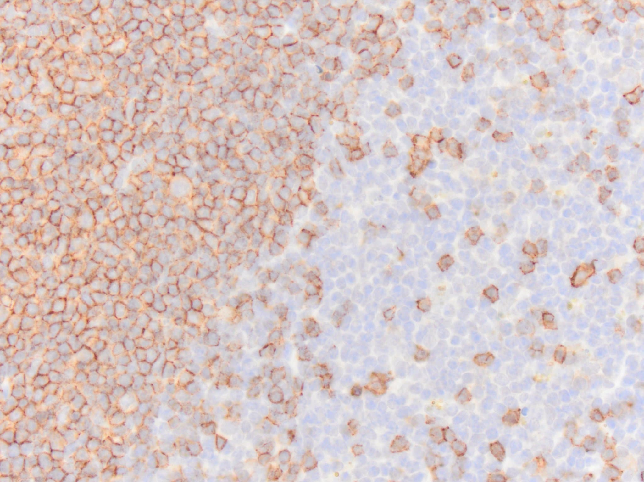
Immunohistochemistry-Paraffin: CD20 Antibody [NBP1-90051] - Staining of human spleen shows strong membranous positivity in cells in white pulp.![Immunohistochemistry-Paraffin: CD20 Antibody [NBP1-90051]](https://resources.rndsystems.com/images/products/CD20-Antibody-Immunohistochemistry-Paraffin-NBP1-90051-img0015.jpg "Immunohistochemistry-Paraffin: CD20 Antibody [NBP1-90051]")
Immunohistochemistry-Paraffin: CD20 Antibody [NBP1-90051]
Immunohistochemistry-Paraffin: CD20 Antibody [NBP1-90051] - Staining of human cerebral cortex shows low expression as expected.![Immunohistochemistry-Paraffin: CD20 Antibody [NBP1-90051]](https://resources.rndsystems.com/images/products/CD20-Antibody-Immunohistochemistry-Paraffin-NBP1-90051-img0021.jpg "Immunohistochemistry-Paraffin: CD20 Antibody [NBP1-90051]")
Immunohistochemistry-Paraffin: CD20 Antibody [NBP1-90051]
Immunohistochemistry-Paraffin: CD20 Antibody [NBP1-90051] - Staining of human spleen using Anti-MS4A1 antibody NBP1-90051.![Immunohistochemistry-Paraffin: CD20 Antibody [NBP1-90051]](https://resources.rndsystems.com/images/products/CD20-Antibody-Immunohistochemistry-Paraffin-NBP1-90051-img0012.jpg "Immunohistochemistry-Paraffin: CD20 Antibody [NBP1-90051]")
Immunohistochemistry-Paraffin: CD20 Antibody [NBP1-90051]
Immunohistochemistry-Paraffin: CD20 Antibody [NBP1-90051] - Staining of human testis.![Immunohistochemistry-Paraffin: CD20 Antibody [NBP1-90051]](https://resources.rndsystems.com/images/products/CD20-Antibody-Immunohistochemistry-Paraffin-NBP1-90051-img0014.jpg "Immunohistochemistry-Paraffin: CD20 Antibody [NBP1-90051]")
Immunohistochemistry-Paraffin: CD20 Antibody [NBP1-90051]
Immunohistochemistry-Paraffin: CD20 Antibody [NBP1-90051] - Staining of human gastrointestinal shows strong membranous positivity in lymphocytes.![Immunohistochemistry-Paraffin: CD20 Antibody [NBP1-90051]](https://resources.rndsystems.com/images/products/CD20-Antibody-Immunohistochemistry-Paraffin-NBP1-90051-img0016.jpg "Immunohistochemistry-Paraffin: CD20 Antibody [NBP1-90051]")
Immunohistochemistry-Paraffin: CD20 Antibody [NBP1-90051]
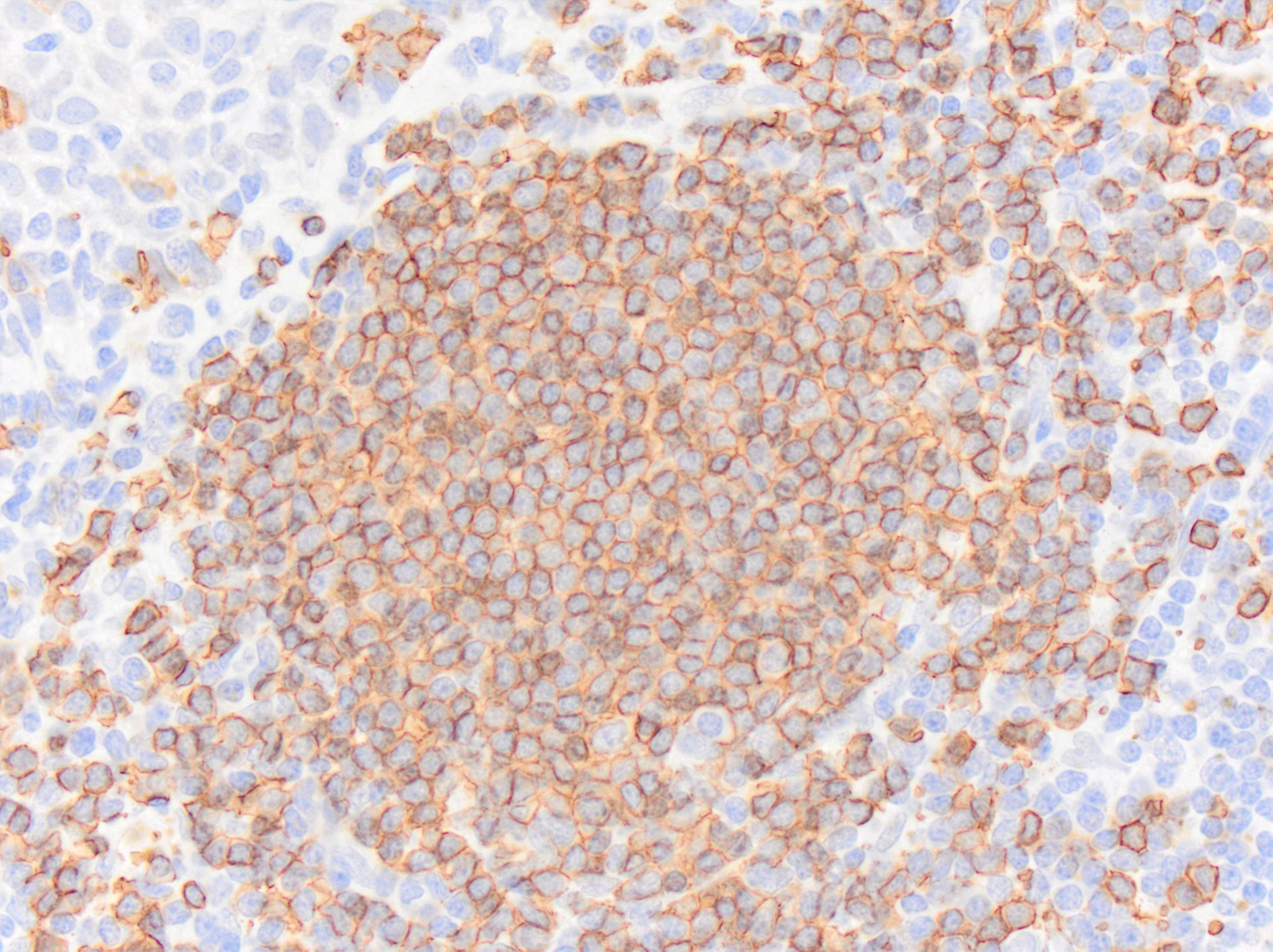
Immunohistochemistry-Paraffin: CD20 Antibody [NBP1-90051] - Staining of human tonsil shows strong membranous positivity in germinal center cells.![Immunohistochemistry-Paraffin: CD20 Antibody [NBP1-90051]](https://resources.rndsystems.com/images/products/CD20-Antibody-Immunohistochemistry-Paraffin-NBP1-90051-img0017.jpg "Immunohistochemistry-Paraffin: CD20 Antibody [NBP1-90051]")
Immunohistochemistry-Paraffin: CD20 Antibody [NBP1-90051]
Immunohistochemistry-Paraffin: CD20 Antibody [NBP1-90051] - Staining of human lymph node shows strong membranous positivity in germinal center cells.![Immunohistochemistry-Paraffin: CD20 Antibody [NBP1-90051]](https://resources.rndsystems.com/images/products/CD20-Antibody-Immunohistochemistry-Paraffin-NBP1-90051-img0019.jpg "Immunohistochemistry-Paraffin: CD20 Antibody [NBP1-90051]")
Immunohistochemistry-Paraffin: CD20 Antibody [NBP1-90051]
Immunohistochemistry-Paraffin: CD20 Antibody [NBP1-90051] - Staining of human cerebral cortex using Anti-MS4A1 antibody NBP1-90051.![Immunohistochemistry-Paraffin: CD20 Antibody [NBP1-90051]](https://resources.rndsystems.com/images/products/CD20-Antibody-Immunohistochemistry-Paraffin-NBP1-90051-img0020.jpg "Immunohistochemistry-Paraffin: CD20 Antibody [NBP1-90051]")
Immunohistochemistry-Paraffin: CD20 Antibody [NBP1-90051]
Immunohistochemistry-Paraffin: CD20 Antibody [NBP1-90051] - Staining of human lymph node using Anti-MS4A1 antibody NBP1-90051.![CD20 Antibody - BSA Free Western Blot: CD20 Antibody - BSA Free [NBP1-90051]](https://resources.rndsystems.com/images/products/nbp1-90051_rabbit-polyclonal-cd20-antibody-30520257483213.jpg "Western Blot: CD20 Antibody - BSA Free [NBP1-90051]")
![CD20 Antibody - BSA Free Immunocytochemistry/ Immunofluorescence: CD20 Antibody [NBP1-90051]](https://resources.rndsystems.com/images/products/nbp1-90051_-immunocytochemistry-immunofluorescence-639174076805621702.jpg "Immunocytochemistry/ Immunofluorescence: CD20 Antibody [NBP1-90051]")
Immunocytochemistry/ Immunofluorescence: CD20 Antibody [NBP1-90051]
Staining of human cell line REH shows localization to nucleus & plasma membrane.Applications for CD20 Antibody - BSA Free
Application
Recommended Usage
Immunocytochemistry/ Immunofluorescence
0.25-2 ug/ml
Immunohistochemistry
1:1000 - 1:2500
Immunohistochemistry-Paraffin
1:1000 - 1:2500
Western Blot
0.04 - 0.4 ug/ml
Application Notes
IHC-Paraffin, HIER pH 6 retrieval is recommended. ICC/IF, Fixation/Permeabilization: PFA/Triton X-100
Reviewed Applications
Read 3 reviews rated 5 using NBP1-90051 in the following applications:
Formulation, Preparation, and Storage
Purification
Affinity purified
Formulation
PBS (pH 7.2) and 40% Glycerol
Format
BSA Free
Preservative
0.02% Sodium Azide
Concentration
Concentrations vary lot to lot. See vial label for concentration. If unlisted please contact technical services.
Shipping
The product is shipped with polar packs. Upon receipt, store it immediately at the temperature recommended below.
Stability & Storage
Store at 4C short term. Aliquot and store at -20C long term. Avoid freeze-thaw cycles.
Background: CD20
References
1. Pavlasova G, Mraz M. The regulation and function of CD20: an "enigma" of B-cell biology and targeted therapy. Haematologica. 2020; 105(6):1494-1506. https://doi.org/10.3324/haematol.2019.243543
2. Payandeh Z, Bahrami AA, Hoseinpoor R, et al. The applications of anti-CD20 antibodies to treat various B cells disorders. Biomed Pharmacother. 2019; 109:2415-2426. https://doi.org/10.1016/j.biopha.2018.11.121
3. Margoni M, Preziosa P, Filippi M, Rocca MA. Anti-CD20 therapies for multiple sclerosis: current status and future perspectives. J Neurol. 2022; 269(3):1316-1334. https://doi.org/10.1007/s00415-021-10744-x
4. Klein C, Jamois C, Nielsen T. Anti-CD20 treatment for B-cell malignancies: current status and future directions. Expert Opin Biol Ther. 2021; 21(2):161-181. https://doi.org/10.1080/14712598.2020.1822318
5. Sharman JP. Targeting CD20: teaching an old dog new tricks. Hematology Am Soc Hematol Educ Program. 2019; 2019(1):273-278. https://doi.org/10.1182/hematology.2019000031
Long Name
Cluster of Differentiation 20
Alternate Names
B1, Bp35, CD20, LEU-16, Ly-44, MS4A1, S7
Gene Symbol
MS4A1
Additional CD20 Products
Product Documents for CD20 Antibody - BSA Free
Certificate of Analysis
To download a Certificate of Analysis, please enter a lot or batch number in the search box below.
Product Specific Notices for CD20 Antibody - BSA Free
This product is for research use only and is not approved for use in humans or in clinical diagnosis. Primary Antibodies are guaranteed for 1 year from date of receipt.
Customer Reviews for CD20 Antibody - BSA Free (3)
5 out of 5
3 Customer Ratings
Have you used CD20 Antibody - BSA Free?
Submit a review and receive an Amazon gift card!
$25/€18/£15/$25CAN/¥2500 Yen for a review with an image
$10/€7/£6/$10CAN/¥1110 Yen for a review without an image
Submit a review
Customer Images

Showing
1
-
3 的
3 reviews
Showing All
Filter By:
-
Application: Immunohistochemistry-ParaffinSample Tested: SpleenSpecies: MouseVerified Customer | Posted 05/08/2024CD20 immunoreactivity in an FFPE section of mouse spleen. NBP1 90051 was diluted to 250ng per mL and was left on tissue sections for 30m at room temperature. Secondary was horse anti rabbit HRP polymer.Section underwent heat-induced epitope retrieval for 20m in a vegetable steamer using Target Retrieval Solution.
 Bio-Techne ResponseThis review was submitted through the legacy Novus Innovators Program, reflecting a new species or application tested on a primary antibody.
 -
Application: Immunohistochemistry-ParaffinSample Tested: TonsilSpecies: HumanVerified Customer | Posted 05/07/2024CD20 immunoreactivity in an FFPE section of human tonsil. NBP1-90051 was diluted to 250ng per mL and was left on tissue sections for 30m at room temperature. Secondary was horse anti rabbit HRP polymer.Section underwent heat induced epitope retrieval in a vegetable steamer for 20m using Target Retrieval Solution.
-
Application: Immunohistochemistry-ParaffinSample Tested: SpleenSpecies: HorseVerified Customer | Posted 05/07/2024CD20 immunoreactivity in an FFPE section of horse spleen. NBP1-90051 was diluted to 250ng per mL and was left on tissue sections for 30m at room temperature. Secondary was horse anti rabbit HRP polymer.Section underwent heat induced epitope retrieval for 20min in a vegetable steamer using Target Retrieval Solution.
Bio-Techne ResponseThis review was submitted through the legacy Novus Innovators Program, reflecting a new species or application tested on a primary antibody.
There are no reviews that match your criteria.
Protocols
Find general support by application which include: protocols, troubleshooting, illustrated assays, videos and webinars.
- Antigen Retrieval Protocol (PIER)
- Antigen Retrieval for Frozen Sections Protocol
- Appropriate Fixation of IHC/ICC Samples
- Cellular Response to Hypoxia Protocols
- Chromogenic IHC Staining of Formalin-Fixed Paraffin-Embedded (FFPE) Tissue Protocol
- Chromogenic Immunohistochemistry Staining of Frozen Tissue
- ClariTSA™ Fluorophore Kits
- Detection & Visualization of Antibody Binding
- Fluorescent IHC Staining of Frozen Tissue Protocol
- Graphic Protocol for Heat-induced Epitope Retrieval
- Graphic Protocol for the Preparation and Fluorescent IHC Staining of Frozen Tissue Sections
- Graphic Protocol for the Preparation and Fluorescent IHC Staining of Paraffin-embedded Tissue Sections
- Graphic Protocol for the Preparation of Gelatin-coated Slides for Histological Tissue Sections
- ICC Cell Smear Protocol for Suspension Cells
- ICC Immunocytochemistry Protocol Videos
- ICC for Adherent Cells
- IHC Sample Preparation (Frozen sections vs Paraffin)
- Immunocytochemistry (ICC) Protocol
- Immunocytochemistry Troubleshooting
- Immunofluorescence of Organoids Embedded in Cultrex Basement Membrane Extract
- Immunofluorescent IHC Staining of Formalin-Fixed Paraffin-Embedded (FFPE) Tissue Protocol
- Immunohistochemistry (IHC) and Immunocytochemistry (ICC) Protocols
- Immunohistochemistry Frozen Troubleshooting
- Immunohistochemistry Paraffin Troubleshooting
- Preparing Samples for IHC/ICC Experiments
- Preventing Non-Specific Staining (Non-Specific Binding)
- Primary Antibody Selection & Optimization
- Protocol for Heat-Induced Epitope Retrieval (HIER)
- Protocol for Making a 4% Formaldehyde Solution in PBS
- Protocol for VisUCyte™ HRP Polymer Detection Reagent
- Protocol for the Fluorescent ICC Staining of Cell Smears - Graphic
- Protocol for the Fluorescent ICC Staining of Cultured Cells on Coverslips - Graphic
- Protocol for the Preparation & Fixation of Cells on Coverslips
- Protocol for the Preparation and Chromogenic IHC Staining of Frozen Tissue Sections
- Protocol for the Preparation and Chromogenic IHC Staining of Frozen Tissue Sections - Graphic
- Protocol for the Preparation and Chromogenic IHC Staining of Paraffin-embedded Tissue Sections
- Protocol for the Preparation and Chromogenic IHC Staining of Paraffin-embedded Tissue Sections - Graphic
- Protocol for the Preparation and Fluorescent ICC Staining of Cells on Coverslips
- Protocol for the Preparation and Fluorescent ICC Staining of Non-adherent Cells
- Protocol for the Preparation and Fluorescent ICC Staining of Stem Cells on Coverslips
- Protocol for the Preparation and Fluorescent IHC Staining of Frozen Tissue Sections
- Protocol for the Preparation and Fluorescent IHC Staining of Paraffin-embedded Tissue Sections
- Protocol for the Preparation of Gelatin-coated Slides for Histological Tissue Sections
- Protocol for the Preparation of a Cell Smear for Non-adherent Cell ICC - Graphic
- R&D Systems Quality Control Western Blot Protocol
- TUNEL and Active Caspase-3 Detection by IHC/ICC Protocol
- The Importance of IHC/ICC Controls
- Troubleshooting Guide: Immunohistochemistry
- Troubleshooting Guide: Western Blot Figures
- Western Blot Conditions
- Western Blot Protocol
- Western Blot Protocol for Cell Lysates
- Western Blot Troubleshooting
- Western Blot Troubleshooting Guide
- View all Protocols, Troubleshooting, Illustrated assays and Webinars
Loading...
Associated Pathways

