![Flow Cytometry: DRAQ7 (TM) [NBP2-81126]](https://resources.rndsystems.com/images/products/DRAQ7-TM-Flow-Cytometry-NBP2-81126-img0003.jpg "Flow Cytometry: DRAQ7 (TM) [NBP2-81126]")
Loading...
Key Product Details
Applications
Flow Cytometry, Immunocytochemistry/ Immunofluorescence, Microscopy
Product Summary for DRAQ7 (TM)
Key Features of DRAQ7 (TM) :
Far-red viability dye used for investigating dead or membrane compromised cells, notably for dead cell exclusion in flow cytometry, single-cell RNAseq and GWAS
Does not enter intact, live cells and acts as an ideal replacement for propidium iodide (PI) and 7-AAD
Serves as a component of apoptosis assays, for any fluorescence-based cell analysis platform, and in cell health / in vitro toxicity assays
Rapid staining and easy to use, without the need for a wash step
Non-toxic and optimal for long-term imaging studies, up to several days
Minimal photobleaching
Spectrally compatible with GFP and FITC labels.
DRAQ7 (TM) is supplied as a blue aqueous solution and shipped at ambient temperature, but on receipt packs should be stored at 2-8C. DO NOT FREEZE.
DRAQ7 (TM) is supplied as a blue aqueous solution and shipped at ambient temperature, but on receipt packs should be stored at 2-8C. DO NOT FREEZE.
Loading...
Product Specifications
Applications
Flow Cytometry (1:100)
Immunocytochemistry/ Immunofluorescence (1:60)
Live Imaging Microscopy (1:100)
Fluorescence Imaging (1:60 - 1:100)
Immunocytochemistry/ Immunofluorescence (1:60)
Live Imaging Microscopy (1:100)
Fluorescence Imaging (1:60 - 1:100)
Application Notes
DRAQ7 (TM) is supplied at a concentration of 0.3mM: the 250 ul size allows for 50 Flow Cytometry assays and 250 Cell Health assays whereas the 1ml size allows for 200 Flow Cytometry assays and 1,000 Cell Health assays. DRAQ7 (TM) can be diluted in culture media (e.g. RPMI 1640) and physiological buffers (eg PBS, Hankss, etc.) and mixed with fixatives such as formaldehyde. DRAQ7 (TM) has many applications in imaging, cytometry and screening and is highly compatible with existing protocols across a wide range of instrumentation platforms.
Spectra Viewer
Plan Your Experiments
Use our spectra viewer to interactively plan your experiments, assessing multiplexing options. View the excitation and emission spectra for our fluorescent dye range and other commonly used dyes.
Spectra Viewer
Scientific Data Images for DRAQ7 (TM)
Flow Cytometry: DRAQ7 (TM) [NBP2-81126]
Flow Cytometry: DRAQ7 (TM) [NBP2-81126] - Lymphoma cells treated with increasing quantities of staurosporine (STS). reports STS-induced apoptosis and cell death in dose-dependent manner with clear separation of positive and negative events.![Live Imaging Microscopy: DRAQ7 (TM) [NBP2-81126]](https://resources.rndsystems.com/images/products/DRAQ7-TM-Live-Imaging-Microscopy-NBP2-81126-img0005.jpg "Live Imaging Microscopy: DRAQ7 (TM) [NBP2-81126]")
Live Imaging Microscopy: DRAQ7 (TM) [NBP2-81126]
Live Imaging Microscopy: DRAQ7 (TM) [NBP2-81126] - DRAQ7(TM) was added directly to unfixed THP-1 cells in RPMI + 20% FBS culture media at 3 uM (1:100) for 30 minutes at room temperature and protected from light. Imaging was done immediately after staining without washing the cells.![Immunocytochemistry/ Immunofluorescence: DRAQ7 (TM) [NBP2-81126]](https://resources.rndsystems.com/images/products/DRAQ7-TM-Immunocytochemistry-Immunofluorescence-NBP2-81126-img0004.jpg "Immunocytochemistry/ Immunofluorescence: DRAQ7 (TM) [NBP2-81126]")
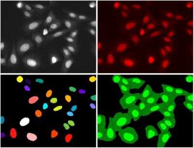
Immunocytochemistry/ Immunofluorescence: DRAQ7 (TM) [NBP2-81126]
Immunocytochemistry/Immunofluorescence: DRAQ7 (TM) [NBP2-81126] - Formaldehyde-fixed U2OS cells labelled with (red, nuclei+D8 and AlexaFluor 488 antibody to beta-tubulin (green).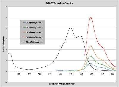
DRAQ7 (TM) [NBP2-81126] - Spectral properties of DRAQ7 (TM) - spectral compatibility with UV-excited and most vis. Range fluorochromes for multi-colour analysis. Detection from blue excitation is achievable only by flow cytometry.
Fluorescence Imaging: DRAQ7 (TM) [NBP2-81126] - montage.
 [NBP2-81126] -")
DRAQ7 (TM) [NBP2-81126] -
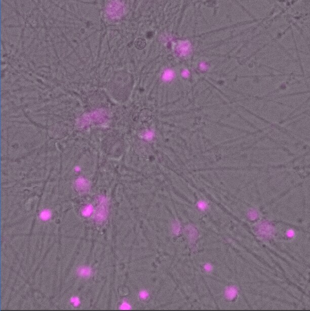
DRAQ7 (TM) [NBP2-81126] - In vitro, Cortical neurons are used to assess the glutamate toxicity using DRAQ7 dye. Image from verified customer review.Formulation, Preparation, and Storage
Purification
>97%
Concentration
Please see the protocols for proper use of this product. If no protocol is available, contact technical services for assistance.
Shipping
The product is shipped at ambient temperature. Upon receipt, store it immediately at the temperature recommended below.
Storage
Store at 4C in the dark. Do not freeze.
Background: DRAQ7 (TM)
The excitation wavelength maxima of DRAQ7 (TM) are 600 nm / 646 nm but it can also be suboptimally excited by the 488nm (blue) laser for flow cytometry. The emission wavelength maximum of this viability dye is 697 nm when bound to dsDNA. The fluorescence signal is proportional and stoichiometric to the amount of bound DNA.
DRAQ7 (TM) is a registered trademark of BioStatus Limited.
Additional DRAQ7 (TM) Products
Product Documents for DRAQ7 (TM)
Certificate of Analysis
To download a Certificate of Analysis, please enter a lot or batch number in the search box below.
Product Specific Notices for DRAQ7 (TM)
DRAQ7 (TM) is a registered trademark of BioStatus Limited.
This product is for research use only and is not approved for use in humans or in clinical diagnosis. Support products are guaranteed for 6 months from date of receipt.
Citations for DRAQ7 (TM)
 Powered by Bioz
Customer Reviews for DRAQ7 (TM) (2)
5 out of 5
2 Customer Ratings
Have you used DRAQ7 (TM)?
Submit a review and receive an Amazon gift card!
$25/€18/£15/$25CAN/¥2500 Yen for a review with an image
$10/€7/£6/$10CAN/¥1110 Yen for a review without an image
Submit a review
Customer Images
Showing
1
-
2 的
2 reviews
Showing All
Filter By:
-
Verified Customer | Posted 06/06/2023Assessing glutamate toxicity at 250 micromolar in vitro.In vitro, Cortical neurons are used to assess the glutamate toxicity using DRAQ7 dye.

-
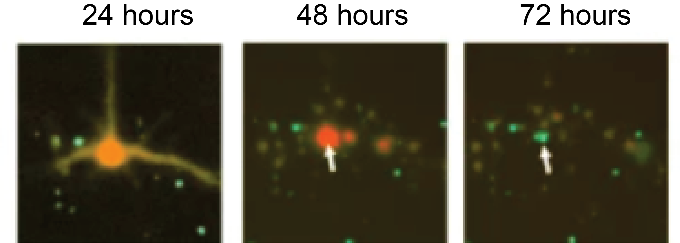
Verified Customer | Posted 10/12/2021Detects cell death very well. Neurons transfected with Tdtomato and undergoing stress for 72 hours. Draq7 labeling is evident at 72 hours.used at 4 micro litre per well of a 24 well dish
There are no reviews that match your criteria.
Protocols
Find general support by application which include: protocols, troubleshooting, illustrated assays, videos and webinars.
- 7-Amino Actinomycin D (7-AAD) Cell Viability Flow Cytometry Protocol
- Appropriate Fixation of IHC/ICC Samples
- Cellular Response to Hypoxia Protocols
- ClariTSA™ Fluorophore Kits
- Detection & Visualization of Antibody Binding
- Extracellular Membrane Flow Cytometry Protocol
- Flow Cytometry Protocol for Cell Surface Markers
- Flow Cytometry Protocol for Staining Membrane Associated Proteins
- Flow Cytometry Staining Protocols
- Flow Cytometry Troubleshooting Guide
- ICC Cell Smear Protocol for Suspension Cells
- ICC Immunocytochemistry Protocol Videos
- ICC for Adherent Cells
- Immunocytochemistry (ICC) Protocol
- Immunocytochemistry Troubleshooting
- Immunofluorescence of Organoids Embedded in Cultrex Basement Membrane Extract
- Immunohistochemistry (IHC) and Immunocytochemistry (ICC) Protocols
- Intracellular Flow Cytometry Protocol Using Alcohol (Methanol)
- Intracellular Flow Cytometry Protocol Using Detergents
- Intracellular Nuclear Staining Flow Cytometry Protocol Using Detergents
- Intracellular Staining Flow Cytometry Protocol Using Alcohol Permeabilization
- Intracellular Staining Flow Cytometry Protocol Using Detergents to Permeabilize Cells
- Preparing Samples for IHC/ICC Experiments
- Preventing Non-Specific Staining (Non-Specific Binding)
- Primary Antibody Selection & Optimization
- Propidium Iodide Cell Viability Flow Cytometry Protocol
- Protocol for Liperfluo
- Protocol for VisUCyte™ HRP Polymer Detection Reagent
- Protocol for the Characterization of Human Th22 Cells
- Protocol for the Characterization of Human Th9 Cells
- Protocol for the Fluorescent ICC Staining of Cell Smears - Graphic
- Protocol for the Fluorescent ICC Staining of Cultured Cells on Coverslips - Graphic
- Protocol for the Preparation and Fluorescent ICC Staining of Cells on Coverslips
- Protocol for the Preparation and Fluorescent ICC Staining of Non-adherent Cells
- Protocol for the Preparation and Fluorescent ICC Staining of Stem Cells on Coverslips
- Protocol for the Preparation of a Cell Smear for Non-adherent Cell ICC - Graphic
- Protocol: Annexin V and PI Staining by Flow Cytometry
- Protocol: Annexin V and PI Staining for Apoptosis by Flow Cytometry
- TUNEL and Active Caspase-3 Detection by IHC/ICC Protocol
- The Importance of IHC/ICC Controls
- Troubleshooting Guide: Fluorokine Flow Cytometry Kits
- View all Protocols, Troubleshooting, Illustrated assays and Webinars
Loading...