
Ethenoadenosine Antibody (1G4) - BSA Free
Novus Biologicals | Catalog # NB600-442

![Flow Cytometry: Ethenoadenosine Antibody (1G4) - BSA Free [NB600-442]](https://resources.rndsystems.com/images/products/Ethenoadenosine-Antibody-1G4-Flow-Cytometry-NB600-442-img0002.jpg "Flow Cytometry: Ethenoadenosine Antibody (1G4) - BSA Free [NB600-442]")
Scientific images may be resized, cropped, or adjusted for brightness or contrast for presentation purposes. These updates are limited to presentation and do not affect the underlying scientific interpretation of the data. Where available, additional source data may be provided upon request.
Key Product Details
Species Reactivity
Validated:
Cited:
Applications
Validated:
Cited:
Label
Antibody Source
Format
Product Specifications
Immunogen
Localization
Specificity
Clonality
Host
Isotype
Scientific Data Images for Ethenoadenosine Antibody (1G4) - BSA Free
Flow Cytometry: Ethenoadenosine Antibody (1G4) - BSA Free [NB600-442]
Ethenoadenosine-Antibody-1G4-Flow-Cytometry-NB600-442-img0002.jpg![Flow Cytometry: Ethenoadenosine Antibody (1G4) - BSA Free [NB600-442]](https://resources.rndsystems.com/images/products/Ethenoadenosine-Antibody-1G4-Flow-Cytometry-NB600-442-img0001.jpg "Flow Cytometry: Ethenoadenosine Antibody (1G4) - BSA Free [NB600-442]")
Flow Cytometry: Ethenoadenosine Antibody (1G4) - BSA Free [NB600-442]
Ethenoadenosine-Antibody-1G4-Flow-Cytometry-NB600-442-img0001.jpg![Flow Cytometry: Ethenoadenosine Antibody (1G4) - BSA Free [NB600-442]](https://resources.rndsystems.com/images/products/Ethenoadenosine-Antibody-1G4-Flow-Cytometry-NB600-442-img0003.jpg "Flow Cytometry: Ethenoadenosine Antibody (1G4) - BSA Free [NB600-442]")
Flow Cytometry: Ethenoadenosine Antibody (1G4) - BSA Free [NB600-442]
Ethenoadenosine-Antibody-1G4-Flow-Cytometry-NB600-442-img0003.jpgApplications for Ethenoadenosine Antibody (1G4) - BSA Free
ELISA
Flow Cytometry
Immunocytochemistry/ Immunofluorescence
Immunoprecipitation
Western Blot
Reviewed Applications
Read 1 review rated 5 using NB600-442 in the following applications:
Flow Cytometry Panel Builder
Bio-Techne Knows Flow Cytometry
Save time and reduce costly mistakes by quickly finding compatible reagents using the Panel Builder Tool.
Advanced Features
- Spectra Viewer - Custom analysis of spectra from multiple fluorochromes
- Spillover Popups - Visualize the spectra of individual fluorochromes
- Antigen Density Selector - Match fluorochrome brightness with antigen density

Formulation, Preparation, and Storage
Purification
Formulation
Format
Preservative
Concentration
Shipping
Stability & Storage
Background: Ethenoadenosine
Alternate Names
Additional Ethenoadenosine Products
Product Documents for Ethenoadenosine Antibody (1G4) - BSA Free
Certificate of Analysis
To download a Certificate of Analysis, please enter a lot or batch number in the search box below.
Product Specific Notices for Ethenoadenosine Antibody (1G4) - BSA Free
This product is for research use only and is not approved for use in humans or in clinical diagnosis. Primary Antibodies are guaranteed for 1 year from date of receipt.
Citations for Ethenoadenosine Antibody (1G4) - BSA Free
 Powered by Bioz
Customer Reviews for Ethenoadenosine Antibody (1G4) - BSA Free (1)
Have you used Ethenoadenosine Antibody (1G4) - BSA Free?
Submit a review and receive an Amazon gift card!
$25/€18/£15/$25CAN/¥2500 for a review with an image
$10/€7/£6/$10CAN/¥1110 for a review without an image
Submit a review
Customer Images
-
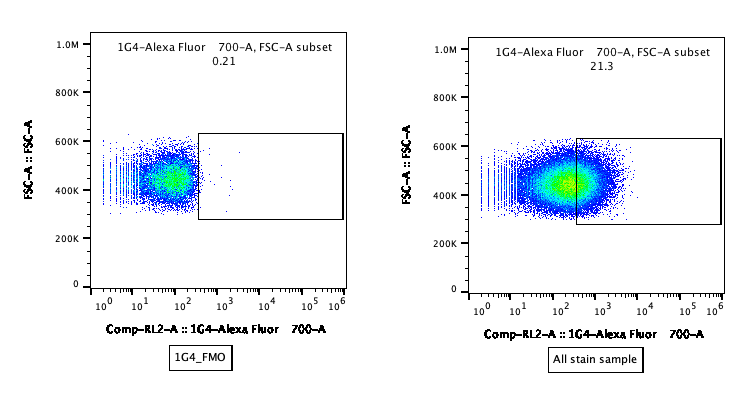
Application: Flow CytometrySample Tested: Mouse spleenocytesSpecies: MouseVerified Customer | Posted 08/03/20211G4-AF700 (1:600)

There are no reviews that match your criteria.
Protocols
View specific protocols for Ethenoadenosine Antibody (1G4) - BSA Free (NB600-442):
ICoating of Plates
DNA coating: DNA is dissolved in PBS at appropriate concentration. 0.1 ml is added/well and plates put in 37 degrees Celsius incubator to evaporate overnight. Alternatively, plates can be coated with a 2-fold higher concentration of DNA for 2 hrs at 37 degrees Celsius then used. Column 1 is not coated. These well will not be used for the assay (no blocking, no antibody and no secondary antibody) but will have substrate added for blanking the reader. Plates are stored in the refrigerator.
Protein coating: Proteins are dissolved in PBS at the appropriate concentration. 0.1 ml is added/well and plates put in 37 degrees Celsius incubator to evaporate overnight. Column 1 is again not coated. Plates are stored in the refrigerator.
An alternate protein coating condition is to dissolve the protein in 0.1 M sodium carbonate buffer pH 9.6. 0.1 ml is added/well and the plates are refrigerated for several hours or overnight. They cannot be used after 3 days.
1 M solution 1.59 g Na2CO3 + 2.93 g NaHCO3/100ml
II Assay
1. Label assay sheet and determine which rows are to be used. Row 1 (A-H) is not used; it will be used to blank the spectrophotometer. Avoid using the outer rows if possible (i.e.12A-H, H 1-12 and A 1-12.
2. Wash plate with wash buffer containing PBS-Tween and NaN3 3 x on each side (right side up and upside down). Shake out onto paper towel.
3. Add 0.2 ml/well of 1% FCS in wash buffer to block non specific binding. Solution of FCS should be made fresh.
4. Incubate 1 hr.
5. Preparation of inhibitor series (during incubation of plate with FCS). Calculate appropriate concentrations to give desired fmol/well=fmol/0.05 ml. Make serial dilutions by adding PBS or CT DNA to tubes followed by competitor.
6. Prepare antibody in 1% FCS washing buffer.
7. At end of incubation period, shake out solution from plate and tap onto paper towel to dry.
8. Add 0.05 ml of competitor to each well followed by 0.05 ml of diluted antibody. Be sure to run all controls including zero (no competitor), minus Ab (no antigen specific antibody but secondary antisera) and positive and negative controls.
9. Incubate for 90 min at 37 degrees Celsius.
10. Wash the plate with washing buffer 3 times on each side. Tap onto paper towels.
11. Secondary antisera - Use goat anti-mouse IgG-alkaline phosphatase for monoclonals and anti rabbit for polyclonals. Dilute as appropriate and add 0.1 ml/well.
12. Incubate for 90 min at 37 degrees Celsius.
13. Wash with wash buffer 3 x each side. Tap onto paper towel.
14. Wash plate 2 times with 0.01 M diethanolamine using the was bottle and covering the well completely each time. Tap onto paper towel. This step removes phosphate buffer which inhibits alkaline phosphatase activity.
15. Prepare the substrate - 2 tablets 95 mg/tablet) Sigma 104 in 10 ml 1 M diethanolamine, pH 8.6. Final concentration 1 mg/ml. Avoid physical contact of skin with the tablets since skin contains alkaline phosphatase. Add 0.1 ml/well
16. Incubate at 37 degrees Celsius and read absorbance at 405 nm. The absorbance of the 0 fmol standard should be between 0.5 and 1. Values above 2 are not usable since the reader may not be linear in this range.
Rinse water - One liter of H2O + 2 ml 10% NaN3
Wash buffer - One liter of 1 x PBS + 500 ul Tween 20 + 2 ml 10% NaN3
Blocking buffer - Wash buffer + 1% FCS
Find general support by application which include: protocols, troubleshooting, illustrated assays, videos and webinars.
- 7-Amino Actinomycin D (7-AAD) Cell Viability Flow Cytometry Protocol
- Appropriate Fixation of IHC/ICC Samples
- Cellular Response to Hypoxia Protocols
- ClariTSA™ Fluorophore Kits
- Detection & Visualization of Antibody Binding
- ELISA Sample Preparation & Collection Guide
- ELISA Troubleshooting Guide
- Extracellular Membrane Flow Cytometry Protocol
- Flow Cytometry Protocol for Cell Surface Markers
- Flow Cytometry Protocol for Staining Membrane Associated Proteins
- Flow Cytometry Staining Protocols
- Flow Cytometry Troubleshooting Guide
- How to Run an R&D Systems DuoSet ELISA
- How to Run an R&D Systems Quantikine ELISA
- How to Run an R&D Systems Quantikine™ QuicKit™ ELISA
- ICC Cell Smear Protocol for Suspension Cells
- ICC Immunocytochemistry Protocol Videos
- ICC for Adherent Cells
- Immunocytochemistry (ICC) Protocol
- Immunocytochemistry Troubleshooting
- Immunofluorescence of Organoids Embedded in Cultrex Basement Membrane Extract
- Immunohistochemistry (IHC) and Immunocytochemistry (ICC) Protocols
- Immunoprecipitation Protocol
- Intracellular Flow Cytometry Protocol Using Alcohol (Methanol)
- Intracellular Flow Cytometry Protocol Using Detergents
- Intracellular Nuclear Staining Flow Cytometry Protocol Using Detergents
- Intracellular Staining Flow Cytometry Protocol Using Alcohol Permeabilization
- Intracellular Staining Flow Cytometry Protocol Using Detergents to Permeabilize Cells
- Preparing Samples for IHC/ICC Experiments
- Preventing Non-Specific Staining (Non-Specific Binding)
- Primary Antibody Selection & Optimization
- Propidium Iodide Cell Viability Flow Cytometry Protocol
- Protocol for Liperfluo
- Protocol for VisUCyte™ HRP Polymer Detection Reagent
- Protocol for the Characterization of Human Th22 Cells
- Protocol for the Characterization of Human Th9 Cells
- Protocol for the Fluorescent ICC Staining of Cell Smears - Graphic
- Protocol for the Fluorescent ICC Staining of Cultured Cells on Coverslips - Graphic
- Protocol for the Preparation and Fluorescent ICC Staining of Cells on Coverslips
- Protocol for the Preparation and Fluorescent ICC Staining of Non-adherent Cells
- Protocol for the Preparation and Fluorescent ICC Staining of Stem Cells on Coverslips
- Protocol for the Preparation of a Cell Smear for Non-adherent Cell ICC - Graphic
- Protocol: Annexin V and PI Staining by Flow Cytometry
- Protocol: Annexin V and PI Staining for Apoptosis by Flow Cytometry
- Quantikine HS ELISA Kit Assay Principle, Alkaline Phosphatase
- Quantikine HS ELISA Kit Principle, Streptavidin-HRP Polymer
- R&D Systems Quality Control Western Blot Protocol
- Sandwich ELISA (Colorimetric) – Biotin/Streptavidin Detection Protocol
- Sandwich ELISA (Colorimetric) – Direct Detection Protocol
- TUNEL and Active Caspase-3 Detection by IHC/ICC Protocol
- The Importance of IHC/ICC Controls
- Troubleshooting Guide: ELISA
- Troubleshooting Guide: Fluorokine Flow Cytometry Kits
- Troubleshooting Guide: Western Blot Figures
- Western Blot Conditions
- Western Blot Protocol
- Western Blot Protocol for Cell Lysates
- Western Blot Troubleshooting
- Western Blot Troubleshooting Guide
- View all Protocols, Troubleshooting, Illustrated assays and Webinars