HIF-2 alpha/EPAS1 Antibody - BSA Free
Novus Biologicals | Catalog # NB100-122

Key Product Details
Validated by
Knockout/Knockdown, Orthogonal Validation, Biological Validation
Species Reactivity
Validated:
Human, Mouse, Rat, Fish, Hamster, Primate, Rabbit, Reptile, Sheep
Cited:
Human, Mouse, Rat, Fish - Danio rerio (Zebrafish), Hamster, Ovine, Rabbit, Reptile
Applications
Validated:
Knockout Validated, Immunohistochemistry, Immunohistochemistry-Paraffin, Immunohistochemistry-Frozen, Western Blot, Immunoblotting, ELISA, Flow Cytometry, Dual RNAscope ISH-IHC, Immunocytochemistry/ Immunofluorescence, Simple Western, Immunoprecipitation, Chromatin Immunoprecipitation (ChIP), In vitro assay, Gel Super Shift Assays, Knockdown Validated, SDS-Page
Cited:
Knockout Validated, Immunohistochemistry, Immunohistochemistry-Paraffin, Immunohistochemistry-Frozen, Western Blot, Immunoblotting, ELISA, Block/Neutralize, Flow Cytometry, Immunocytochemistry, Immunocytochemistry/ Immunofluorescence, Immunoprecipitation, Chromatin Immunoprecipitation (ChIP), Chemotaxis, Chip Cytometry, CoIP, In vivo assay, Gel Supershift Assay, IF/IHC, Knockdown Validated
Label
Unconjugated
Antibody Source
Polyclonal Rabbit IgG
Format
BSA Free
Loading...
Product Specifications
Immunogen
This HIF-2 alpha/EPAS1 Antibody was developed against a peptide derived from the C-terminus of mouse/human HIF-2 alpha protein.
Reactivity Notes
Use in Mouse reported in scientific literature (PMID:33758176).
Specificity
This HIF-2 alpha/EPAS1 Antibody is specific for HIF-2 alpha/EPAS, and does not cross-react with HIF-1 alpha.
Clonality
Polyclonal
Host
Rabbit
Isotype
IgG
Theoretical MW
96.5 kDa.
Disclaimer note: The observed molecular weight of the protein may vary from the listed predicted molecular weight due to post translational modifications, post translation cleavages, relative charges, and other experimental factors.
Disclaimer note: The observed molecular weight of the protein may vary from the listed predicted molecular weight due to post translational modifications, post translation cleavages, relative charges, and other experimental factors.
Scientific Data Images for HIF-2 alpha/EPAS1 Antibody - BSA Free


Dual RNAscope ISH-IHC Analysis of HIF-2 alpha/EPAS1 in Human Placenta
Formalin-fixed paraffin-embedded tissue sections of human placenta were probed for HIF-2 alpha/EPAS1 mRNA (ACD RNAScope Probe, catalog #410598; Fast Red chromogen, ACD catalog # 322750). Adjacent tissue section was processed for immunohistochemistry using Rabbit Polyclonal (Novus Biologicals catalog # NB100-122) at 1:100 dilution with one-hour incubation at room temperature followed by incubation with anti-rabbit IgG VisUCyte HRP Polymer Antibody (Catalog # VC003) and DAB chromogen (yellow-brown). Tissue was counterstained with hematoxylin (blue). Specific staining was localized to trophoblastic cells.
Immunohistological Staining of HIF-2 alpha/EPAS1 in Normal and Diseased Lung Tissue
HIF-2-alpha-EPAS1-Antibody-Immunohistochemistry-NB100-122-img0028.jpg
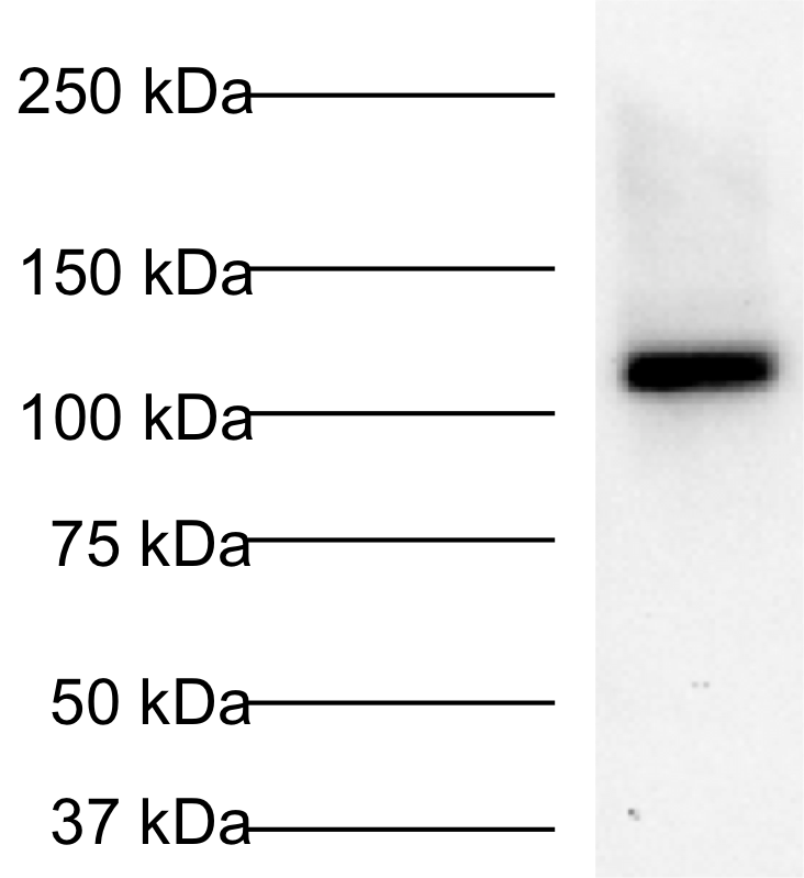
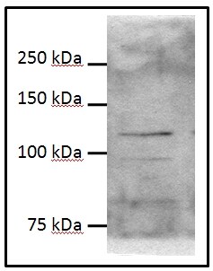
Simple Western Analysis of HIF-2 alpha in HeLa Cell Lysate
Image shows a specific band for HIF-2 alpha in 0.5 mg/mL of hypoxic HeLa lysate. This experiment was performed under reducing conditions using the 12-230 kDa separation system.


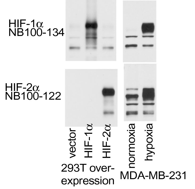
Chromatin Immunoprecipitation in MDA-MB-231 Cells Using HIF-2 alpha/EPAS1 Antibody
MDA-MB-231 cells were exposed to 20% or 1% O2 for 16 hours, and chromatin immunoprecipitation (ChIP) was performed with the indicated antibody (Ab). Primers flanking the HIF binding site were used for qPCR. ChIP image submitted by a verified customer review.

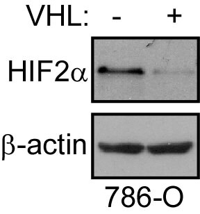
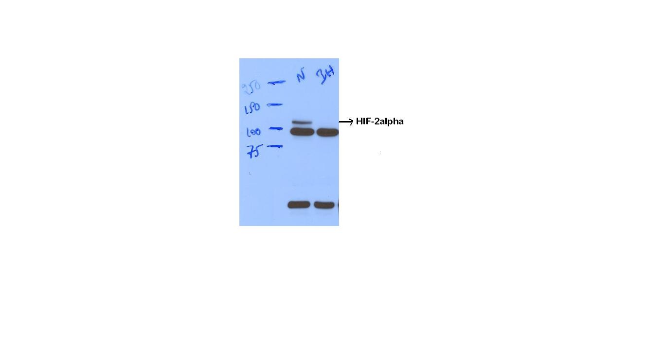
Western Blot Detection of HIF-2 alpha/EPAS1 in 786-O Cells With or Without VHL Overexpression
786-O cells without or with VHL overexpression. Image from verified customer review.

Immunohistological Staining of HIF-2 alpha/EPAS1 in Paraffin Embedded Human Cardiac Myocytes
HIF-2 alpha immunoreactivity in human cardiac myocytes stained with NB100-122.
Immunohistological Analysis of HIF-1 alpha, HIF-2 alpha/EPAS1 and HAF in ccRCC
Expression and correlation of 3 antibodies: HIF-1 alpha, HIF-2 alpha and HAF in ccRCC. (A, C & E) Positive nuclear staining to the 3 antibodies: HIF-1 alpha, HIF-2 alpha and HAF, in primary ccRCCs at high magnification (100X). (B, D & F) Heterogenous nuclear staining of 3 antibodies in a tumor at low magnification (20X). (G, H and I). Correlation of 3 antibodies with the Fuhrman grade.Citation: Ambrosetti D, Dufies M, Dadone B, Durand M, Borchiellini D, Amiel J, et al. (2018) The two glycolytic markers GLUT1 and MCT1 correlate with tumor grade and survival in clear-cell renal cell carcinoma. PLoS ONE 13(2): e0193477. https://doi.org/10.1371/journal.pone.0193477
Knockdown Validation of HIF-2 alpha/EPAS1 Antibody in 786-O Cells
Reduction of HIF-2alpha levels leads to protection in UV-triggered apoptosis, but not for apoptosis caused by glucose and serum starvation in 786-O cells. Parental 786-O or those either stably expressing wild-type VHLp19 or stably infected with a control vector (pSuperRetro) or a pool of two HIF-2alpha shRNAs vectors [21] were grown to confluence and lysed. Cell alpha-tubulin.
Knockdown Validated: HIF-2 alpha/EPAS1 Antibody - BSA Free [NB100-122] -
Sp1 and HIFs contribute to the synergistic activation of multiple genes in OVSAYO cells under SSH.b) Activation of multiple genes under hypoxia is dependent on HIFs. Western blotting is also shown for HIFs. Data shown are the mean (n = 3) ± SD.
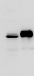
Western Blot: Rabbit Polyclonal HIF-2 alpha/EPAS1 Antibody [NB100-122] -
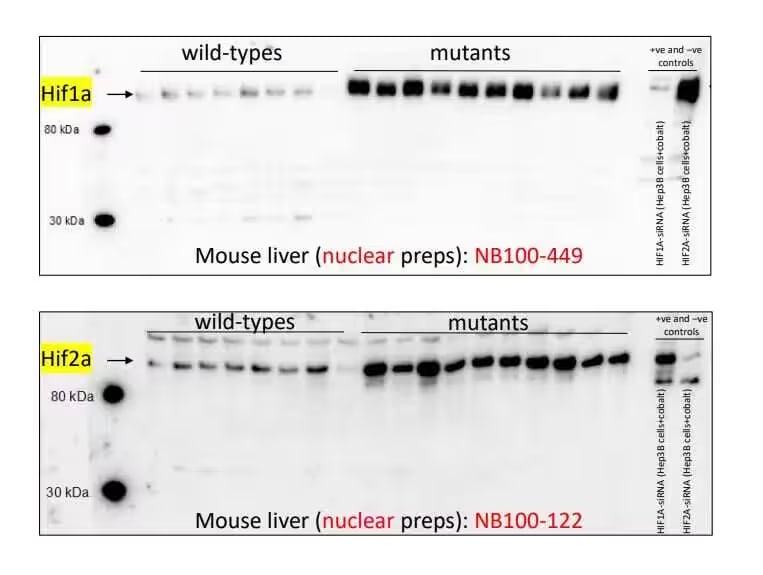
Western Blot: Rabbit Polyclonal HIF-2 alpha/EPAS1 Antibody [NB100-122] - Analysis of HIF-2 alpha/EPAS1 antibody on mouse liver nuclear extracts. Image from a verified customer review.
Immunocytochemistry/ Immunofluorescence: HIF-2 alpha/EPAS1 Antibody - BSA Free [NB100-122] -
Immunocytochemistry/ Immunofluorescence: HIF-2 alpha/EPAS1 Antibody - BSA Free [NB100-122] - Muscle repair following eccentric contraction–induced injury is concomitant with dynamic alterations of HIF2A & HIF1A expression in SCs.(A–C) Representative images of EDL myofibers from injured muscles at various time points (n >50 myofibers from 3 mice/group/time point) & stained for Pax7, DAPI, & EdU (A), HIF2A (B), or HIF1A (C). Scale bars: 20 μm. Arrowheads indicate SCs. (D) Number of Pax7+ SCs per myofiber at various time points. (E) Percentage of EdU+ SCs at various time points. (F) Percentage of HIF2A+ SCs at various time points. (G) Percentage of HIF1A+ SCs at various time points. Data represent the mean ± SEM. Image collected & cropped by CiteAb from the following publication (https://www.jci.org/articles/view/96208), licensed under a CC-BY license. Not internally tested by Novus Biologicals.
Immunocytochemistry/ Immunofluorescence: HIF-2 alpha/EPAS1 Antibody - BSA Free [NB100-122] -
Immunocytochemistry/ Immunofluorescence: HIF-2 alpha/EPAS1 Antibody - BSA Free [NB100-122] - Genetic ablation of HIF2A in QSCs leads to transient activation, proliferation, & differentiation of SCs.(B) Representative images of myofibers from SC-HIF2AKO mice & control littermates (n >50 myofibers from 5 mice/group; 10 dpr). Immunofluorescence of Pax7 (red), HIF2A (green), MyoD (purple), & DAPI (blue) staining revealed HIF2A–MyoD+ & HIF2A+MyoD– SCs (arrowheads) in SC-HIF2AKO & control mice, respectively. Scale bar: 10 μm. Image collected & cropped by CiteAb from following publication (https://www.jci.org/articles/view/96208), licensed under a CC-BY license. Not internally tested by Novus Biologicals.
Immunocytochemistry/ Immunofluorescence: HIF-2 alpha/EPAS1 Antibody - BSA Free [NB100-122] -
Immunocytochemistry/ Immunofluorescence: HIF-2 alpha/EPAS1 Antibody - BSA Free [NB100-122] - EpEX & EpCAM activate a STAT3-HIF2 alpha signal for EpEX/EpCAM-mediated iPSC formation.(A) iPSCs were infected with two different EpCAM shRNAs (two clones, #1 & #2). The protein expressions of EpCAM & HIF2 alpha were detected by Western blotting. (B) MEFs were stimulated by EpEX (1 μg/mL) at the indicated times. Nuclear-translocation was detected with a specific antibody against HIF2 alpha (n = 3). (C) Immunofluorescence staining was performed to detect subcellular localization of HIF2 alpha. Nuclei were stained with DAPI. Scale bar: 10 μm. (D) MEFs were treated with STAT3 inhibitor (WP1066, 10 μM), & then stimulated with EpEX for 30 min. The nuclear-translocation of HIF2 alpha was detected by Western blotting with anti-HIF2 alpha antibody (n = 3). (E) iPSC morphology was observed at day 20 after induction. Reprogramming of Oct4-GFP MEFs was induced by transfection of OSKM, OE + EpEX, & KE + EpEX with or without STAT3 inhibitor WP1066, or HIF2 alpha shRNA (n = 3). Scale bar: 50 μm. Image collected & cropped by CiteAb from the following publication (https://www.nature.com/articles/srep41852), licensed under a CC-BY license. Not internally tested by Novus Biologicals.
Gel Super Shift Assays: HIF-2 alpha/EPAS1 Antibody - BSA Free [NB100-122] -
Gel Super Shift Assays: HIF-2 alpha/EPAS1 Antibody - BSA Free [NB100-122] - Manipulation of DNA methylation at HRE sequences alters HIF binding. (A) The Hypoxia Response Element (HRE) contains a CpG site that can be methylated which prevents HIF1 & HIF2 binding in vitro. EMSA of in vitro–translated HIF1 & HIF2 binding to 32P-labelled unmethylated & methylated HRE probes. Competition with 250X molar excess of unlabelled/unmethylated HRE probe (lanes 3, 7,12, 16) or unlabelled control HRE-free probe (lanes 4, 8, 13, 17). HIF1 & HIF2 complexes bound to unmethylated HRE or methylated HRE supershifted with anti-HIF1 alpha (lanes 9 & 18) & anti-HIF2 alpha (lanes 5 & 14). (B) RCC4-VHL cells were grown in normoxia (NT) or treated with 25 µM decitabine (5aza) or grown in hypoxia (1%) for 48 hrs & with the addition of 25 µM decitabine (1% + 5aza). QPCR was performed using primers specific to VEGF, uPAR, TGF alpha, GUS, U1AsnRNP1. Samples were normalised to relative expression of the housekeeping gene, ACTIN. Image collected & cropped by CiteAb from the following publication (https://www.nature.com/articles/s41598-018-21524-5), licensed under a CC-BY license. Not internally tested by Novus Biologicals.
Immunocytochemistry/ Immunofluorescence: HIF-2 alpha/EPAS1 Antibody - BSA Free [NB100-122] -
Immunocytochemistry/ Immunofluorescence: HIF-2 alpha/EPAS1 Antibody - BSA Free [NB100-122] - HIF-1 alpha /2 alpha expression in myeloid-specific KO mice targeting the HIF pathway. (A) Images of the colon of wild-type (WT), myeloid-specific Hif-1a KO (hMRP8 Hif-1a KO) or von Hippel Lindau (Vhl) KO (hMRP8 Vhl KO) mice, immunostained for MRP8 (green) & the DNA-binding regions of Hif-1a mRNA (red). Mice were fed with 5% DSS for 4 days prior to immunostaining analyses. Note that there were no MRP8-positive cells that were positive for Hif-1a mRNA in hMRP8 Hif-1a KO (middle column) mice, but we observed many cells that were double positive for MRP8 & Hif-1a mRNA in hMRP8 Vhl KO mice (right column). (B) Images of the colon of hMRP8 Vhl KO mice fed with 5% DSS as in A, immunostained for MRP8 (green) & HIF-1 alpha (red, upper row) or HIF-2 alpha (red, bottom row). DAPI-stained nuclei are shown in blue. White boxes in A & B indicate the regions magnified in the lower or right images, respectively. Yellow arrowheads in A & B indicate cells positive for both markers. Scale bars: 100 μm. Image collected & cropped by CiteAb from the following publication (https://pubmed.ncbi.nlm.nih.gov/29967068), licensed under a CC-BY license. Not internally tested by Novus Biologicals.
Immunocytochemistry/ Immunofluorescence: HIF-2 alpha/EPAS1 Antibody - BSA Free [NB100-122] -
Immunocytochemistry/ Immunofluorescence: HIF-2 alpha/EPAS1 Antibody - BSA Free [NB100-122] - HIF2 alpha stabilization results in exocrine cell atrophy & expansion of duct-like tubular structures. (A) Body weight (left panel), pancreas weight (middle panel) & body/pancreas weight ratio (right panel) in Pdx1-Cre;HIF2dPA & control mice at 2 & 8 weeks of age. Data are presented as mean ± SD. (B) HIF2 alpha accumulation in Pdx1-Cre;HIF2dPA analyzed by Western blot with anti-HA antibody. Two independent two-week-old control & mutant mice are shown. beta -actin protein was used for loading control. Full-length blots are presented in Supplementary Fig. 2. (D) Immunofluorescence analysis of HIF2 alpha in two-week-old control pancreata. Endogenous HIF2 alpha expression is observed in islets (marked by an white asterisk) but not in exocrine tissue. (D) Robust HIF2 alpha accumulation in the pancreas of two-week-old Pdx1-Cre;HIF2dPA mice. Hematoxylin/Eosin-stained pancreatic sections from P0 (E,F), two- (I,J) & eight-week-old (M,N) Pdx1-Cre;HIF2dPA & control mice. Inset in N shows an area with adipose tissue in Pdx1-Cre;HIF2dPA pancreata. Immunofluorescence of amylase & KRT19 shows no differences between Pdx1-Cre;HIF2dPA & control mice at P0 (G,H). Duct-like tubular structures & loss of amylase immunoreactivity in two- (K,L) & eight-week-old (O,P) Pdx1-Cre;HIF2dPA mice compared to control mice. Note areas with normal acini in 8-week-old Pdx1-Cre;HIF2dPA mice (white asterisk in O). Insets in (H,L & P) show higher magnification pictures. DAPI staining is shown in blue in (C,D,G,H,K,L,O & P). Scale bars = 50 µm for (C,D); 100 µm for (E–P). ***P < 0.001. Image collected & cropped by CiteAb from the following publication (https://pubmed.ncbi.nlm.nih.gov/30209343), licensed under a CC-BY license. Not internally tested by Novus Biologicals.
Immunocytochemistry/ Immunofluorescence: HIF-2 alpha/EPAS1 Antibody - BSA Free [NB100-122] -
Immunocytochemistry/ Immunofluorescence: HIF-2 alpha/EPAS1 Antibody - BSA Free [NB100-122] - QSCs are hypoxic in the niche & express HIF2A, but not HIF1A.(A) Timeline of in vivo pimonidazole labeling in SC-INTACT mice & representative confocal images of uninjured/resting EDL myofibers (n >50 myofibers from n = 3 mice) showing that nmGFP+ QSCs were pimonidazole+. Scale bars: 50 μm & 10 μm (insets). Inset images show that pimonidazole signals were relatively enriched in the cytoplasm of QSCs. Arrowheads indicate a QSC; asterisks indicate a myonucleus. (B) Percentage of pimonidazole+ QSCs. (C) Timeline of in vivo CCI-103F labeling in C57BL/6 mice & representative images of uninjured/resting EDL myofibers (n >50 myofibers from 3 mice) showing that nmGFP+ QSCs were CCI-103F+. Arrowheads indicate a QSC; asterisks indicate a myonucleus. Scale bar: 20 μm. (D) Percentage of CCI-103F+ QSCs. (E & F) Representative images of uninjured/resting EDL myofibers from C57BL/6 mice (n >50 myofibers from 6 mice/group) showing that most QSCs were HIF2A+, but HIF1A–. Scale bars: 10 μm. (G) Percentage of HIF1A+ & HIF2A+ QSCs. Data represent the mean ± SEM. Image collected & cropped by CiteAb from the following publication (https://www.jci.org/articles/view/96208), licensed under a CC-BY license. Not internally tested by Novus Biologicals.Applications for HIF-2 alpha/EPAS1 Antibody - BSA Free
Application
Recommended Usage
Chromatin Immunoprecipitation (ChIP)
1:10-1:500
ELISA
1:100 - 1:2000
Gel Super Shift Assays
reported in scientific literature (PMID 15184875)
Immunoblotting
reported in scientific literature (PMID 28115701)
Immunocytochemistry/ Immunofluorescence
1:100
Immunohistochemistry
1:100
Immunohistochemistry-Paraffin
1:100
Immunoprecipitation
5 ug / 1 mg lysate
In vitro assay
reported in scientific literature (PMID 24998849)
Knockdown Validated
reported in scientific literature (PMID 31061092)
Knockout Validated
reported in scientific literature (PMID 26861754)
Simple Western
1:50
Western Blot
1 - 2 ug/mL
Application Notes
In WB, this antibody recognizes a band at 118 kDa representing HIF-2 alpha.
See Simple Western Antibody Database for Simple Western validation: tested in hypoxic HeLa lysate (0.5 mg/ml); separated by Size- Wes/Sally Sue/Peggy Sue; antibody dilution of 1:50; apparent MW in kDa on Simple Western was 110kDa; matrix was 12-230 kDa.
See Simple Western Antibody Database for Simple Western validation: tested in hypoxic HeLa lysate (0.5 mg/ml); separated by Size- Wes/Sally Sue/Peggy Sue; antibody dilution of 1:50; apparent MW in kDa on Simple Western was 110kDa; matrix was 12-230 kDa.
Reviewed Applications
Read 39 reviews rated 4.4 using NB100-122 in the following applications:
Flow Cytometry Panel Builder
Bio-Techne Knows Flow Cytometry
Save time and reduce costly mistakes by quickly finding compatible reagents using the Panel Builder Tool.
Advanced Features
- Spectra Viewer - Custom analysis of spectra from multiple fluorochromes
- Spillover Popups - Visualize the spectra of individual fluorochromes
- Antigen Density Selector - Match fluorochrome brightness with antigen density

Formulation, Preparation, and Storage
Purification
Immunogen affinity purified
Formulation
PBS
Format
BSA Free
Preservative
0.05% Sodium Azide
Concentration
1.0 mg/ml
Shipping
The product is shipped with polar packs. Upon receipt, store it immediately at the temperature recommended below.
Stability & Storage
Store at -20 °C.
Background: HIF-2 alpha/EPAS1
HIF-1 or hypoxia inducible factor 1, is a transcription factor commonly referred to as a "master regulator of the hypoxic response" for its central role in the regulation of cellular adaptations to hypoxia. Similarly, HIF-2 alpha plays a role in cellular responses to hypoxia, but whereas HIF-1 alpha is ubiquitously expressed, HIF-2 alpha is predominantly expressed in the vascular endothelium at embryonic stages and after birth in select cells and tissue types (e.g., fibroblasts, hepatocytes and myocytes at 96kDa) (4). Following a similar mechanism to HIF-1 alpha, HIF-2 alpha is stabilized under hypoxic conditions by the formation of a heterodimer with an ARNT/HIF-1 beta subunit. Stable HIF-2 alpha-ARNT/HIF-1 beta heterodimers engage p300/CBP in the nucleus for binding to hypoxic response elements (HREs), inducing transcription, and thus regulation of genes (e.g., EPO, VEGFA). HIF-1 predominantly transactivates genes involved in glycolytic control and pro- apoptotic genes (e.g., LDHA and BNIP3), and HIF-2 regulates the expression of genes involved in invasion and stemness (e.g., MMP2, and OCT4). Common gene targets for HIF-1 and HIF-2 include VEGFA and GLUT1 (5).
The HIF-2 alpha subunit is rapidly targeted and degraded by the ubiquitin proteasome system under normoxic conditions. This process is mediated by oxygen-sensing enzymes, prolyl hydroxylase domain enzymes (PHDs), which catalyze the hydroxylation of key proline residues (Pro-405 and Pro-531) within the oxygen-dependent degradation domain of HIF-2 alpha (5). Once hydroxylated, HIF-2 alpha binds the von Hippel-Lindau tumor suppressor protein (pVHL) for subsequent ubiquitination and proteasomal degradation (5,6).
References
1. Semenza, G. L., Agani, F., Feldser, D., Iyer, N., Kotch, L., Laughner, E., & Yu, A. (2000). Hypoxia, HIF-1, and the pathophysiology of common human diseases. Advances in Experimental Medicine and Biology.
2.Muz, B., de la Puente, P., Azab, F., & Azab, A. K. (2015). The role of hypoxia in cancer progression, angiogenesis, metastasis, and resistance to therapy. Hypoxia. https://doi.org/10.2147/hp.s93413
3. Huang, Y., Lin, D., & Taniguchi, C. M. (2017). Hypoxia inducible factor (HIF) in the tumor microenvironment: friend or foe? Science China Life Sciences. https://doi.org/10.1007/s11427-017-9178-y
4. Hu, C.-J., Wang, L.-Y., Chodosh, L. A., Keith, B., & Simon, M. C. (2003). Differential Roles of Hypoxia-Inducible Factor 1 (HIF-1) and HIF-2 in Hypoxic Gene Regulation. Molecular and Cellular Biology. https://doi.org/10.1128/mcb.23.24.9361-9374.2003
5. Koh, M. Y., & Powis, G. (2012). Passing the baton: The HIF switch. Trends in Biochemical Sciences. https://doi.org/10.1016/j.tibs.2012.06.004
6. Koyasu, S., Kobayashi, M., Goto, Y., Hiraoka, M., & Harada, H. (2018). Regulatory mechanisms of hypoxia-inducible factor 1 activity: Two decades of knowledge. Cancer Science. https://doi.org/10.1111/cas.13483
Long Name
Hypoxia-inducible Transcription Factor 2 alpha
Alternate Names
EPAS1, HIF 2A, HIF2 alpha, HIF2A, HLF, MOP2, anti hif 2 alpha, anti-HIF-2 alpha
Gene Symbol
EPAS1
Additional HIF-2 alpha/EPAS1 Products
Product Documents for HIF-2 alpha/EPAS1 Antibody - BSA Free
Certificate of Analysis
To download a Certificate of Analysis, please enter a lot or batch number in the search box below.
Product Specific Notices for HIF-2 alpha/EPAS1 Antibody - BSA Free
This product is for research use only and is not approved for use in humans or in clinical diagnosis. Primary Antibodies are guaranteed for 1 year from date of receipt.
Citations for HIF-2 alpha/EPAS1 Antibody - BSA Free
 Powered by Bioz
Customer Reviews for HIF-2 alpha/EPAS1 Antibody - BSA Free (39)
4.4 out of 5
39 Customer Ratings
Have you used HIF-2 alpha/EPAS1 Antibody - BSA Free?
Submit a review and receive an Amazon gift card!
$25/€18/£15/$25CAN/¥2500 Yen for a review with an image
$10/€7/£6/$10CAN/¥1110 Yen for a review without an image
Submit a review
Customer Images


-(01-ml)_NB100-122_10076.gif)
-(01-ml)_NB100-122_9741.jpg)
-(01-ml)_NB100-122_7241.jpg)
-(01-ml)_NB100-122_6841.jpg)
Showing
1
-
5 的
39 reviews
Showing All
Filter By:
-
Application: Western BlotSample Tested: mouse liverSpecies: MouseVerified Customer | Posted 01/11/2024Must be nuclear extracts!Nuclear extracts using Nuclear and Cytoplasmic Extraction Reagents

-
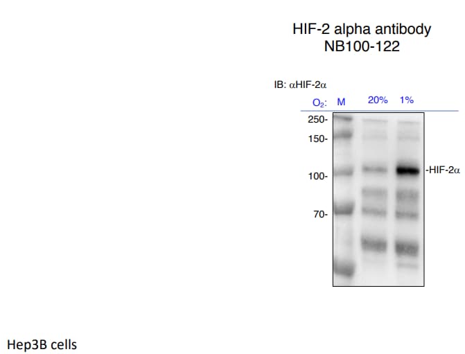
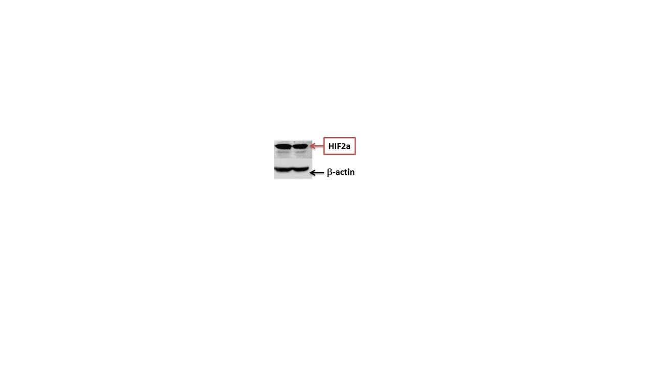
Application: Western BlotSample Tested: Hep3B human hepatocellular carcinoma cell lineSpecies: HumanVerified Customer | Posted 10/05/2022HIF-2alpha expression in normoxia vs hypoxia
-
Application: Western BlotSample Tested: Hep3B human hepatocellular carcinoma cell lineSpecies: HumanVerified Customer | Posted 03/15/2021HIF-2alpha expression in Hep3B cells exposed to 24 hours of hypoxia
-
Application: Western BlotSample Tested: Human cancer cell linesSpecies: HumanVerified Customer | Posted 07/05/2019
-
Application: Western BlotSample Tested: HBE-16Species: HumanVerified Customer | Posted 04/16/2019HIF2a
-
Application: Western BlotSample Tested: cultured cellsSpecies: HumanVerified Customer | Posted 02/04/2019
-
Application: Western BlotSample Tested: 786O cell lysatesSpecies: HumanVerified Customer | Posted 02/02/2019
-
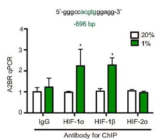
Application: Chromatin ImmunoprecipitationSample Tested: MCF7 cell lineSpecies: HumanVerified Customer | Posted 12/02/2018MCF7 were exposed to 20% or 1% O2 for 16h, and ChIP assays were performed using IgG or antibodies against HIF-1a, HIF-2a, HIF-1b.
-
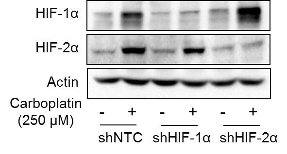
Application: Western BlotSample Tested: Human Cancer CellsSpecies: HumanVerified Customer | Posted 09/18/2018This antibody was used to detect HIF-2alpha expression in control (NTC) and HIF-2alpha knockdown subclone in response to carboplatin treatment in human breast cancer cell line by western blot.
-
Application: Western BlotSample Tested: 293T HEK cell lysateSpecies: HumanVerified Customer | Posted 05/11/2018Immunoblot showing HIF2 expression in HIF2 over-expressed HEK 293T cells.Antibody dilution- 1:500 in 5% BSA- overnight 4 degerees
-
Application: Western BlotSample Tested: transfected U2OS cell lysateSpecies: MouseVerified Customer | Posted 01/12/2017
-
Application: Western BlotSample Tested: cultured cellsSpecies: HumanVerified Customer | Posted 01/12/2017
-
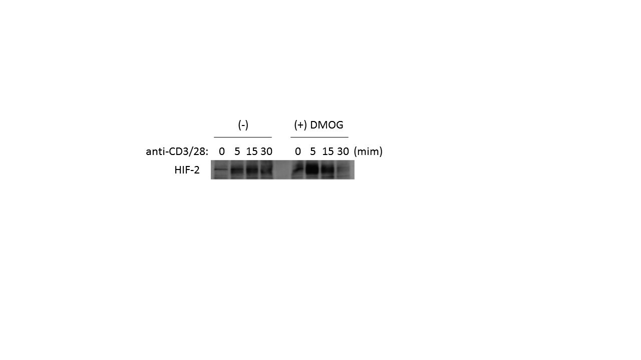
Application: Western BlotSample Tested: Mouse CD4+ T CellsSpecies: MouseVerified Customer | Posted 10/18/2016Dilution (1:1000)
-
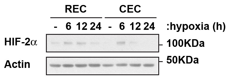
Application: Western BlotSample Tested: Retinal and Choroidal primary endotheliaSpecies: HumanVerified Customer | Posted 07/11/2016Endogenous HIF-2alpha in Retinal and Choroidal Endothelial Cells
-
Application: Western BlotSample Tested: HCT116 cell line lysateSpecies: HumanVerified Customer | Posted 03/31/2016
-
Application: Western BlotSample Tested: cytoplasmic and nuclear protein fractions of U266 cellsSpecies: HumanVerified Customer | Posted 11/10/2015HIF2-Alpha Expression in cytoplasmic and nuclear fractions of U266 cells
-
Application: Western BlotSample Tested: Primary Epithelial CellsSpecies: HumanVerified Customer | Posted 08/12/2015Human Epithelial Cells treated in hypoxia (0.5%)
-
Application: Western BlotSample Tested: MDA-MB-231 Cell LysateSpecies: HumanVerified Customer | Posted 06/01/2015overexpression and endogenous HIF1a & HIF2a
-
Application: Western BlotSample Tested: 786O cell lysatesSpecies: HumanVerified Customer | Posted 03/27/2015
-
Application: Chromatin ImmunoprecipitationSample Tested: See PMID:24248342Species: HumanVerified Customer | Posted 12/12/2014
-
Application: Western BlotSample Tested: See PMID:24308012Species: HumanVerified Customer | Posted 12/12/2014
-
Application: Western BlotSample Tested: HEK293 cell lysateSpecies: HumanVerified Customer | Posted 09/12/2014HEK293
-
Application: Western BlotSample Tested: Clear cell renal cell carcinoma samplesSpecies: HumanVerified Customer | Posted 08/30/2014Hif2a protein expression in clear cell renal cell carcinoma samples
-
Application: Western BlotSample Tested: PC12 whole cell lysateSpecies: OtherVerified Customer | Posted 07/26/2014
-
Application: Western BlotSample Tested: Whole cell lysateSpecies: HumanVerified Customer | Posted 06/30/2014
-
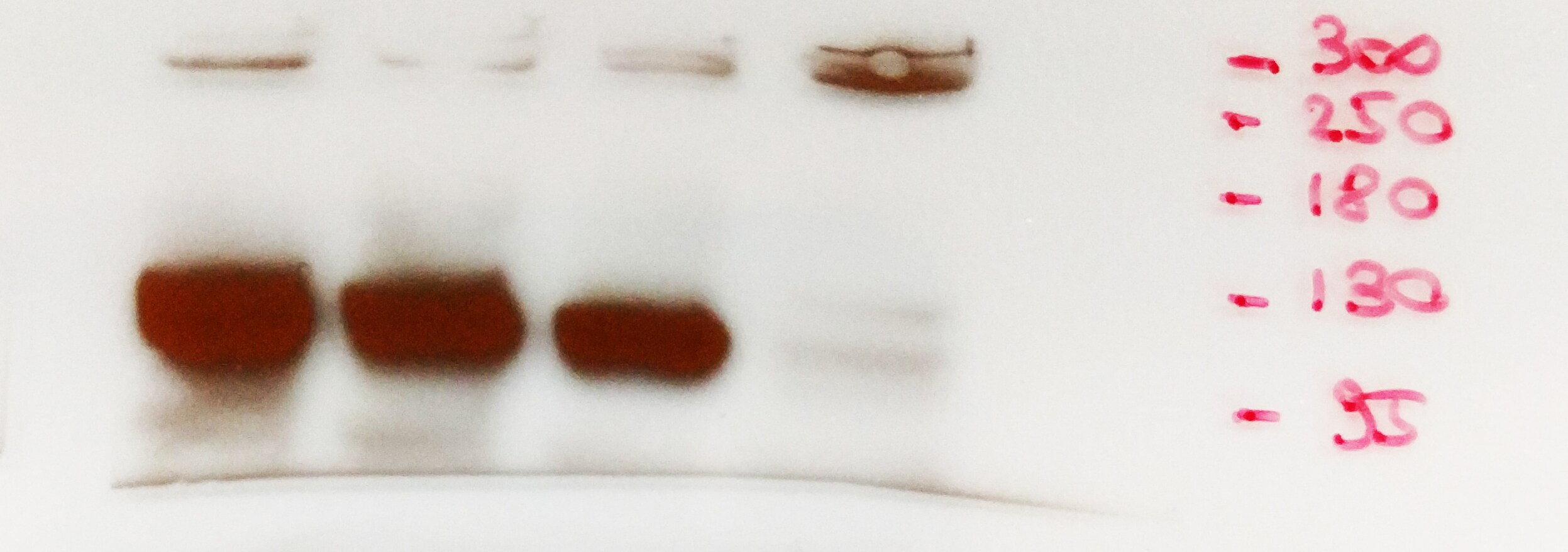
Application: Western BlotSample Tested: whole cell lysate U87MG (human glioblastoma cells)Species: HumanVerified Customer | Posted 04/30/2014Western blot detection HIF-2a (U87MG cells)
-
Application: Immunohistochemistry-ParaffinSample Tested: Mouse IntestineSpecies: MouseVerified Customer | Posted 04/11/2014Intestinal epithelium of a mouse
-
Application: Western BlotSample Tested: SH-SY5Y whole cell lysateSpecies: OtherVerified Customer | Posted 04/04/2014
-
Application: Western BlotSample Tested:Species: HumanVerified Customer | Posted 04/03/2014Western blot of 786-O cells without or with VHL overexpression
-
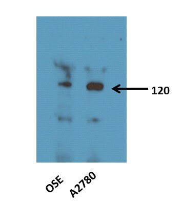
Application: Western BlotSample Tested: UNTREATED OVARIAN SURFACE EPITHELIUM CELLS (OSE) AND A2780 CELLS LYSATES 25ugSpecies: HumanVerified Customer | Posted 01/24/2014Western Blot: HIF-2 alpha Antibody [NB100-122]
-
Application: Western BlotSample Tested: Human Glioma cells, whole cell lysatesSpecies: HumanVerified Customer | Posted 11/20/2013
-
Application: ImmunofluorescenceSample Tested:Species: MouseVerified Customer | Posted 04/15/2013IF for HIF-2alpha(green)/DAPI(blue) in mouse ES cell-derived embryoid bodies
-
Application: Chromatin ImmunoprecipitationSample Tested:Species: RatVerified Customer | Posted 04/02/2013
-
Application: Western BlotVerified Customer | Posted 09/19/2012
-
Application: Western BlotSample Tested: MouseSpecies: MouseVerified Customer | Posted 12/08/2011
-
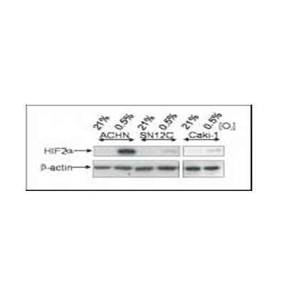
Application: Western BlotSample Tested: ACHN, SN12C, Caki1 whole cell lysates, Sample Amount: 30ugSpecies: HumanVerified Customer | Posted 06/15/2011
-
Application: Western BlotSample Tested: Colon Cancer Cell llines, Sample Amount: 50ugSpecies: OtherVerified Customer | Posted 03/11/2010
-
Application: Western BlotSample Tested: head and neck squamous cell carcinoma cells, Sample Amount: 35 ugSpecies: OtherVerified Customer | Posted 02/09/2009
-
Application: Immunohistochemistry-ParaffinSample Tested: human melanomaSpecies: HumanVerified Customer | Posted 02/02/2009
There are no reviews that match your criteria.
Protocols
View specific protocols for HIF-2 alpha/EPAS1 Antibody - BSA Free (NB100-122):
HIF-2 alpha/EPAS1 Antibody:
Immunocytochemistry Protocol
Culture cells to appropriate density in 35 mm culture dishes or 6-well plates.
1. Remove culture medium and wash the cells briefly in PBS. Add 10% formalin to the dish and fix at room temperature for 10 minutes.
2. Remove the formalin and wash the cells in PBS.
3. Permeablize the cells with 0.1% Triton X100 or other suitable detergent for 10 min.
4. Remove the permeablization buffer and wash three times for 10 minutes each in PBS. Be sure to not let the specimen dry out.
5. To block nonspecific antibody binding, incubate in 10% normal goat serum from 1 hour to overnight at room temperature.
6. Add primary antibody at appropriate dilution and incubate overnight at 4C.
7. Remove primary antibody and replace with PBS. Wash three times for 10 minutes each.
8. Add secondary antibody at appropriate dilution. Incubate for 1 hour at room temperature.
9. Remove secondary antibody and replace with PBS. Wash three times for 10 minutes each.
10. Counter stain DNA with DAPi if required.
General considerations for Western blot analysis of HIF-alpha proteins
1. HIF-2alpha is degraded under normoxic conditions and it is stabilized at O2 concentrations below 5% or with treatment using certain agents (CoCl2, DFO, etc.).
2. Positive and negative controls should always be run side by side in a Western blot to accurately identify the protein band upregulated in the hypoxic sample.
3. (HepG2 Hypoxic (CoCl2)/Normoxic Cell Lysate: NBP2-36451; HepG2 Hypoxic/Normoxic Cell Lysate: NBP2-36453).
4. To accurately compare treated and untreated samples and to ensure equal loading of samples the expression of a loading control should be evaluated.
(alpha Tubulin Antibody (DM1A): NB100-690)
5. The fully post-translationally modified form of HIF-2alpha is ~118 kDa, or larger.
6. HIF-2alpha may form a heterodimer with HIF-1beta. However, this is not typically seeing under denaturing conditions.
Western Blot Protocol
Materials
1x Laemmli Sample Buffer: 2% SDS, 2.5% 2-mercaptoethanol (bME), 25% glycerol, 0.01% bromophenol blue, 62.5 mM Tris HC, pH 6.8
1X Running Buffer: 25 mM Tris-base, 192 mM glycine, 0.1% SDS. Adjust to pH 8.3
1X Transfer buffer (wet): 25 mM Tris-base, 192 mM glycine, 20% methanol.
1X TBS
TBST (1X TBS with 0.1% Tween-20)
Blocking solution: TBST with 5% non-fat dry milk
Rabbit polyclonal anti-HIF-2 alpha primary antibody (NB100-122) in blocking solution (~1-2 ug/mL)
Methods
Whole-Cell Lysates
1. Load samples of treated and untreated cell lysates, 10-40 mg of total protein per lane on a 7.5% polyacrylamide gel (SDS-PAGE). Alternatively, gradient gels can be used for better resolution of lower molecular weight loading controls.
2. Resolve proteins by electrophoresis as required.
3. Transfer proteins to 0.45 mm PVDF membrane for 1 hour at 100V or equivalent.
4. Stain the blot using Ponceau S for 1-2 minutes to confirm efficient protein transfer onto the membrane.
5. Rinse the blot in distilled water to remove excess stain and mark the lanes and locations of molecular weight markers using a pencil.
6. Block the membrane using Blocking solution for 1 hour.
7. Dilute the rabbit anti-HIF-2 alpha primary antibody (NB100-122) in blocking solution (1-2 ug/ml) and incubate 1 hour at room temperature or overnight at 4oC.
8. Wash the membrane 3X 10 min in TBST.
9. Incubate in the appropriate diluted rabbit-IgG HRP-conjugated secondary antibody in blocking solution (as per manufacturer's instructions) for 1 hour at room temperature.
10. Wash the membrane 3X10 min in TBST.
11. Apply the detection reagent of choice in accordance with the manufacturer's instructions (e.g., ECL, ECL Plus).
Image blot.
Find general support by application which include: protocols, troubleshooting, illustrated assays, videos and webinars.
- 7-Amino Actinomycin D (7-AAD) Cell Viability Flow Cytometry Protocol
- Antigen Retrieval Protocol (PIER)
- Antigen Retrieval for Frozen Sections Protocol
- Appropriate Fixation of IHC/ICC Samples
- Cellular Response to Hypoxia Protocols
- ChIP Protocol Video
- Chromatin Immunoprecipitation (ChIP) Protocol
- Chromatin Immunoprecipitation Protocol
- Chromogenic IHC Staining of Formalin-Fixed Paraffin-Embedded (FFPE) Tissue Protocol
- Chromogenic Immunohistochemistry Staining of Frozen Tissue
- ClariTSA™ Fluorophore Kits
- Detection & Visualization of Antibody Binding
- ELISA Sample Preparation & Collection Guide
- ELISA Troubleshooting Guide
- Extracellular Membrane Flow Cytometry Protocol
- Flow Cytometry Protocol for Cell Surface Markers
- Flow Cytometry Protocol for Staining Membrane Associated Proteins
- Flow Cytometry Staining Protocols
- Flow Cytometry Troubleshooting Guide
- Fluorescent IHC Staining of Frozen Tissue Protocol
- Graphic Protocol for Heat-induced Epitope Retrieval
- Graphic Protocol for the Preparation and Fluorescent IHC Staining of Frozen Tissue Sections
- Graphic Protocol for the Preparation and Fluorescent IHC Staining of Paraffin-embedded Tissue Sections
- Graphic Protocol for the Preparation of Gelatin-coated Slides for Histological Tissue Sections
- How to Run an R&D Systems DuoSet ELISA
- How to Run an R&D Systems Quantikine ELISA
- How to Run an R&D Systems Quantikine™ QuicKit™ ELISA
- ICC Cell Smear Protocol for Suspension Cells
- ICC Immunocytochemistry Protocol Videos
- ICC for Adherent Cells
- IHC Sample Preparation (Frozen sections vs Paraffin)
- ISH-IHC Protocol for Chromogenic Detection on Formalin Fixed Paraffin Embedded (FFPE) Tissue
- Immunocytochemistry (ICC) Protocol
- Immunocytochemistry Troubleshooting
- Immunofluorescence of Organoids Embedded in Cultrex Basement Membrane Extract
- Immunofluorescent IHC Staining of Formalin-Fixed Paraffin-Embedded (FFPE) Tissue Protocol
- Immunohistochemistry (IHC) and Immunocytochemistry (ICC) Protocols
- Immunohistochemistry Frozen Troubleshooting
- Immunohistochemistry Paraffin Troubleshooting
- Immunoprecipitation Protocol
- Intracellular Flow Cytometry Protocol Using Alcohol (Methanol)
- Intracellular Flow Cytometry Protocol Using Detergents
- Intracellular Nuclear Staining Flow Cytometry Protocol Using Detergents
- Intracellular Staining Flow Cytometry Protocol Using Alcohol Permeabilization
- Intracellular Staining Flow Cytometry Protocol Using Detergents to Permeabilize Cells
- Preparing Samples for IHC/ICC Experiments
- Preventing Non-Specific Staining (Non-Specific Binding)
- Primary Antibody Selection & Optimization
- Propidium Iodide Cell Viability Flow Cytometry Protocol
- Protocol for Heat-Induced Epitope Retrieval (HIER)
- Protocol for Liperfluo
- Protocol for Making a 4% Formaldehyde Solution in PBS
- Protocol for VisUCyte™ HRP Polymer Detection Reagent
- Protocol for the Characterization of Human Th22 Cells
- Protocol for the Characterization of Human Th9 Cells
- Protocol for the Fluorescent ICC Staining of Cell Smears - Graphic
- Protocol for the Fluorescent ICC Staining of Cultured Cells on Coverslips - Graphic
- Protocol for the Preparation & Fixation of Cells on Coverslips
- Protocol for the Preparation and Chromogenic IHC Staining of Frozen Tissue Sections
- Protocol for the Preparation and Chromogenic IHC Staining of Frozen Tissue Sections - Graphic
- Protocol for the Preparation and Chromogenic IHC Staining of Paraffin-embedded Tissue Sections
- Protocol for the Preparation and Chromogenic IHC Staining of Paraffin-embedded Tissue Sections - Graphic
- Protocol for the Preparation and Fluorescent ICC Staining of Cells on Coverslips
- Protocol for the Preparation and Fluorescent ICC Staining of Non-adherent Cells
- Protocol for the Preparation and Fluorescent ICC Staining of Stem Cells on Coverslips
- Protocol for the Preparation and Fluorescent IHC Staining of Frozen Tissue Sections
- Protocol for the Preparation and Fluorescent IHC Staining of Paraffin-embedded Tissue Sections
- Protocol for the Preparation of Gelatin-coated Slides for Histological Tissue Sections
- Protocol for the Preparation of a Cell Smear for Non-adherent Cell ICC - Graphic
- Protocol: Annexin V and PI Staining by Flow Cytometry
- Protocol: Annexin V and PI Staining for Apoptosis by Flow Cytometry
- Quantikine HS ELISA Kit Assay Principle, Alkaline Phosphatase
- Quantikine HS ELISA Kit Principle, Streptavidin-HRP Polymer
- R&D Systems Quality Control Western Blot Protocol
- Sandwich ELISA (Colorimetric) – Biotin/Streptavidin Detection Protocol
- Sandwich ELISA (Colorimetric) – Direct Detection Protocol
- TUNEL and Active Caspase-3 Detection by IHC/ICC Protocol
- The Importance of IHC/ICC Controls
- Troubleshooting Guide: ELISA
- Troubleshooting Guide: Fluorokine Flow Cytometry Kits
- Troubleshooting Guide: Immunohistochemistry
- Troubleshooting Guide: Western Blot Figures
- Western Blot Conditions
- Western Blot Protocol
- Western Blot Protocol for Cell Lysates
- Western Blot Troubleshooting
- Western Blot Troubleshooting Guide
- View all Protocols, Troubleshooting, Illustrated assays and Webinars
FAQs for HIF-2 alpha/EPAS1 Antibody - BSA Free
Showing
1
-
5 的
10 FAQs
Showing All
-
Q: Could you tell us if these antibodies need to use cell lysate treated with special extraction for WB? Do you have the recommended preparation method of cell lysate?
A:
Usually HIF1 alpha and HIF2 alpha are hard to detect endogenously and require some inducement. The most common is hypoxic treatment of the cells. This can be also be chemically induced by CoCl2 treatment as well. Our recommended protocols and lysate preparations can be found in our white paper here
-
Q: For paraffin sections, do you recommend the antigen retrieval?
A: Unless specially noted in the protocol section we always recommend using citrate buffer pH 6 for antigen retrieval.
-
Q: I am working on a WB using the antibody against mouse HIF1a and HIF2a (catalog numbers NB100-105 and NB100-122). I tried several anti-mouse and anti-rabbit HRP secondary antibodies (from another company) and found they gave different patterns. Please provide the secondary antibody info which was used in the WB showed on the datasheet.
A: We use catalog numbers NB7570 and NB7183 as our secondary antibodies for testing these antibodies in WB.
-
Q: Is this antibody useful for EMSA-supershift?
A:
Unfortunately we have not tested if this antibody can be used in EMSA-supershift. If you would be interested in testing this novel application, please take a look at our Innovator's Reward program.
-
Q: We are using HIF2 Antibody from Novus to detect HIF2 in hypoxic HUVEC cells. What washing buffer do you use for Western Blot with HIF2? Are you using PBS or TBS, 0.3 or 0.1% Tween to solute the 5% milk (NFDM)? Do you solute the antibody 1:500 or 1:1000?
A: Please see our specific protocol for Hif2 here. We recommend 0.5 to 1.0% Tween as a washing buffer and we recommend a dilution of 1:200 to 1:1000. This is depending on your sample and you will have to optimize this accordingly.
-
Q: We've purchased NB100-122 for Western blot and IHC. I'd like to know what would be good positive control for this antibody in case of IHC.
A: For HIF2 alpha positive control tissues, we recommend the following: Kidney, Ling, Brain, Heart. For western blot, we recommend using a positive and negative control such as normoxic/hypoxic or treated/untreated with CoCl2. We sell a CoCl2 treated/untreated Cos7 lysate: View Cos7 lysate datasheet.
-
Q: What do you recommend for blocking for a Western blot with this antibody?
A: We recommend to block membranes for 1.5 hours with 1X western wash buffer containing 5% non-fat dry milk (NFDM). For more information, a product specific WB protocol can be found on the datasheet if you click on the "Protocols & FAQs" tab.
-
Q: What is the sequence for the peptide used to purify this antibody?
A: The exact sequence of the immunogen is proprietary, but I can tell you the range is within 625-695.
-
Q: What is the synthetic peptide of NB100-122, corresponding to which amino acids? Can this antibody cross-react with chicken or not?
A: Unfortunately we cannot provide any further information on this epitope as it is considered proprietary. The immunogen is a peptide derived from the C-terminus of mouse/human HIF-2 alpha protein. I have carried out a blast for the % homology between human HIF-2 alpha and Chicken HIF-2 alpha. The % homology is 75% and our lab only recommends use when the homology is 85% or above.
-
Q: Will the hif protein be degraded when preparing the nuclear extract?
A:
Working with Hif-2 can be tricky at times and the possibility of degradation is a very real one. Please see this link for a specific protocol for using NB100-122. The nuclear extract protocol can be found using this reference: Wang and Semenza. Purification and Characterization of Hypoxia-Inducible Factor 1. Journal of Biological Chemistry. 270(3): 1230-1237, 1995. I would also recommend that you use a positive control; a nuclear extract of hypoxia induced cell lines (293, Hep3B, COS7, Hepa) are all excellent choices. You can purchase the hypoxic COS7 lysate from us to make it more convenient - the catalog number is NB800-PC26. Please also see our Hypoxia support page for suggestions.
-
Q: Could you tell us if these antibodies need to use cell lysate treated with special extraction for WB? Do you have the recommended preparation method of cell lysate?
A:
Usually HIF1 alpha and HIF2 alpha are hard to detect endogenously and require some inducement. The most common is hypoxic treatment of the cells. This can be also be chemically induced by CoCl2 treatment as well. Our recommended protocols and lysate preparations can be found in our white paper here
-
Q: For paraffin sections, do you recommend the antigen retrieval?
A: Unless specially noted in the protocol section we always recommend using citrate buffer pH 6 for antigen retrieval.
-
Q: I am working on a WB using the antibody against mouse HIF1a and HIF2a (catalog numbers NB100-105 and NB100-122). I tried several anti-mouse and anti-rabbit HRP secondary antibodies (from another company) and found they gave different patterns. Please provide the secondary antibody info which was used in the WB showed on the datasheet.
A: We use catalog numbers NB7570 and NB7183 as our secondary antibodies for testing these antibodies in WB.
-
Q: Is this antibody useful for EMSA-supershift?
A:
Unfortunately we have not tested if this antibody can be used in EMSA-supershift. If you would be interested in testing this novel application, please take a look at our Innovator's Reward program.
-
Q: We are using HIF2 Antibody from Novus to detect HIF2 in hypoxic HUVEC cells. What washing buffer do you use for Western Blot with HIF2? Are you using PBS or TBS, 0.3 or 0.1% Tween to solute the 5% milk (NFDM)? Do you solute the antibody 1:500 or 1:1000?
A: Please see our specific protocol for Hif2 here. We recommend 0.5 to 1.0% Tween as a washing buffer and we recommend a dilution of 1:200 to 1:1000. This is depending on your sample and you will have to optimize this accordingly.
-
Q: We've purchased NB100-122 for Western blot and IHC. I'd like to know what would be good positive control for this antibody in case of IHC.
A: For HIF2 alpha positive control tissues, we recommend the following: Kidney, Ling, Brain, Heart. For western blot, we recommend using a positive and negative control such as normoxic/hypoxic or treated/untreated with CoCl2. We sell a CoCl2 treated/untreated Cos7 lysate: View Cos7 lysate datasheet.
-
Q: What do you recommend for blocking for a Western blot with this antibody?
A: We recommend to block membranes for 1.5 hours with 1X western wash buffer containing 5% non-fat dry milk (NFDM). For more information, a product specific WB protocol can be found on the datasheet if you click on the "Protocols & FAQs" tab.
-
Q: What is the sequence for the peptide used to purify this antibody?
A: The exact sequence of the immunogen is proprietary, but I can tell you the range is within 625-695.
-
Q: What is the synthetic peptide of NB100-122, corresponding to which amino acids? Can this antibody cross-react with chicken or not?
A: Unfortunately we cannot provide any further information on this epitope as it is considered proprietary. The immunogen is a peptide derived from the C-terminus of mouse/human HIF-2 alpha protein. I have carried out a blast for the % homology between human HIF-2 alpha and Chicken HIF-2 alpha. The % homology is 75% and our lab only recommends use when the homology is 85% or above.
-
Q: Will the hif protein be degraded when preparing the nuclear extract?
A:
Working with Hif-2 can be tricky at times and the possibility of degradation is a very real one. Please see this link for a specific protocol for using NB100-122. The nuclear extract protocol can be found using this reference: Wang and Semenza. Purification and Characterization of Hypoxia-Inducible Factor 1. Journal of Biological Chemistry. 270(3): 1230-1237, 1995. I would also recommend that you use a positive control; a nuclear extract of hypoxia induced cell lines (293, Hep3B, COS7, Hepa) are all excellent choices. You can purchase the hypoxic COS7 lysate from us to make it more convenient - the catalog number is NB800-PC26. Please also see our Hypoxia support page for suggestions.
Loading...
Associated Pathways

