5'-Nucleotidase (also [ecto]-5'-nucleotidase/5'-NT, designated CD73) is a variably glycosylated, 69-73 kDa member of the 5'-Nucleotidase family of enzymes. It is expressed on multiple cell types, including vascular endothelium, transitional and nonkeratinized epithelium, cardiomyocytes, small intestine epithelium, FoxP3+ Treg lymphocytes, FDCs and B cells. 5'-Nucleotidase hydrolyzes AMP to adenosine and phosphate. This creates diffusible nucleosides necessary for cell homeostasis, and a ligand for cell membrane adenosine receptors. Mature human 5'-Nucleotidase is a 523 amino acid (aa) GPI-linked protein (aa 27-549). It contains a large
Zn‑dependent nucleotidase catalytic region (aa 28-532) and a C-terminal substrate binding site (aa 500-506). On the cell surface it exists as a disulfide-linked homodimer. Two splice variants are reported. One shows a deletion of aa 405-454, and a second possesses a 12 aa substitution for aa 253-574. Over aa 1-511, human 5'‑Nucleotidase shares 88% aa identity with both mouse and rat 5'-Nucleotidase.
Human 5'-Nucleotidase/CD73 Antibody
R&D Systems | Catalog # AF5795

Key Product Details
Species Reactivity
Validated:
Human
Cited:
Human
Applications
Validated:
Immunohistochemistry, Western Blot, Dual RNAscope ISH-IHC Compatible, Immunocytochemistry, Simple Western
Cited:
Western Blot, Flow Cytometry
Label
Unconjugated
Antibody Source
Polyclonal Sheep IgG
Loading...
Product Specifications
Immunogen
Chinese hamster ovary cell line CHO-derived recombinant human 5'‑Nucleotidase/CD73
Trp27-Lys547
Accession # AAH65937
Trp27-Lys547
Accession # AAH65937
Specificity
Detects human 5'‑Nucleotidase/CD73 in direct ELISAs and Western blots. In direct ELISAs, approximately 50% cross‑reactivity with recombinant mouse CD73 is observed.
Clonality
Polyclonal
Host
Sheep
Isotype
IgG
Scientific Data Images for Human 5'-Nucleotidase/CD73 Antibody
Detection of Human 5'-Nucleotidase/CD73 by Western Blot.
Western blot shows lysates of U266 human myeloma cell line. PVDF membrane was probed with 1 µg/mL of Sheep Anti-Human 5'-Nucleotidase/CD73 Antigen Affinity-purified Polyclonal Antibody (Catalog # AF5795) followed by HRP-conjugated Anti-Sheep IgG Secondary Antibody (Catalog # HAF016). A specific band was detected for 5'-Nucleotidase/CD73 at approximately 70 kDa (as indicated). This experiment was conducted under reducing conditions and using Immunoblot Buffer Group 8.
Detection of Human 5'‑Nucleotidase/CD73 by Simple WesternTM.
Simple Western shows lysates of Exosome Standards (HT‑29) (NBP3-11685), Exosome Standards (U‑87 MG) (NBP2-49844) and A431 human epithelial carcinoma cell line, loaded at 0.5 mg/ml. A specific band was detected for 5'‑Nucleotidase/CD73 at approximately 86 kDa (as indicated) using 10 µg/mL of Sheep Anti-Human 5'‑Nucleotidase/CD73 Antigen Affinity-purified Polyclonal Antibody (Catalog # AF5795). This experiment was conducted under reducing conditions and using the 12-230kDa separation system.
5'‑Nucleotidase/CD73 in A431 Human Cell Line.
5'-Nucleotidase/CD73 was detected in immersion fixed A431 human epithelial carcinoma cell line using Sheep Anti-Human 5'-Nucleotidase/CD73 Antigen Affinity-purified Polyclonal Antibody (Catalog # AF5795) at 5 µg/mL for 3 hours at room temperature. Cells were stained using the NorthernLights™ 557-conjugated Anti-Sheep IgG Secondary Antibody (red; Catalog # NL010) and counterstained with DAPI (blue). Specific staining was localized to plasma membrane. View our protocol for Fluorescent ICC Staining of Cells on Coverslips.
5'‑Nucleotidase/CD73 in Human Cervical Cancer Tissue.
5'-Nucleotidase/CD73 was detected in immersion fixed paraffin-embedded sections of human cervical cancer tissue using Sheep Anti-Human 5'-Nucleotidase/CD73 Antigen Affinity-purified Polyclonal Antibody (Catalog # AF5795) at 1 µg/mL overnight at 4 °C. Tissue was stained using the Anti-Sheep HRP-DAB Cell & Tissue Staining Kit (brown; Catalog # CTS019) and counterstained with hematoxylin (blue). Specific staining was localized to plasma membranes in cancer cells. View our protocol for Chromogenic IHC Staining of Paraffin-embedded Tissue Sections.
Detection of Human 5'‑Nucleotidase/CD73 by Simple WesternTM.
Simple Western lane view shows lysates of U-87 MG human glioblastoma/astrocytoma cell line, U-251 MG human glioblastoma cell line, and A431 human epithelial carcinoma cell line, loaded at 0.2 mg/mL. A specific band was detected for 5'-Nucleotidase/CD73 at approximately 86 kDa (as indicated) using 10 µg/mL of Sheep Anti-Human 5'-Nucleotidase/CD73 Antigen Affinity-purified Polyclonal Antibody (Catalog # AF5795) followed by 1:50 dilution of HRP-conjugated Anti-Sheep IgG Secondary Antibody (Catalog # HAF016). This experiment was conducted under reducing conditions and using the 12-230 kDa separation system. Non-specific interaction with the 230 kDa Simple Western standard may be seen with this antibody.
Detection of 5'‑Nucleotidase/CD73 in Human Colon Cancer.
Formalin-fixed paraffin-embedded tissue sections of human colon cancer were probed for NT5E mRNA (ACD RNAScope Probe, catalog #437931; Fast Red chromogen, ACD catalog # 322750). Adjacent tissue section was processed for immunohistochemistry using sheep anti-human NT5E polyclonal antibody (R&D Systems catalog # AF5795) at 1ug/mL with overnight incubation at 4 degrees Celsius followed by incubation with anti-sheep IgG VisUCyte HRP Polymer Antibody (Catalog # VC006) and DAB chromogen (yellow-brown). Tissue was counterstained with hematoxylin (blue). Specific staining was localized to glandular cells.Applications for Human 5'-Nucleotidase/CD73 Antibody
Application
Recommended Usage
Dual RNAscope ISH-IHC Compatible
5-15 µg/mL
Sample: Immersion fixed paraffin-embedded sections of human colon cancer
Sample: Immersion fixed paraffin-embedded sections of human colon cancer
Immunocytochemistry
5-15 µg/mL
Sample: Immersion fixed A431 human epithelial carcinoma cell line
Sample: Immersion fixed A431 human epithelial carcinoma cell line
Immunohistochemistry
1-15 µg/mL
Sample: Immersion fixed paraffin-embedded sections of human cervical cancer tissue
Sample: Immersion fixed paraffin-embedded sections of human cervical cancer tissue
Simple Western
10 µg/mL
Sample: Exosome Standards (HT-29) (Catalog # NBP3-11685), Exosome Standards (U-87 MG) (Catalog # NBP2-49844) and A431 human epithelial carcinoma cell line
Sample: Exosome Standards (HT-29) (Catalog # NBP3-11685), Exosome Standards (U-87 MG) (Catalog # NBP2-49844) and A431 human epithelial carcinoma cell line
Western Blot
1 µg/mL
Sample: U266 human myeloma cell line
Sample: U266 human myeloma cell line
Reviewed Applications
Read 1 review rated 5 using AF5795 in the following applications:
Formulation, Preparation, and Storage
Purification
Antigen Affinity-purified
Reconstitution
Reconstitute at 0.2 mg/mL in sterile PBS. For liquid material, refer to CoA for concentration.
Loading...
Formulation
Lyophilized from a 0.2 μm filtered solution in PBS with Trehalose. See Certificate of Analysis for details.
*Small pack size (-SP) is supplied either lyophilized or as a 0.2 µm filtered solution in PBS.
*Small pack size (-SP) is supplied either lyophilized or as a 0.2 µm filtered solution in PBS.
Shipping
Lyophilized product is shipped at ambient temperature. Liquid small pack size (-SP) is shipped with polar packs. Upon receipt, store immediately at the temperature recommended below.
Stability & Storage
Use a manual defrost freezer and avoid repeated freeze-thaw cycles.
- 12 months from date of receipt, -20 to -70 °C as supplied.
- 1 month, 2 to 8 °C under sterile conditions after reconstitution.
- 6 months, -20 to -70 °C under sterile conditions after reconstitution.
Calculators
Background: 5'-Nucleotidase/CD73
Alternate Names
5-NT, CD73, E5NT, eNT, NT5E, NTE
Gene Symbol
NT5E
UniProt
Additional 5'-Nucleotidase/CD73 Products
Product Documents for Human 5'-Nucleotidase/CD73 Antibody
Certificate of Analysis
To download a Certificate of Analysis, please enter a lot or batch number in the search box below.
Note: Certificate of Analysis not available for kit components.
Product Specific Notices for Human 5'-Nucleotidase/CD73 Antibody
For research use only
Citations for Human 5'-Nucleotidase/CD73 Antibody
 Powered by Bioz
Customer Reviews for Human 5'-Nucleotidase/CD73 Antibody (1)
5 out of 5
1 Customer Rating
Have you used Human 5'-Nucleotidase/CD73 Antibody?
Submit a review and receive an Amazon gift card!
$25/€18/£15/$25CAN/¥2500 Yen for a review with an image
$10/€7/£6/$10CAN/¥1110 Yen for a review without an image
Submit a review
Customer Images
Showing
1
-
1 的
1 review
Showing All
Filter By:
-
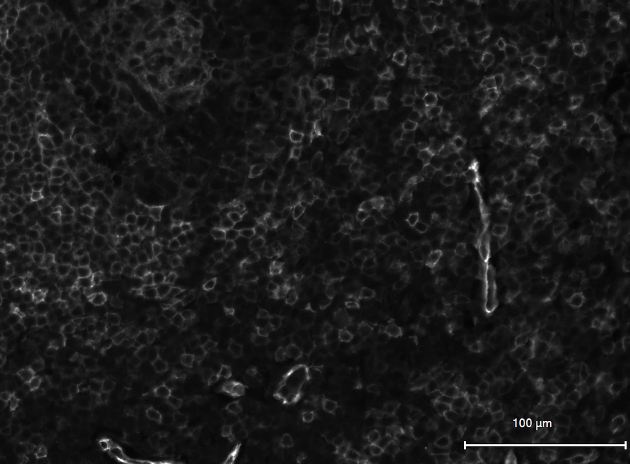
Application: immunofluorescence - paraffinSample Tested: human tonsilSpecies: HumanVerified Customer | Posted 05/02/2019pH 9 heat-induced antigen retrieval

There are no reviews that match your criteria.
Protocols
Find general support by application which include: protocols, troubleshooting, illustrated assays, videos and webinars.
- Antigen Retrieval Protocol (PIER)
- Antigen Retrieval for Frozen Sections Protocol
- Appropriate Fixation of IHC/ICC Samples
- Cellular Response to Hypoxia Protocols
- Chromogenic IHC Staining of Formalin-Fixed Paraffin-Embedded (FFPE) Tissue Protocol
- Chromogenic Immunohistochemistry Staining of Frozen Tissue
- ClariTSA™ Fluorophore Kits
- Detection & Visualization of Antibody Binding
- Fluorescent IHC Staining of Frozen Tissue Protocol
- Graphic Protocol for Heat-induced Epitope Retrieval
- Graphic Protocol for the Preparation and Fluorescent IHC Staining of Frozen Tissue Sections
- Graphic Protocol for the Preparation and Fluorescent IHC Staining of Paraffin-embedded Tissue Sections
- Graphic Protocol for the Preparation of Gelatin-coated Slides for Histological Tissue Sections
- ICC Cell Smear Protocol for Suspension Cells
- ICC Immunocytochemistry Protocol Videos
- ICC for Adherent Cells
- IHC Sample Preparation (Frozen sections vs Paraffin)
- ISH-IHC Protocol for Chromogenic Detection on Formalin Fixed Paraffin Embedded (FFPE) Tissue
- Immunocytochemistry (ICC) Protocol
- Immunocytochemistry Troubleshooting
- Immunofluorescence of Organoids Embedded in Cultrex Basement Membrane Extract
- Immunofluorescent IHC Staining of Formalin-Fixed Paraffin-Embedded (FFPE) Tissue Protocol
- Immunohistochemistry (IHC) and Immunocytochemistry (ICC) Protocols
- Immunohistochemistry Frozen Troubleshooting
- Immunohistochemistry Paraffin Troubleshooting
- Preparing Samples for IHC/ICC Experiments
- Preventing Non-Specific Staining (Non-Specific Binding)
- Primary Antibody Selection & Optimization
- Protocol for Heat-Induced Epitope Retrieval (HIER)
- Protocol for Making a 4% Formaldehyde Solution in PBS
- Protocol for VisUCyte™ HRP Polymer Detection Reagent
- Protocol for the Fluorescent ICC Staining of Cell Smears - Graphic
- Protocol for the Fluorescent ICC Staining of Cultured Cells on Coverslips - Graphic
- Protocol for the Preparation & Fixation of Cells on Coverslips
- Protocol for the Preparation and Chromogenic IHC Staining of Frozen Tissue Sections
- Protocol for the Preparation and Chromogenic IHC Staining of Frozen Tissue Sections - Graphic
- Protocol for the Preparation and Chromogenic IHC Staining of Paraffin-embedded Tissue Sections
- Protocol for the Preparation and Chromogenic IHC Staining of Paraffin-embedded Tissue Sections - Graphic
- Protocol for the Preparation and Fluorescent ICC Staining of Cells on Coverslips
- Protocol for the Preparation and Fluorescent ICC Staining of Non-adherent Cells
- Protocol for the Preparation and Fluorescent ICC Staining of Stem Cells on Coverslips
- Protocol for the Preparation and Fluorescent IHC Staining of Frozen Tissue Sections
- Protocol for the Preparation and Fluorescent IHC Staining of Paraffin-embedded Tissue Sections
- Protocol for the Preparation of Gelatin-coated Slides for Histological Tissue Sections
- Protocol for the Preparation of a Cell Smear for Non-adherent Cell ICC - Graphic
- R&D Systems Quality Control Western Blot Protocol
- TUNEL and Active Caspase-3 Detection by IHC/ICC Protocol
- The Importance of IHC/ICC Controls
- Troubleshooting Guide: Immunohistochemistry
- Troubleshooting Guide: Western Blot Figures
- Western Blot Conditions
- Western Blot Protocol
- Western Blot Protocol for Cell Lysates
- Western Blot Troubleshooting
- Western Blot Troubleshooting Guide
- View all Protocols, Troubleshooting, Illustrated assays and Webinars
Loading...