CCL4/MIP-1 beta is a beta chemokine that is secreted at sites of inflammation by activated leukocytes, lymphocytes, vascular endothelial cells, and pulmonary smooth muscle cells. It attracts a variety of immune cells to sites of microbial infection as well as to other pathologic inflammation such as allergic asthma and ischemic myocardium. CCL4 is secreted from activated monocytes as a heterodimer with CCL3/MIP-1 alpha. It signals through CCR5, and an N-terminally trimmed form additionally interacts with CCR1 and CCR2. In humans, the ability of CCL4 to bind CCR5 inhibits the cellular entry of M-tropic HIV-1 which utilizes CCR5 as a coreceptor.
Human CCL4/MIP-1 beta Antibody
R&D Systems | Catalog # AF-271-NA

Key Product Details
Species Reactivity
Validated:
Human
Cited:
Human, Primate - Macaca mulatta (Rhesus Macaque)
Applications
Validated:
Immunohistochemistry, Western Blot, Neutralization, Immunocytochemistry
Cited:
Immunohistochemistry, Immunohistochemistry-Frozen, Neutralization, Immunocytochemistry, Functional Assay, Lateral Flow Assay
Label
Unconjugated
Antibody Source
Polyclonal Goat IgG
Loading...
Product Specifications
Immunogen
S. frugiperda insect ovarian cell line Sf 21-derived recombinant human CCL4/MIP-1 beta
Specificity
Detects human CCL4/MIP-1 beta in direct ELISAs and Western blots.
Clonality
Polyclonal
Host
Goat
Isotype
IgG
Endotoxin Level
<0.10 EU per 1 μg of the antibody by the LAL method.
Scientific Data Images for Human CCL4/MIP-1 beta Antibody
Chemotaxis Induced by CCL4/MIP‑1 beta and Neutral-ization by Human CCL4/MIP‑1 beta Antibody.
Recombinant Human CCL4/MIP-1 beta (271-BME) chemoattracts the BaF3 mouse pro-B cell line transfected with human CCR5 in a dose-dependent manner (orange line). The amount of cells that migrated through to the lower chemotaxis chamber was measured by Resazurin (AR002). Chemotaxis elicited by Recombinant Human CCL4/MIP-1 beta (40 ng/mL) is neutralized (green line) by increasing concentrations of Goat Anti-Human CCL4/MIP-1 beta Antigen Affinity-purified Polyclonal Antibody (Catalog # AF-271-NA). The ND50 is typically 0.3-1.5 µg/mL.
CCL4/MIP‑1 beta in Human Alzheimer's Disease Brain.
CCL4/MIP-1 beta was detected in immersion fixed paraffin-embedded sections of human Alzheimer's disease brain (hippocampus) using 15 µg/mL Goat Anti-Human CCL4/MIP-1 beta Antigen Affinity-purified Polyclonal Antibody (Catalog # AF-271-NA) overnight at 4 °C. Tissue was stained with the Anti-Goat HRP-AEC Cell & Tissue Staining Kit (red; CTS009) and counterstained with hematoxylin (blue). View our protocol for Chromogenic IHC Staining of Paraffin-embedded Tissue Sections.
CCL4/MIP‑1 beta in Human PBMCs.
CCL4/MIP-1 beta was detected in immersion fixed human peripheral blood mononuclear cells (PBMCs) stimulated with PHA and monensin using Goat Anti-Human CCL4/MIP-1 beta Antigen Affinity-purified Polyclonal Antibody (Catalog # AF-271-NA) at 10 µg/mL for 3 hours at room temperature. Cells were stained using the NorthernLights™ 557-conjugated Anti-Goat IgG Secondary Antibody (yellow; NL001) and counterstained with DAPI (blue). View our protocol for Fluorescent ICC Staining of Non-adherent Cells.
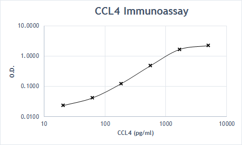
Human CCL4 / MIP-1 beta ELISA Standard Curve
Recombinant Human CCL4/MIP‑1 beta (Catalog # 271-BME) was serially diluted and captured by Mouse Anti-Human CCL4/MIP‑1 beta Monoclonal Antibody (Catalog # MAB271) coated on a Clear Polystyrene Microplate (Catalog # DY990). Goat Anti-Human CCL4/MIP‑1 beta Antigen Affinity-purified Polyclonal Antibody (Catalog # AF-271-NA) was biotinylated and incubated with the protein captured on the plate. Detection of the standard curve was achieved by incubating Streptavidin-HRP (Catalog # DY998)Applications for Human CCL4/MIP-1 beta Antibody
Application
Recommended Usage
Immunocytochemistry
5-15 µg/mL
Sample: Immersion fixed human peripheral blood mononuclear cells stimulated with PHA and monensin
Sample: Immersion fixed human peripheral blood mononuclear cells stimulated with PHA and monensin
Immunohistochemistry
5-15 µg/mL
Sample: Immersion fixed paraffin-embedded sections of human Alzheimer's disease brain (hippocampus)
Sample: Immersion fixed paraffin-embedded sections of human Alzheimer's disease brain (hippocampus)
Western Blot
0.1 µg/mL
Sample: Recombinant Human CCL4/MIP‑1 beta (Catalog # 271-BME)
Sample: Recombinant Human CCL4/MIP‑1 beta (Catalog # 271-BME)
Neutralization
Measured by its ability to neutralize CCL4/MIP‑1 beta -induced chemotaxis in the BaF3 mouse pro‑B cell line transfected with human CCR5. The Neutralization Dose (ND50) is typically 0.3-1.5 µg/mL in the presence of 40 ng/mL Recombinant Human CCL4/MIP‑1 beta.
Reviewed Applications
Read 1 review rated 5 using AF-271-NA in the following applications:
Formulation, Preparation, and Storage
Purification
Antigen Affinity-purified
Reconstitution
Reconstitute at 0.2 mg/mL in sterile PBS. For liquid material, refer to CoA for concentration.
Loading...
Formulation
Lyophilized from a 0.2 μm filtered solution in PBS with Trehalose. *Small pack size (SP) is supplied either lyophilized or as a 0.2 µm filtered solution in PBS.
Shipping
Lyophilized product is shipped at ambient temperature. Liquid small pack size (-SP) is shipped with polar packs. Upon receipt, store immediately at the temperature recommended below.
Stability & Storage
Use a manual defrost freezer and avoid repeated freeze-thaw cycles.
- 12 months from date of receipt, -20 to -70 °C as supplied.
- 1 month, 2 to 8 °C under sterile conditions after reconstitution.
- 6 months, -20 to -70 °C under sterile conditions after reconstitution.
Calculators
Background: CCL4/MIP-1 beta
Alternate Names
Exodus-3, MIP-1 beta, MIP1 beta
Entrez Gene IDs
Gene Symbol
CCL4
Additional CCL4/MIP-1 beta Products
Product Documents for Human CCL4/MIP-1 beta Antibody
Certificate of Analysis
To download a Certificate of Analysis, please enter a lot or batch number in the search box below.
Note: Certificate of Analysis not available for kit components.
Product Specific Notices for Human CCL4/MIP-1 beta Antibody
For research use only
Citations for Human CCL4/MIP-1 beta Antibody
 Powered by Bioz
Customer Reviews for Human CCL4/MIP-1 beta Antibody (1)
5 out of 5
1 Customer Rating
Have you used Human CCL4/MIP-1 beta Antibody?
Submit a review and receive an Amazon gift card!
$25/€18/£15/$25CAN/¥2500 Yen for a review with an image
$10/€7/£6/$10CAN/¥1110 Yen for a review without an image
Submit a review
Customer Images
Showing
1
-
1 的
1 review
Showing All
Filter By:
-
Application: ELISASample Tested: SerumSpecies: HumanVerified Customer | Posted 08/19/2019

There are no reviews that match your criteria.
Protocols
Find general support by application which include: protocols, troubleshooting, illustrated assays, videos and webinars.
- Antigen Retrieval Protocol (PIER)
- Antigen Retrieval for Frozen Sections Protocol
- Appropriate Fixation of IHC/ICC Samples
- Cellular Response to Hypoxia Protocols
- Chromogenic IHC Staining of Formalin-Fixed Paraffin-Embedded (FFPE) Tissue Protocol
- Chromogenic Immunohistochemistry Staining of Frozen Tissue
- ClariTSA™ Fluorophore Kits
- Detection & Visualization of Antibody Binding
- Fluorescent IHC Staining of Frozen Tissue Protocol
- Graphic Protocol for Heat-induced Epitope Retrieval
- Graphic Protocol for the Preparation and Fluorescent IHC Staining of Frozen Tissue Sections
- Graphic Protocol for the Preparation and Fluorescent IHC Staining of Paraffin-embedded Tissue Sections
- Graphic Protocol for the Preparation of Gelatin-coated Slides for Histological Tissue Sections
- ICC Cell Smear Protocol for Suspension Cells
- ICC Immunocytochemistry Protocol Videos
- ICC for Adherent Cells
- IHC Sample Preparation (Frozen sections vs Paraffin)
- Immunocytochemistry (ICC) Protocol
- Immunocytochemistry Troubleshooting
- Immunofluorescence of Organoids Embedded in Cultrex Basement Membrane Extract
- Immunofluorescent IHC Staining of Formalin-Fixed Paraffin-Embedded (FFPE) Tissue Protocol
- Immunohistochemistry (IHC) and Immunocytochemistry (ICC) Protocols
- Immunohistochemistry Frozen Troubleshooting
- Immunohistochemistry Paraffin Troubleshooting
- Preparing Samples for IHC/ICC Experiments
- Preventing Non-Specific Staining (Non-Specific Binding)
- Primary Antibody Selection & Optimization
- Protocol for Heat-Induced Epitope Retrieval (HIER)
- Protocol for Making a 4% Formaldehyde Solution in PBS
- Protocol for VisUCyte™ HRP Polymer Detection Reagent
- Protocol for the Fluorescent ICC Staining of Cell Smears - Graphic
- Protocol for the Fluorescent ICC Staining of Cultured Cells on Coverslips - Graphic
- Protocol for the Preparation & Fixation of Cells on Coverslips
- Protocol for the Preparation and Chromogenic IHC Staining of Frozen Tissue Sections
- Protocol for the Preparation and Chromogenic IHC Staining of Frozen Tissue Sections - Graphic
- Protocol for the Preparation and Chromogenic IHC Staining of Paraffin-embedded Tissue Sections
- Protocol for the Preparation and Chromogenic IHC Staining of Paraffin-embedded Tissue Sections - Graphic
- Protocol for the Preparation and Fluorescent ICC Staining of Cells on Coverslips
- Protocol for the Preparation and Fluorescent ICC Staining of Non-adherent Cells
- Protocol for the Preparation and Fluorescent ICC Staining of Stem Cells on Coverslips
- Protocol for the Preparation and Fluorescent IHC Staining of Frozen Tissue Sections
- Protocol for the Preparation and Fluorescent IHC Staining of Paraffin-embedded Tissue Sections
- Protocol for the Preparation of Gelatin-coated Slides for Histological Tissue Sections
- Protocol for the Preparation of a Cell Smear for Non-adherent Cell ICC - Graphic
- R&D Systems Quality Control Western Blot Protocol
- TUNEL and Active Caspase-3 Detection by IHC/ICC Protocol
- The Importance of IHC/ICC Controls
- Troubleshooting Guide: Immunohistochemistry
- Troubleshooting Guide: Western Blot Figures
- Western Blot Conditions
- Western Blot Protocol
- Western Blot Protocol for Cell Lysates
- Western Blot Troubleshooting
- Western Blot Troubleshooting Guide
- View all Protocols, Troubleshooting, Illustrated assays and Webinars

