
Estrogen-related receptor alpha (ERR-alpha, ERR1, ESRL1; NR3B1) is a member of the orphan nuclear receptor family. ERR-alpha is expressed in the liver, kidney, brain, heart, uterus, vagina and cervix. ERR-alpha takes part in the cell cycle and the fatty acid metabolizing of the musculus skeleti. ERR-alpha was shown to interact with ER-alpha. ERR-beta (ERR2; NR3B2) is also expressed in the liver, brain, uterus, vagina, and cervix. ERR-beta has a role in early placental development. ERR-gamma (ESRRG; NR3B3) is expressed in the brain, kidney, testis, lung, adrenal gland, pancreas, placenta and bone marrow. ERR-gamma was shown to be located in the critical region for Usher's syndrome.
Human ERR beta/NR3B2 Antibody (H6705)
R&D Systems | Catalog # PP-H6705-00


Scientific images may be resized, cropped, or adjusted for brightness or contrast for presentation purposes. These updates are limited to presentation and do not affect the underlying scientific interpretation of the data. Where available, additional source data may be provided upon request.
Loading...
Key Product Details
Species Reactivity
Validated:
Human
Cited:
Human, Mouse
Applications
Validated:
Immunohistochemistry, Western Blot, Immunoprecipitation
Cited:
Immunohistochemistry, Western Blot, Immunocytochemistry, Immunoprecipitation, Chromatin Immunoprecipitation (ChIP), Bioassay
Label
Unconjugated
Antibody Source
Monoclonal Mouse IgG2A Clone # H6705
Loading...
Product Specifications
Clonality
Monoclonal
Host
Mouse
Isotype
IgG2A
Scientific Data Images for Human ERR beta/NR3B2 Antibody (H6705)
Detection of Mouse ERR beta/NR3B2 by Immunocytochemistry/Immunofluorescence
Immediate firing motoneurons, but not delayed firing motoneurons, express ERR beta.(A) Examples of Err beta. staining (red) in neurobiotin (green) filled motoneurons. The arrowhead in the first row points to the nucleus (Err beta −positive) of the recorded motoneuron. The asterisk in the second row indicates that the nucleus of the recorded motoneuron was Err beta −negative. (B) Plot of the AHP relaxation time constants against the rheobases for labelled motoneurons. All immediate firing motoneurons were Err beta −positive whereas all delayed firing motoneurons were Err beta −negative. Arrowheads point to the motoneurons illustrated in A.DOI:https://dx.doi.org/10.7554/eLife.04046.008 Image collected and cropped by CiteAb from the following publication (https://elifesciences.org/articles/04046), licensed under a CC-BY license. Not internally tested by R&D Systems.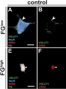
Detection of ERR beta/NR3B2 by Immunocytochemistry/ Immunofluorescence
Generation of distinguishable gamma motor neurons in the absence of ERR2/3.(A-I) Imaris 3D reconstruction: VGLUT1+ synaptic varicosities (green) associated with large FGlow, NeuNhigh motor neurons in control (A, B) and ERR2/3cko mice (C, D) (scale bar: 20 μm). (E-H) Absence of VGLUT1+ puncta on FGhigh, NeuNlow motor neurons in control (E, F) and ERR2/3cko mice (G, H) (scale bar: 20 μm). (I) Significant difference in VGLUT1+ varicosities associated with FGhigh, NeuNlow and FGlow, NeuNhigh motor neurons from control mice. Lack of significant differences in VGLUT1+ synaptic varicosities associated FGlow, NeuNhigh motor neurons in control (20 ± 3.48, n = 9) versus ERR2/3cko mice (23.1 ± 4.06, n = 10). Lack of significant differences in VGLUT1+ varicosities between control FGhigh, NeuNlow (1.1 ± 0.36, n = 10) and ERR2/3cko FGhigh, NeuNlow motor neurons (2.4 ± 0.78, n = 10). (J-O) P300 mouse extensor digitorum longus (EDL) muscle spindles of control (J-L) and ERR2/3cko(M-O) mice. (J-L) Distribution of Ia sensory annulospiral endings in the central spindle segment (visualized by VGLUT1, green), motor innervation (VACHT, magenta) and their postsynaptic sites (arrowheads) on intrafusal fibers (BTX, alpha bungarotoxin, grey) (scale bar: 100 μm). (K-O) Normal appearance of ERR2/3cko muscle spindles, including Ia sensory annulospiral endings and motor innervation of BTX+ postsynaptic sites (arrowheads) on intrafusal fibers (scale bar: 100 μm). Data are presented as mean ± SEM. n = # of neurons. Statistically significant differences control and ERR2/3cko motor neurons are indicated as: *p < 0.05, **p < 0.01, ***p < 0.001, n.s. = not significant, Student t test). Data for Fig 4I can be found in S2 Data. Image collected and cropped by CiteAb from the following open publication (https://pubmed.ncbi.nlm.nih.gov/36542664), licensed under a CC-BY license. Not internally tested by R&D Systems.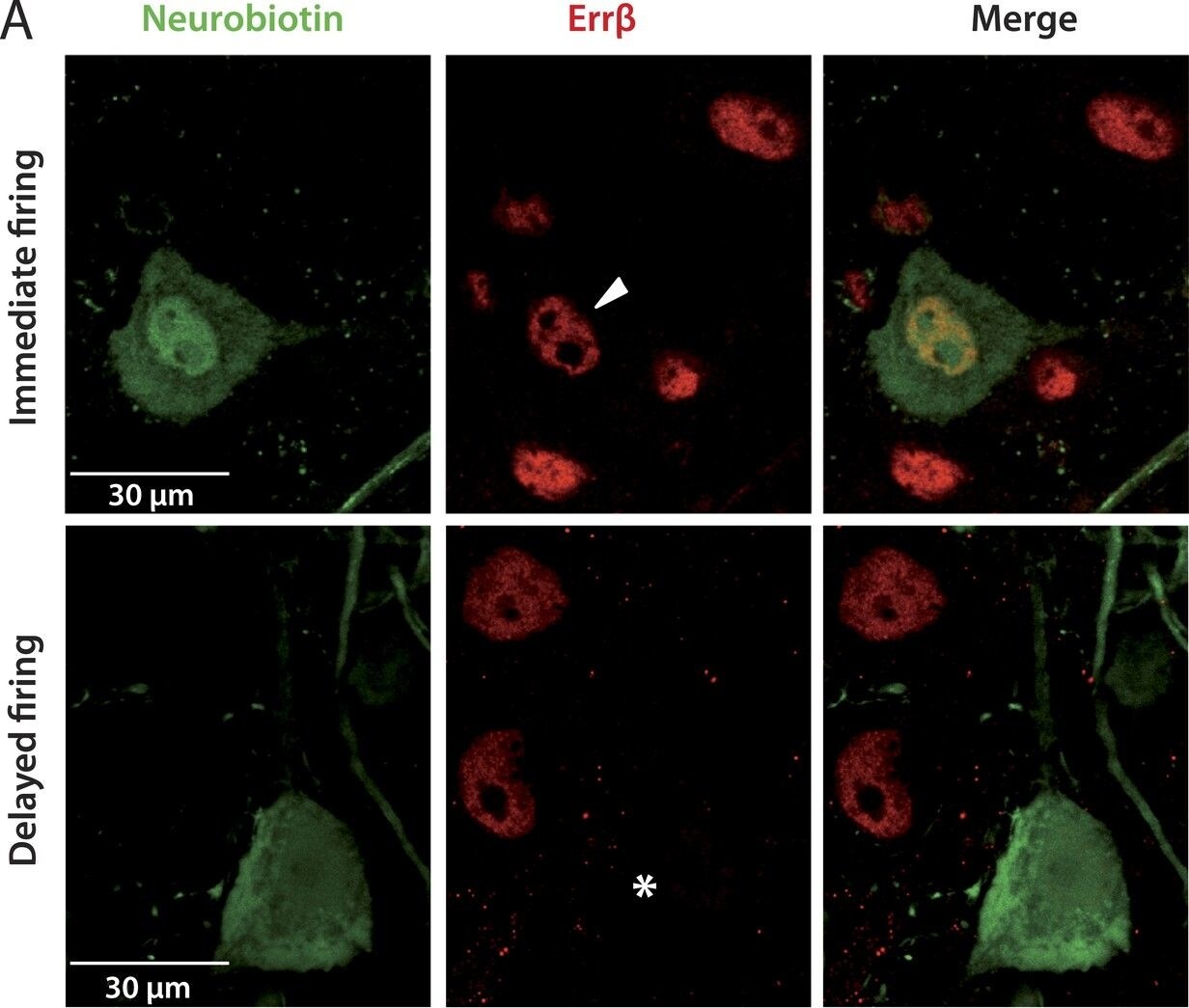
Detection of ERR beta/NR3B2 by Immunohistochemistry
Immediate firing motoneurons, but not delayed firing motoneurons, express ERR beta.(A) Examples of Err beta. staining (red) in neurobiotin (green) filled motoneurons. The arrowhead in the first row points to the nucleus (Err beta −positive) of the recorded motoneuron. The asterisk in the second row indicates that the nucleus of the recorded motoneuron was Err beta −negative. (B) Plot of the AHP relaxation time constants against the rheobases for labelled motoneurons. All immediate firing motoneurons were Err beta −positive whereas all delayed firing motoneurons were Err beta −negative. Arrowheads point to the motoneurons illustrated in A.DOI:http://dx.doi.org/10.7554/eLife.04046.008. Image collected and cropped by CiteAb from the following open publication (https://pubmed.ncbi.nlm.nih.gov/25313866), licensed under a CC-BY license. Not internally tested by R&D Systems.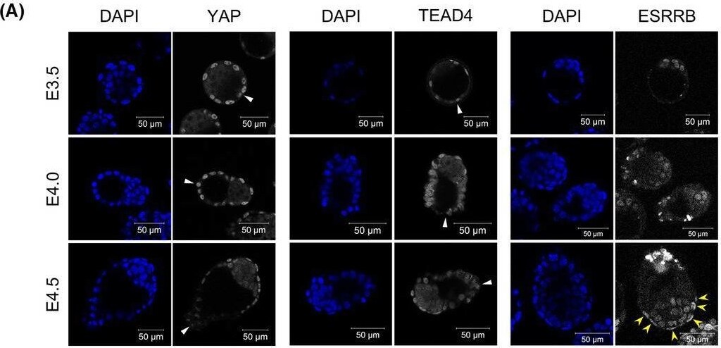
Detection of ERR beta/NR3B2 by Immunocytochemistry/ Immunofluorescence
Relationship between CDX2 downregulation and Hippo signaling activity in implantation‐stage blastocysts. (A) Immunofluorescence analysis of YAP, TEAD4, and ESRRB in E3.5, 4.0, and 4.5 blastocysts. White arrowheads indicate the tips of mural TE. Yellow arrowheads indicate the ESRRB‐positive TE. Scale bar: 50 μm. (B) Immunofluorescence analysis of CDX2 in DMSO‐ (control) or verteporfin (VP)‐treated blastocysts. CDX2 expression in TE was found to be weaker in VP‐treated blastocysts than in control blastocysts. Scale bar: 100 μm. (C) Comparison of CDX2 fluorescence intensity in the TE of control and VP‐treated blastocysts. Statistical significance was determined by the Student's t‐test (*p < 0.01). n indicates the number of nuclei analyzed. Bars indicate mean ± S.D. Image collected and cropped by CiteAb from the following open publication (https://pubmed.ncbi.nlm.nih.gov/35386376), licensed under a CC-BY license. Not internally tested by R&D Systems.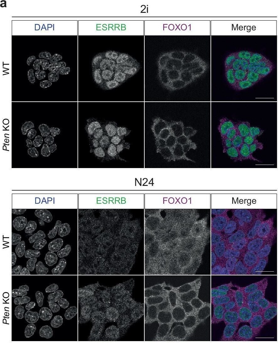
Detection of ERR beta/NR3B2 by Immunocytochemistry/ Immunofluorescence
FoxO TFs translocate into the nucleus upon exit from naïve pluripotency.a Confocal microscopy images after IF, showing FOXO1 (purple) and ESRRB (green) in WT and Pten KOs in 2i and at N24. DAPI staining is shown in blue. One representative image from n = 3 independent experiments is shown. Scale bar = 20 μM. b Quantification of FOXO1 and ESRRB nuclear intensity (in arbitrary unit, arb. units) measured from confocal images of WT cells in 2i (n = 135) and 8 h (N8, n = 136), 16 h (N16, n = 88) and 24 h (N24, n = 127) after 2i withdrawal. Data were obtained from n = 2 independent experiments. p values are derived from a two-tailed Wilcoxon rank sum test. c Quantification of FOXO1 nuclear intensity (arb. units) based on images of WT (n = 189 [2i] and n = 164 [N24]) and Pten KO (n = 100 [2i] and n = 153 [N24]), as in (a). Data were obtained from n = 2 independent experiments. p values were calculated as in (b). d Confocal microscopy images after IF showing FOXO1 (white), SOX2 (green), GATA4 (red), or OTX2 (red) in WT E4.5, E4.75 and E5.5 embryos. Hoechst staining is shown in cyan. One representative image of n = 3 independent experiments is shown. Scale bar = 100 μM. e Quantification of FOXO1 nuclear intensity (arb. units) measured from confocal images of WT epiblast from E4.5 (n = 12), E4.75 (n = 12) and E5.5 (n = 8) embryos, as in (d). Data were obtained from n = 3 independent experiments. Indicated p values are derived from a two-tailed Wilcoxon rank sum test. Image collected and cropped by CiteAb from the following open publication (https://pubmed.ncbi.nlm.nih.gov/39251582), licensed under a CC-BY license. Not internally tested by R&D Systems.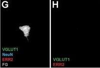
Detection of ERR beta/NR3B2 by Immunocytochemistry/ Immunofluorescence
Generation of distinguishable gamma motor neurons in the absence of ERR2/3.(A-I) Imaris 3D reconstruction: VGLUT1+ synaptic varicosities (green) associated with large FGlow, NeuNhigh motor neurons in control (A, B) and ERR2/3cko mice (C, D) (scale bar: 20 μm). (E-H) Absence of VGLUT1+ puncta on FGhigh, NeuNlow motor neurons in control (E, F) and ERR2/3cko mice (G, H) (scale bar: 20 μm). (I) Significant difference in VGLUT1+ varicosities associated with FGhigh, NeuNlow and FGlow, NeuNhigh motor neurons from control mice. Lack of significant differences in VGLUT1+ synaptic varicosities associated FGlow, NeuNhigh motor neurons in control (20 ± 3.48, n = 9) versus ERR2/3cko mice (23.1 ± 4.06, n = 10). Lack of significant differences in VGLUT1+ varicosities between control FGhigh, NeuNlow (1.1 ± 0.36, n = 10) and ERR2/3cko FGhigh, NeuNlow motor neurons (2.4 ± 0.78, n = 10). (J-O) P300 mouse extensor digitorum longus (EDL) muscle spindles of control (J-L) and ERR2/3cko(M-O) mice. (J-L) Distribution of Ia sensory annulospiral endings in the central spindle segment (visualized by VGLUT1, green), motor innervation (VACHT, magenta) and their postsynaptic sites (arrowheads) on intrafusal fibers (BTX, alpha bungarotoxin, grey) (scale bar: 100 μm). (K-O) Normal appearance of ERR2/3cko muscle spindles, including Ia sensory annulospiral endings and motor innervation of BTX+ postsynaptic sites (arrowheads) on intrafusal fibers (scale bar: 100 μm). Data are presented as mean ± SEM. n = # of neurons. Statistically significant differences control and ERR2/3cko motor neurons are indicated as: *p < 0.05, **p < 0.01, ***p < 0.001, n.s. = not significant, Student t test). Data for Fig 4I can be found in S2 Data. Image collected and cropped by CiteAb from the following open publication (https://pubmed.ncbi.nlm.nih.gov/36542664), licensed under a CC-BY license. Not internally tested by R&D Systems.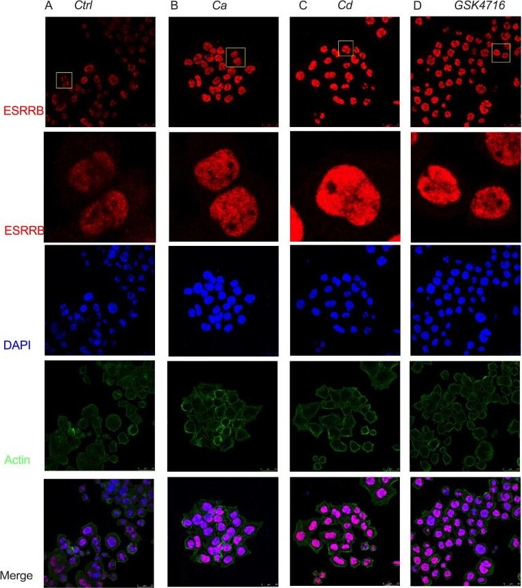
Detection of ERR beta/NR3B2 by Immunocytochemistry/ Immunofluorescence
Effects of calcium and cadmium on ESRRB localization in MDA-MB-453 cells. (A–D), Immunofluorescence representative images of ESRRB in control (A), calcium (B), cadmium (C) and GSK4716 (D) treated cells. The second row of panels contains the zoomed-in images marked in yellow boxes in the top panels; red is ESRRB staining; blue is nuclear staining with DAPI; green is actin staining with phalloidin; bottom panels are merged IF pictures; the scale bar represents 25 μm. Image collected and cropped by CiteAb from the following open publication (https://www.mdpi.com/1422-0067/27/1/231), licensed under a CC-BY license. Not internally tested by R&D Systems.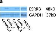
Detection of ERR beta/NR3B2 by Western Blot
Characterization of model cell line. a Expression of Esrrb is confirmed by Western blot. Two replicates of Esrrb transfected cells showed successful expression of Esrrb protein and b 1 % Hh-CM treated NIH3T3-pc3.1 showed increased concentration of Gli1 protein. Image collected and cropped by CiteAb from the following open publication (https://pubmed.ncbi.nlm.nih.gov/26597826), licensed under a CC-BY license. Not internally tested by R&D Systems.Applications for Human ERR beta/NR3B2 Antibody (H6705)
Application
Recommended Usage
Immunohistochemistry
5-15 µg/mL
Sample: Perfusion fixed paraffin-embedded sections of rat kidney
Sample: Perfusion fixed paraffin-embedded sections of rat kidney
Immunoprecipitation
Optimal dilutions of this antibody should be experimentally determined.
Western Blot
Optimal dilutions of this antibody should be experimentally determined.
Background: ERR beta/NR3B2
Long Name
Estrogen-related Receptor beta
Alternate Names
ERR2, ESRL2, ESRRB, NR3B2
Entrez Gene IDs
2103 (Human)
Gene Symbol
ESRRB
Additional ERR beta/NR3B2 Products
Product Documents for Human ERR beta/NR3B2 Antibody (H6705)
Certificate of Analysis
To download a Certificate of Analysis, please enter a lot or batch number in the search box below.
Note: Certificate of Analysis not available for kit components.
Product Specific Notices for Human ERR beta/NR3B2 Antibody (H6705)
For research use only
Related Research Areas
Citations for Human ERR beta/NR3B2 Antibody (H6705)
 Powered by Bioz
Customer Reviews for Human ERR beta/NR3B2 Antibody (H6705)
There are currently no reviews for this product. Be the first to review Human ERR beta/NR3B2 Antibody (H6705) and earn rewards!
Have you used Human ERR beta/NR3B2 Antibody (H6705)?
Submit a review and receive an Amazon gift card!
$25/€18/£15/$25CAN/¥2500 for a review with an image
$10/€7/£6/$10CAN/¥1110 for a review without an image
Submit a review
Protocols
Find general support by application which include: protocols, troubleshooting, illustrated assays, videos and webinars.
- Antigen Retrieval Protocol (PIER)
- Antigen Retrieval for Frozen Sections Protocol
- Appropriate Fixation of IHC/ICC Samples
- Cellular Response to Hypoxia Protocols
- Chromogenic IHC Staining of Formalin-Fixed Paraffin-Embedded (FFPE) Tissue Protocol
- Chromogenic Immunohistochemistry Staining of Frozen Tissue
- ClariTSA™ Fluorophore Kits
- Detection & Visualization of Antibody Binding
- Fluorescent IHC Staining of Frozen Tissue Protocol
- Graphic Protocol for Heat-induced Epitope Retrieval
- Graphic Protocol for the Preparation and Fluorescent IHC Staining of Frozen Tissue Sections
- Graphic Protocol for the Preparation and Fluorescent IHC Staining of Paraffin-embedded Tissue Sections
- Graphic Protocol for the Preparation of Gelatin-coated Slides for Histological Tissue Sections
- IHC Sample Preparation (Frozen sections vs Paraffin)
- Immunofluorescent IHC Staining of Formalin-Fixed Paraffin-Embedded (FFPE) Tissue Protocol
- Immunohistochemistry (IHC) and Immunocytochemistry (ICC) Protocols
- Immunohistochemistry Frozen Troubleshooting
- Immunohistochemistry Paraffin Troubleshooting
- Immunoprecipitation Protocol
- Preparing Samples for IHC/ICC Experiments
- Preventing Non-Specific Staining (Non-Specific Binding)
- Primary Antibody Selection & Optimization
- Protocol for Heat-Induced Epitope Retrieval (HIER)
- Protocol for Making a 4% Formaldehyde Solution in PBS
- Protocol for VisUCyte™ HRP Polymer Detection Reagent
- Protocol for the Preparation & Fixation of Cells on Coverslips
- Protocol for the Preparation and Chromogenic IHC Staining of Frozen Tissue Sections
- Protocol for the Preparation and Chromogenic IHC Staining of Frozen Tissue Sections - Graphic
- Protocol for the Preparation and Chromogenic IHC Staining of Paraffin-embedded Tissue Sections
- Protocol for the Preparation and Chromogenic IHC Staining of Paraffin-embedded Tissue Sections - Graphic
- Protocol for the Preparation and Fluorescent IHC Staining of Frozen Tissue Sections
- Protocol for the Preparation and Fluorescent IHC Staining of Paraffin-embedded Tissue Sections
- Protocol for the Preparation of Gelatin-coated Slides for Histological Tissue Sections
- R&D Systems Quality Control Western Blot Protocol
- TUNEL and Active Caspase-3 Detection by IHC/ICC Protocol
- The Importance of IHC/ICC Controls
- Troubleshooting Guide: Immunohistochemistry
- Troubleshooting Guide: Western Blot Figures
- Western Blot Conditions
- Western Blot Protocol
- Western Blot Protocol for Cell Lysates
- Western Blot Troubleshooting
- Western Blot Troubleshooting Guide
- View all Protocols, Troubleshooting, Illustrated assays and Webinars
Loading...