

Human FGF-9 Antibody

R&D Systems | Catalog # AF-273-NA

R&D Systems
Loading...

Key Product Details

Species Reactivity

Validated:

Human

Cited:

Human, Transgenic Mouse

Applications

Validated:

Immunohistochemistry, Western Blot

Cited:

Immunohistochemistry, Immunohistochemistry-Paraffin, Western Blot, Neutralization

Label

Unconjugated

Antibody Source

Polyclonal Goat IgG
Loading...

Product Specifications

Immunogen

S. frugiperda insect ovarian cell line Sf 21-derived recombinant human FGF-9
Met1-Ser208
Accession # P31371

Specificity

Detects human FGF-9 in direct ELISAs and Western blots. In direct ELISAs, less than 1% cross-reactivity with recombinant human (rh) FGF‑4, rhFGF-5, rhFGF-6, rhFGF-7, rhFGF-8, rhFGF-10, rhFGF-17, rhFGF-18, and rhFGF-19 is observed.

Clonality

Polyclonal

Host

Goat

Isotype

IgG

Scientific Data Images for Human FGF-9 Antibody

FGF‑9 antibody in Human Placenta by Immunohistochemistry (IHC-P).

FGF‑9 in Human Placenta.

FGF-9 was detected in immersion fixed paraffin-embedded sections of human placenta using 5 µg/mL Goat Anti-Human FGF-9 Antigen Affinity-purified Polyclonal Antibody (Catalog # AF-273-NA) overnight at 4 °C. Tissue was stained with the Anti-Goat HRP-AEC Cell & Tissue Staining Kit (red; Catalog # CTS009) and counterstained with hematoxylin (blue). View our protocol for Chromogenic IHC Staining of Paraffin-embedded Tissue Sections.
Human FGF-9 ELISA Standard Curve

Human FGF-9 ELISA Standard Curve

Recombinant Human FGF‑9 (Catalog # 273-F9) was serially diluted and captured by Mouse Anti-Human FGF‑9 Monoclonal Antibody (Catalog # MAB273) coated on a Clear Polystyrene Microplate (Catalog # DY990). Goat Anti-Human FGF‑9 Antigen Affinity-purified Polyclonal Antibody (Catalog # AF-273-NA) was biotinylated and incubated with the protein captured on the plate. Detection of the standard curve was achieved by incubating Streptavidin-HRP (Catalog # DY998)
Detection of FGF-9 by Western Blot

Detection of FGF-9 by Western Blot

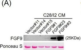
Fibroblast growth factor 9 (FGF9) regulates NRF2/GPX3 expression in chondrocytes. a) Western blot analysis of FGF9 levels in conditioned media (CM) from C28/I2 cells. b) Western blot analysis of NRF2, and KEAP1 in normal primary chondrocytes (NP-Chon) cultured with FGF9-CM; beta -actin was used as a loading control. c) Western blot analysis of NRF2 in NP-Chon and osteoarthritis (OA) primary chondrocytes (OAP-Chon). d) Left: Quantitative reverse transcription polymerase chain reaction (qRT-PCR) analysis of FGF9 messenger RNA (mRNA) expression in C28/I2 cells infected with shFGF9-D1. Right: qRT-PCR analysis of GPX1–4 expression in FGF9-depleted C28/I2 cells. e) qRT-PCR analysis of Fgf9 (left) and Gpx3 (right) expression in ATDC5 cells infected with lentiviral control or short hairpin RNA (shRNA). f) Left: qRT-PCR analysis of GPX3 mRNA expression in NP-Chon cells treated with vector control or FGF9-conditioned medium (FGF9-CM). Right: Western blot analysis of GPX3 protein expression in NP-Chon cells treated with vector control or FGF9-CM. g) Western blot analysis of GPX3 protein expression in NP-Chon and OAP-Chon cells. h) and i) qRT-PCR analysis of NFE2L2 and GPX3 expression levels in NP-Chon (n = 71) and OAP-Chon (n = 195) samples, presented as violin plots. Error bars represent mean (SD). **p < 0.01, ***p < 0.001, independent-samples t-test. Image collected and cropped by CiteAb from the following open publication (https://boneandjoint.org.uk/doi/10.1302/2046-3758.156.BJR-2025-0365.R1), licensed under a CC-BY license. Not internally tested by R&D Systems.
Detection of FGF-9 by Western Blot

Detection of FGF-9 by Western Blot

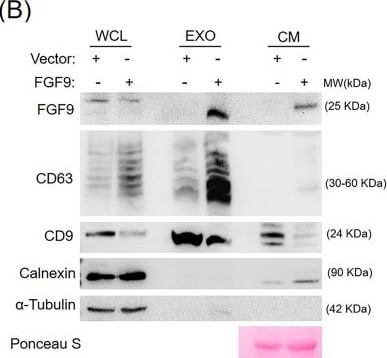
Fibroblast growth factor 9 (FGF9)-exosome attenuates the senescence of primary chondrocytes. a) Nanoparticle tracking analysis (NTA) of exosomes isolated from C28/I2 chondrocytes stably expressing vector control or FGF9. b) Western blot analysis of FGF9, exosomal markers (CD63, CD9), and negative marker (calnexin) in whole cell lysates (WCL), exosomes (EXO), and conditioned media (CM). alpha -Tubulin was used as a loading control. c) Senescence-associated beta -galactosidase (SA-beta -Gal) staining of primary chondrocytes treated with vector or FGF9-exosomes (7 × 10⁹ particles) for one week. Representative images of senescent cells (blue staining, left panel) and quantification of SA-beta -Gal–positive cells (right panel) are shown. Error bars represent mean (SD). **p < 0.01, independent-samples t-test. Image collected and cropped by CiteAb from the following open publication (https://boneandjoint.org.uk/doi/10.1302/2046-3758.156.BJR-2025-0365.R1), licensed under a CC-BY license. Not internally tested by R&D Systems.

Applications for Human FGF-9 Antibody

Application
Recommended Usage

Immunohistochemistry

5-15 µg/mL
Sample: Immersion fixed paraffin-embedded sections of human placenta

Western Blot

0.1 µg/mL
Sample: Recombinant Human FGF‑9 (Catalog # 273-F9)

Reviewed Applications

Read 1 review rated 5 using AF-273-NA in the following applications:

Formulation, Preparation, and Storage

Purification

Antigen Affinity-purified

Reconstitution

Reconstitute at 0.2 mg/mL in sterile PBS. For liquid material, refer to CoA for concentration.


Loading...

Formulation

Lyophilized from a 0.2 μm filtered solution in PBS with Trehalose. *Small pack size (SP) is supplied either lyophilized or as a 0.2 µm filtered solution in PBS.

Shipping

Lyophilized product is shipped at ambient temperature. Liquid small pack size (-SP) is shipped with polar packs. Upon receipt, store immediately at the temperature recommended below.

Stability & Storage

Use a manual defrost freezer and avoid repeated freeze-thaw cycles.
  • 12 months from date of receipt, -20 to -70 °C as supplied.
  • 1 month, 2 to 8 °C under sterile conditions after reconstitution.
  • 6 months, -20 to -70 °C under sterile conditions after reconstitution.

Calculators

The reconstitution calculator allows you to quickly calculate the volume of a reagent to reconstitute your vial. Simply enter the mass of reagent and the target concentration and the calculator will determine the rest.

=
÷

Background: FGF-9

The FGF family is comprised of at least nine polypeptides that show a variety of biological activities toward cells of mesenchymal, neuronal, and epithelial origin. All FGFs have two conserved cysteine residues and share 30-50% sequence identity at the amino acid level. FGF-9, also named glia-activating factor, was originally identified and purified from the supernatant of a human glioma cell line as a heparin-binding mitogenic growth factor for glial cells. FGF-9 has also been shown to stimulate the proliferation of oligodendrocyte type 2 astrocyte progenitor cells, Balb/c3T3 fibroblasts, and PC-12 cells. However, unlike FGF acidic and basic, FGF-9 is not a mitogen for human umbilical vein endothelial cells.

The human FGF-9 cDNA encodes a 208 amino acid residue protein that contains a potential N-linked glycosylation site. The native protein is glycosylated. FGF-9 exhibits approximately 30% sequence similarity to other members of the FGF family. Although FGF-9 lacks a typical secretion signal, the protein is secreted efficiently after synthesis. Rat FGF-9 cDNA has been cloned and shown to be highly homologous to human FGF-9. The two proteins differ only in one amino acid residue. The expression of the FGF-9 transcripts has been shown to be restricted to the brain and the kidney.

References

  1. Naruo, K. et al. (1993) J. Biol. Chem. 268:2857.
  2. Miyamoto, M. et al. (1993) Mol. Cell Biol. 13:4251.

Long Name

Fibroblast Growth Factor 9

Alternate Names

FGF9

Entrez Gene IDs

2254 (Human); 14180 (Mouse)

Gene Symbol

FGF9

UniProt

Additional FGF-9 Products

Product Documents for Human FGF-9 Antibody

Certificate of Analysis

To download a Certificate of Analysis, please enter a lot or batch number in the search box below.

Note: Certificate of Analysis not available for kit components.

Product Specific Notices for Human FGF-9 Antibody

For research use only

Related Research Areas

Citations for Human FGF-9 Antibody

Customer Reviews for Human FGF-9 Antibody (1)

5 out of 5
1 Customer Rating
5 Stars
100%
4 Stars
0%
3 Stars
0%
2 Stars
0%
1 Stars
0%

Have you used Human FGF-9 Antibody?

Submit a review and receive an Amazon gift card!

$25/€18/£15/$25CAN/¥2500 for a review with an image

$10/€7/£6/$10CAN/¥1110 for a review without an image

Submit a review
Amazon Gift Card
Showing  1 - 1 的 1 review Showing All
Filter By:
  • Name: Anonymous
    Application: Immunohistochemistry-Paraffin
    Sample Tested: See PMID 23797050
    Species: Human
    Verified Customer | Posted 01/07/2015

There are no reviews that match your criteria.

Showing  1 - 1 的 1 review Showing All

Protocols

Find general support by application which include: protocols, troubleshooting, illustrated assays, videos and webinars.

FAQs

No product specific FAQs exist for this product.

View all FAQs for Antibodies
Loading...
