
Granulocyte Macrophage Colony Stimulating Factor (GM-CSF) is a growth factor produced by a variety of lymphoid cells. It binds to receptor heterodimers consisting of a GM-CSF-specific alpha chain and the common beta chain that is shared by the high-affinity receptors for IL-3 and IL-5.


Scientific images may be resized, cropped, or adjusted for brightness or contrast for presentation purposes. These updates are limited to presentation and do not affect the underlying scientific interpretation of the data. Where available, additional source data may be provided upon request.
Key Product Details
Species Reactivity
Validated:
Human
Cited:
Human, Mouse
Applications
Validated:
Western Blot, Neutralization, Immunocytochemistry
Cited:
Immunohistochemistry, Immunohistochemistry-Paraffin, Immunohistochemistry-Frozen, Neutralization, Immunodepletion
Label
Unconjugated
Antibody Source
Monoclonal Mouse IgG1 Clone # 3209
Loading...
Product Specifications
Immunogen
E. coli-derived recombinant human GM-CSF
Ala18-Glu144
Accession # P04141
Ala18-Glu144
Accession # P04141
Specificity
Detects human GM-CSF in Western blots.
Clonality
Monoclonal
Host
Mouse
Isotype
IgG1
Endotoxin Level
<0.10 EU per 1 μg of the antibody by the LAL method.
Scientific Data Images for Human GM-CSF Antibody (3209)
GMCSF in Human PBMCs.
Granulocyte Macrophage Colony Stimulating Factor (GM-CSF) was detected in human peripheral blood mononuclear cells (PBMCs) using Human GM-CSF Monoclonal Antibody (Catalog # MAB215) at 10 µg/mL for 3 hours at room temperature. Cells were stained using the NorthernLights™ 557-conjugated Anti-Mouse IgG Secondary Antibody (red; Catalog # NL007) and counterstained with DAPI (blue). View our protocol for Fluorescent ICC Staining of Non-adherent Cells.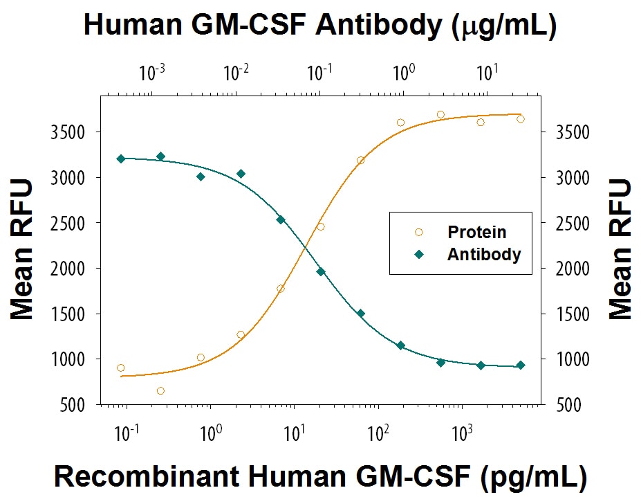
Cell Proliferation Induced by GM‑CSF and Neutralization by Human GM‑CSF Antibody.
Recombinant Human GM-CSF (Catalog # 215-GM) stimulates proliferation in the TF-1 human erythroleukemic cell line in a dose-dependent manner (orange line) as measured by Resazurin (Catalog # AR002). Proliferation elicited by Recombinant Human GM-CSF (0.1 ng/mL) is neutralized (green line) by increasing concentrations of Human GM-CSF Monoclonal Antibody (Catalog # MAB215). The ND50 is typically 0.075-0.45 µg/mL.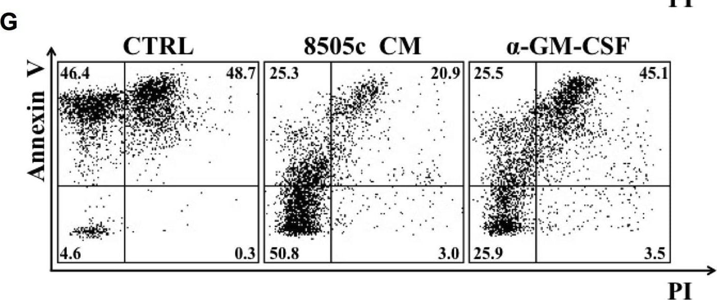
Detection of GM-CSF by Flow Cytometry
TC-derived soluble factors promoted neutrophil survival. A. Neutrophils were cultured in a TC-CM or the control medium. At the indicated time points, live cells were evaluated by flow cytometry with FITC-conjugated annexin V and PI. Results were expressed as percentages of live cells (mean ± SEM of five independent experiments); ***p < 0.005; **p < 0.01; *p < 0.05. B. Representative flow cytometric panels of dot plots of PMNs cultured in a TC-CM or control medium and stained with FITC-conjugated annexin V and propidium iodide (PI) at 24 (upper panels) and 48 (lower panels) hours. C. The GM-CSF release by TPC1 and 8505c cells was evaluated by an ELISA in a TC-CM or in the control medium. Results were expressed as mean ± SEM of seven independent experiments; ****p < 0.001; ***p < 0.005. D-F. Neutrophil survival in a TPC1-derived (D-E) or 8505c-derived (F-G) conditioned medium was evaluated in the presence of an anti-GM-CSF blocking antibody or the relative isotype control (10 μg/ml). At 24 hours, live cells were stained with FITC-conjugated annexin V and PI and analyzed by flow cytometry. Figs E and G illustrate representative flow cytometric panels of one out of five independent experiments. The results were expressed as mean ± SEM of five independent experiments; **p < 0.01; *p < 0.05. Image collected and cropped by CiteAb from the following open publication (https://pubmed.ncbi.nlm.nih.gov/29953504), licensed under a CC-BY license. Not internally tested by R&D Systems.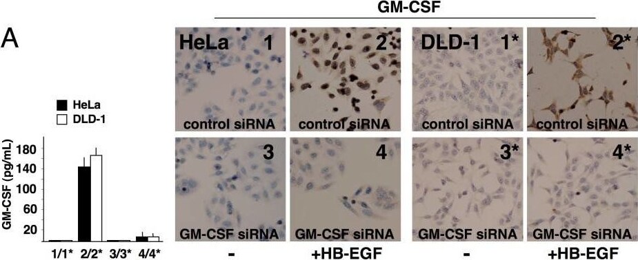
Detection of GM-CSF by Immunohistochemistry
Knockdown of GM-CSF protein levels after siRNA application in cancer cells. HeLa/DLD-1 cells were transfected with control siRNA (1/1*, 2/2*) or GM-CSF siRNA (3/3*, 4/4*) and cultured in the absence or presence of 25 ng/mL HB-EGF. The numbers indicate the culture conditions and the corresponding supernatants (SN) used for ELISA or cell stimulation. (A) Blockade of GM-CSF production in cultures of HeLa/DLD-1 cells transfected with GM-CSF siRNA was confirmed by immunocytochemistry (2/2* vs. 4/4*) and ELISA (left side; 2/2* vs. 4/4*, p < 0.05). (B) SN from GM-CSF-silenced HeLa/DLD-1 did not induce HB-EGF expression in mononuclear phagocytes (Mø), as revealed by flow cytometry (2/2* vs. 4/4*) and ELISA (left side; 2/2* vs. 4/4*, p < 0.05). (C) Mø stimulated with SN from GM-CSF-silenced HeLa/DLD-1 cells released SN less effective at inducing GM-CSF in non-silenced cancer cells, as determined by ELISA (see Methods section; SN2 vs. SN4, p < 0.05). Representative pictures or the means ± SD out of 5 experiments are shown. Image collected and cropped by CiteAb from the following open publication (https://pubmed.ncbi.nlm.nih.gov/20946648), licensed under a CC-BY license. Not internally tested by R&D Systems.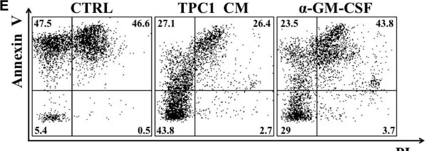
Detection of GM-CSF by Flow Cytometry
TC-derived soluble factors promoted neutrophil survival. A. Neutrophils were cultured in a TC-CM or the control medium. At the indicated time points, live cells were evaluated by flow cytometry with FITC-conjugated annexin V and PI. Results were expressed as percentages of live cells (mean ± SEM of five independent experiments); ***p < 0.005; **p < 0.01; *p < 0.05. B. Representative flow cytometric panels of dot plots of PMNs cultured in a TC-CM or control medium and stained with FITC-conjugated annexin V and propidium iodide (PI) at 24 (upper panels) and 48 (lower panels) hours. C. The GM-CSF release by TPC1 and 8505c cells was evaluated by an ELISA in a TC-CM or in the control medium. Results were expressed as mean ± SEM of seven independent experiments; ****p < 0.001; ***p < 0.005. D-F. Neutrophil survival in a TPC1-derived (D-E) or 8505c-derived (F-G) conditioned medium was evaluated in the presence of an anti-GM-CSF blocking antibody or the relative isotype control (10 μg/ml). At 24 hours, live cells were stained with FITC-conjugated annexin V and PI and analyzed by flow cytometry. Figs E and G illustrate representative flow cytometric panels of one out of five independent experiments. The results were expressed as mean ± SEM of five independent experiments; **p < 0.01; *p < 0.05. Image collected and cropped by CiteAb from the following open publication (https://pubmed.ncbi.nlm.nih.gov/29953504), licensed under a CC-BY license. Not internally tested by R&D Systems.
Human GM-CSF ELISA Standard Curve
Recombinant Human GM‑CSF (Catalog # 215-GM) was serially diluted and captured by Mouse Anti-Human GM‑CSF Monoclonal Antibody (Catalog # MAB615) coated on a Clear Polystyrene Microplate (Catalog # DY990). Mouse Anti-Human GM‑CSF Monoclonal Antibody (Catalog # MAB215) was biotinylated and incubated with the protein captured on the plate. Detection of the standard curve was achieved by incubating Streptavidin-HRP (Catalog # DY998)Applications for Human GM-CSF Antibody (3209)
Application
Recommended Usage
Immunocytochemistry
8-25 µg/mL
Sample: Immersion fixed human peripheral blood mononuclear cells
Sample: Immersion fixed human peripheral blood mononuclear cells
Western Blot
1 µg/mL
Sample: Recombinant Human GM-CSF (Catalog # 215-GM)
under non-reducing conditions only
Sample: Recombinant Human GM-CSF (Catalog # 215-GM)
under non-reducing conditions only
Neutralization
Measured by its ability to neutralize GM‑CSF-induced proliferation in the TF‑1 human erythroleukemic cell line. Kitamura, T. et al. (1989) J. Cell Physiol. 140:323. The Neutralization Dose (ND50) is typically 0.075-0.45 µg/mL in the presence of 0.1 ng/mL Recombinant Human GM‑CSF.
Reviewed Applications
Read 2 reviews rated 4.5 using MAB215 in the following applications:
Formulation, Preparation, and Storage
Purification
Protein A or G purified from hybridoma culture supernatant
Reconstitution
Reconstitute at 0.5 mg/mL in sterile PBS. For liquid material, refer to CoA for concentration.
Loading...
Formulation
Lyophilized from a 0.2 μm filtered solution in PBS with Trehalose. *Small pack size (SP) is supplied either lyophilized or as a 0.2 µm filtered solution in PBS.
Shipping
Lyophilized product is shipped at ambient temperature. Liquid small pack size (-SP) is shipped with polar packs. Upon receipt, store immediately at the temperature recommended below.
Stability & Storage
Use a manual defrost freezer and avoid repeated freeze-thaw cycles.
- 12 months from date of receipt, -20 to -70 °C as supplied.
- 1 month, 2 to 8 °C under sterile conditions after reconstitution.
- 6 months, -20 to -70 °C under sterile conditions after reconstitution.
Calculators
Background: GM-CSF
Long Name
Granulocyte Macrophage Growth Factor
Alternate Names
CSF-2, CSF2, GMCSF, Molgramostim, Sargramostim
Entrez Gene IDs
Gene Symbol
CSF2
UniProt
Additional GM-CSF Products
Product Documents for Human GM-CSF Antibody (3209)
Certificate of Analysis
To download a Certificate of Analysis, please enter a lot or batch number in the search box below.
Note: Certificate of Analysis not available for kit components.
Product Specific Notices for Human GM-CSF Antibody (3209)
For research use only
Related Research Areas
Citations for Human GM-CSF Antibody (3209)
 Powered by Bioz
Customer Reviews for Human GM-CSF Antibody (3209) (2)
4.5 out of 5
2 Customer Ratings
Have you used Human GM-CSF Antibody (3209)?
Submit a review and receive an Amazon gift card!
$25/€18/£15/$25CAN/¥2500 for a review with an image
$10/€7/£6/$10CAN/¥1110 for a review without an image
Submit a review
Customer Images
Showing
1
-
2 的
2 reviews
Showing All
Filter By:
-
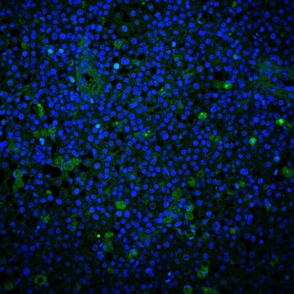
Application: Immunocytochemistry/ImmunofluorescenceSample Tested: Melanoma tissueSpecies: HumanVerified Customer | Posted 11/23/2020

-
Application: Western BlotSample Tested: Pancreatic cancer cellsSpecies: HumanVerified Customer | Posted 05/02/2018
There are no reviews that match your criteria.
Protocols
Find general support by application which include: protocols, troubleshooting, illustrated assays, videos and webinars.
- Appropriate Fixation of IHC/ICC Samples
- Cellular Response to Hypoxia Protocols
- ClariTSA™ Fluorophore Kits
- Detection & Visualization of Antibody Binding
- ICC Cell Smear Protocol for Suspension Cells
- ICC Immunocytochemistry Protocol Videos
- ICC for Adherent Cells
- Immunocytochemistry (ICC) Protocol
- Immunocytochemistry Troubleshooting
- Immunofluorescence of Organoids Embedded in Cultrex Basement Membrane Extract
- Immunohistochemistry (IHC) and Immunocytochemistry (ICC) Protocols
- Preparing Samples for IHC/ICC Experiments
- Preventing Non-Specific Staining (Non-Specific Binding)
- Primary Antibody Selection & Optimization
- Protocol for VisUCyte™ HRP Polymer Detection Reagent
- Protocol for the Fluorescent ICC Staining of Cell Smears - Graphic
- Protocol for the Fluorescent ICC Staining of Cultured Cells on Coverslips - Graphic
- Protocol for the Preparation and Fluorescent ICC Staining of Cells on Coverslips
- Protocol for the Preparation and Fluorescent ICC Staining of Non-adherent Cells
- Protocol for the Preparation and Fluorescent ICC Staining of Stem Cells on Coverslips
- Protocol for the Preparation of a Cell Smear for Non-adherent Cell ICC - Graphic
- R&D Systems Quality Control Western Blot Protocol
- TUNEL and Active Caspase-3 Detection by IHC/ICC Protocol
- The Importance of IHC/ICC Controls
- Troubleshooting Guide: Western Blot Figures
- Western Blot Conditions
- Western Blot Protocol
- Western Blot Protocol for Cell Lysates
- Western Blot Troubleshooting
- Western Blot Troubleshooting Guide
- View all Protocols, Troubleshooting, Illustrated assays and Webinars


