
Key Product Details
Species Reactivity
Validated:
Human
Cited:
Human, Mouse, Porcine
Applications
Validated:
Immunohistochemistry, Western Blot, Neutralization
Cited:
Immunohistochemistry-Paraffin, Western Blot, Neutralization, Flow Cytometry, Immunocytochemistry, Bioassay, Functional Assay, Luminex Development
Label
Unconjugated
Antibody Source
Monoclonal Mouse IgG1 Clone # 24612
Loading...
Product Specifications
Immunogen
S. frugiperda insect ovarian cell line Sf 21-derived recombinant human HGF
Gln32-Ser728 (Asp384Asn, Asp416Asn, Leu622Ser, His645Arg, Val678Ile)
Accession # P14210
Gln32-Ser728 (Asp384Asn, Asp416Asn, Leu622Ser, His645Arg, Val678Ile)
Accession # P14210
Specificity
Detects human HGF in Western blots. In Western blots, no-cross reactivity with recombinant mouse HGF or recombinant canine HGF is observed.
Clonality
Monoclonal
Host
Mouse
Isotype
IgG1
Endotoxin Level
<0.10 EU per 1 μg of the antibody by the LAL method.
Scientific Data Images for Human HGF Antibody (24612)
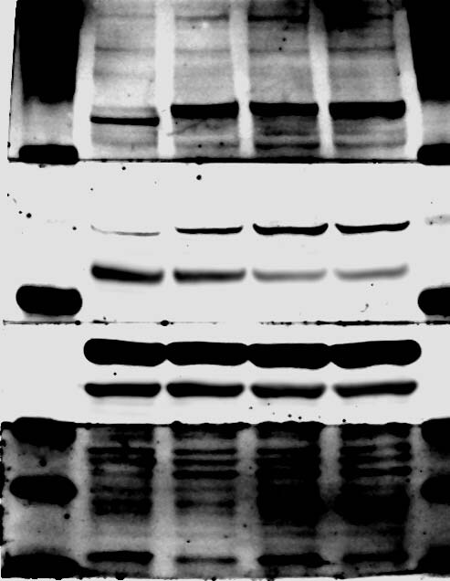
Detection of Recombinant Human, Mouse, and Canine HGF by Western Blot.
Western blot shows 25 ng of Recombinant Human HGF (Catalog # 294-HG), Recombinant Mouse HGF (2207-HG) and Recombinant Canine HGF (3386-HG). PVDF Membrane was probed with 1 µg/mL of Mouse Anti-Human HGF Monoclonal Antibody (Catalog # MAB294) followed by HRP-conjugated Anti-Mouse IgG Secondary Antibody (Catalog # HAF018). A specific band was detected for HGF at approximately 75 kDa (as indicated). This experiment was conducted under non-reducing conditions and using Immunoblot Buffer Group 3.
HGF in Human Lung Adenocarcinoma Tissue.
HGF was detected in immersion fixed paraffin-embedded sections of human lung adenocarcinoma tissue using Mouse Anti-Human HGF Monoclonal Antibody (Catalog # MAB294) at 15 µg/mL overnight at 4 °C. Before incubation with the primary antibody, tissue was subjected to heat-induced epitope retrieval using Antigen Retrieval Reagent-Basic (CTS013). Tissue was stained using the Anti-Mouse IgG VisUCyte™ HRP Polymer Antibody (brown VC001) and counterstained with hematoxylin (blue). Specific staining was localized to cytoplasm in cancer cells. View our protocol for IHC Staining with VisUCyte HRP Polymer Detection Reagents.
Cell Proliferation Induced by HGF and Neutralization by Human HGF Antibody.
Recombinant Human HGF (Catalog # 294-HG) stimulates proliferation in the 4MBr-5 rhesus monkey epithelial cell line in a dose-dependent manner (orange line). Proliferation elicited by Recombinant Human HGF (100 ng/mL) is neutralized (green line) by increasing concentrations of Mouse Anti-Human HGF Monoclonal Antibody (Catalog # MAB294). The ND50 is typically 0.1-0.3 µg/mL.
Detection of HGF by Western Blot
VEGF and HGF cooperate to induce ERK1/2, but not Akt or P38-mediated intracellular signaling.(A–C) – HUVEC were stimulated by 25 ng/ml of VEGF165, HGF or their combination (5 or 15 minutes). Cell lysates were analyzed using western blot with specific antibodies against phosphorylated ERK1/2, Akt or P38 and normalized against beta -actin or Akt to obtain densitometric data and corresponding graphs (right column). Image collected and cropped by CiteAb from the following open publication (https://pubmed.ncbi.nlm.nih.gov/22719942), licensed under a CC-BY license. Not internally tested by R&D Systems.
Detection of HGF by Western Blot
FB2-conditioned medium (CM) (A) and CAF-associated ligands HGF and Nrg1 beta 1 (B) influence the expression of fatty acid metabolism-regulating targets upon ALK signaling perturbation via lorlatinib in H2228 and H3122 cells, as shown by representative western blots of FASN, ACLY, and SREBP-1 in H2228 and H3122 cells. Expression fold changes were normalized to those of beta -actin. ACLY, ATP-citrate lyase; FASN, fatty acid synthase; pSREBP-1, precursor sterol-responsive element binding protein-1; mSREBP-1, mature SREBP-1 Image collected and cropped by CiteAb from the following open publication (https://pubmed.ncbi.nlm.nih.gov/40524253), licensed under a CC-BY license. Not internally tested by R&D Systems.
Detection of HGF by Western Blot
FB2-conditioned medium (CM) (A) and CAF-associated ligands HGF and Nrg1 beta 1 (B) influence the activation of AKT signaling upon ALK signaling perturbation via lorlatinib in H2228 and H3122 cells, as shown by representative western blots of AKT/pAKT and S6K/pS6K in H2228 and H3122 cells. Phosphorylation rates were normalized to those of beta -actin. AKT, protein kinase B; S6K, ribosomal S6 kinase Image collected and cropped by CiteAb from the following open publication (https://pubmed.ncbi.nlm.nih.gov/40524253), licensed under a CC-BY license. Not internally tested by R&D Systems.Applications for Human HGF Antibody (24612)
Application
Recommended Usage
Immunohistochemistry
8-25 µg/mL
Western Blot
1 µg/mL
Sample: Recombinant Human HGF (Catalog # 294-HG)
under non-reducing conditions only
Sample: Recombinant Human HGF (Catalog # 294-HG)
under non-reducing conditions only
Neutralization
Measured by its ability to neutralize HGF-induced proliferation in the 4MBr‑5 rhesus monkey epithelial cell line. The Neutralization Dose (ND50) is typically 0.1-0.3 µg/mL in the presence of 100 ng/mL Recombinant Human HGF.
Reviewed Applications
Read 1 review rated 4 using MAB294 in the following applications:
Formulation, Preparation, and Storage
Purification
Protein A or G purified from hybridoma culture supernatant
Reconstitution
Reconstitute at 0.5 mg/mL in sterile PBS. For liquid material, refer to CoA for concentration.
Loading...
Formulation
Lyophilized from a 0.2 μm filtered solution in PBS with Trehalose. *Small pack size (SP) is supplied either lyophilized or as a 0.2 µm filtered solution in PBS.
Shipping
Lyophilized product is shipped at ambient temperature. Liquid small pack size (-SP) is shipped with polar packs. Upon receipt, store immediately at the temperature recommended below.
Stability & Storage
Use a manual defrost freezer and avoid repeated freeze-thaw cycles.
- 12 months from date of receipt, -20 to -70 °C as supplied.
- 1 month, 2 to 8 °C under sterile conditions after reconstitution.
- 6 months, -20 to -70 °C under sterile conditions after reconstitution.
Calculators
Background: HGF
Long Name
Hepatocyte Growth Factor
Alternate Names
DFNB39, F-TCF, Hepatopoietin A, HGFB, HPTA, SF
Entrez Gene IDs
Gene Symbol
HGF
UniProt
Additional HGF Products
Product Documents for Human HGF Antibody (24612)
Certificate of Analysis
To download a Certificate of Analysis, please enter a lot or batch number in the search box below.
Note: Certificate of Analysis not available for kit components.
Product Specific Notices for Human HGF Antibody (24612)
For research use only
Related Research Areas
Citations for Human HGF Antibody (24612)
 Powered by Bioz
Customer Reviews for Human HGF Antibody (24612) (1)
4 out of 5
1 Customer Rating
Have you used Human HGF Antibody (24612)?
Submit a review and receive an Amazon gift card!
$25/€18/£15/$25CAN/¥2500 Yen for a review with an image
$10/€7/£6/$10CAN/¥1110 Yen for a review without an image
Submit a review
Customer Images
Showing
1
-
1 的
1 review
Showing All
Filter By:
-
Application: Western BlotSample Tested: Cancer cell lysatesSpecies: HumanVerified Customer | Posted 09/08/2017

There are no reviews that match your criteria.
Protocols
Find general support by application which include: protocols, troubleshooting, illustrated assays, videos and webinars.
- Antigen Retrieval Protocol (PIER)
- Antigen Retrieval for Frozen Sections Protocol
- Appropriate Fixation of IHC/ICC Samples
- Cellular Response to Hypoxia Protocols
- Chromogenic IHC Staining of Formalin-Fixed Paraffin-Embedded (FFPE) Tissue Protocol
- Chromogenic Immunohistochemistry Staining of Frozen Tissue
- ClariTSA™ Fluorophore Kits
- Detection & Visualization of Antibody Binding
- Fluorescent IHC Staining of Frozen Tissue Protocol
- Graphic Protocol for Heat-induced Epitope Retrieval
- Graphic Protocol for the Preparation and Fluorescent IHC Staining of Frozen Tissue Sections
- Graphic Protocol for the Preparation and Fluorescent IHC Staining of Paraffin-embedded Tissue Sections
- Graphic Protocol for the Preparation of Gelatin-coated Slides for Histological Tissue Sections
- IHC Sample Preparation (Frozen sections vs Paraffin)
- Immunofluorescent IHC Staining of Formalin-Fixed Paraffin-Embedded (FFPE) Tissue Protocol
- Immunohistochemistry (IHC) and Immunocytochemistry (ICC) Protocols
- Immunohistochemistry Frozen Troubleshooting
- Immunohistochemistry Paraffin Troubleshooting
- Preparing Samples for IHC/ICC Experiments
- Preventing Non-Specific Staining (Non-Specific Binding)
- Primary Antibody Selection & Optimization
- Protocol for Heat-Induced Epitope Retrieval (HIER)
- Protocol for Making a 4% Formaldehyde Solution in PBS
- Protocol for VisUCyte™ HRP Polymer Detection Reagent
- Protocol for the Preparation & Fixation of Cells on Coverslips
- Protocol for the Preparation and Chromogenic IHC Staining of Frozen Tissue Sections
- Protocol for the Preparation and Chromogenic IHC Staining of Frozen Tissue Sections - Graphic
- Protocol for the Preparation and Chromogenic IHC Staining of Paraffin-embedded Tissue Sections
- Protocol for the Preparation and Chromogenic IHC Staining of Paraffin-embedded Tissue Sections - Graphic
- Protocol for the Preparation and Fluorescent IHC Staining of Frozen Tissue Sections
- Protocol for the Preparation and Fluorescent IHC Staining of Paraffin-embedded Tissue Sections
- Protocol for the Preparation of Gelatin-coated Slides for Histological Tissue Sections
- R&D Systems Quality Control Western Blot Protocol
- TUNEL and Active Caspase-3 Detection by IHC/ICC Protocol
- The Importance of IHC/ICC Controls
- Troubleshooting Guide: Immunohistochemistry
- Troubleshooting Guide: Western Blot Figures
- Western Blot Conditions
- Western Blot Protocol
- Western Blot Protocol for Cell Lysates
- Western Blot Troubleshooting
- Western Blot Troubleshooting Guide
- View all Protocols, Troubleshooting, Illustrated assays and Webinars
Loading...
Associated Pathways
