
Matrix metalloproteinases are a family of zinc and calcium dependent endopeptidases with the combined ability to degrade all the components of the extracellular matrix. MMP-10 (stromelysin 2) degrades a broad range of substrates including gelatin, collagen types III, IV and V, fibronectin, aggrecan, and pig cartilage proteoglycan. MMP-10 can activate other MMPs such as MMP-1 and MMP-8. MMP-10 is expressed in keratinocytes, T cells, menstrual endometrium, and a few tumor samples. Structurally, MMP-10 may be divided into four distinct domains: a pro-domain which is cleaved upon activation, a catalytic domain containing the zinc binding site; a short linker region, and a carboxyl terminal hemopexin-like domain.

Scientific images may be resized, cropped, or adjusted for brightness or contrast for presentation purposes. These updates are limited to presentation and do not affect the underlying scientific interpretation of the data. Where available, additional source data may be provided upon request.
Key Product Details
Species Reactivity
Validated:
Human
Cited:
Human
Applications
Validated:
Immunohistochemistry, Western Blot, Neutralization, Immunoprecipitation
Cited:
Immunohistochemistry, Western Blot
Label
Unconjugated
Antibody Source
Monoclonal Mouse IgG2B Clone # 117239
Loading...
Product Specifications
Immunogen
Mouse myeloma cell line NS0-derived recombinant human MMP-10
Tyr18-Cys476
Accession # P09238
Tyr18-Cys476
Accession # P09238
Specificity
Detects the pro and active forms of human MMP-10 in direct ELISAs and Western blots. In direct ELISAs, no cross-reactivity with recombinant human MMP-1, -2, -3, -7, -8, -9, -12, or -13 is observed.
Clonality
Monoclonal
Host
Mouse
Isotype
IgG2B
Endotoxin Level
<0.10 EU per 1 μg of the antibody by the LAL method.
Scientific Data Images for Human MMP-10 Antibody (117239)
Detection of Human MMP-10 by Immunohistochemistry
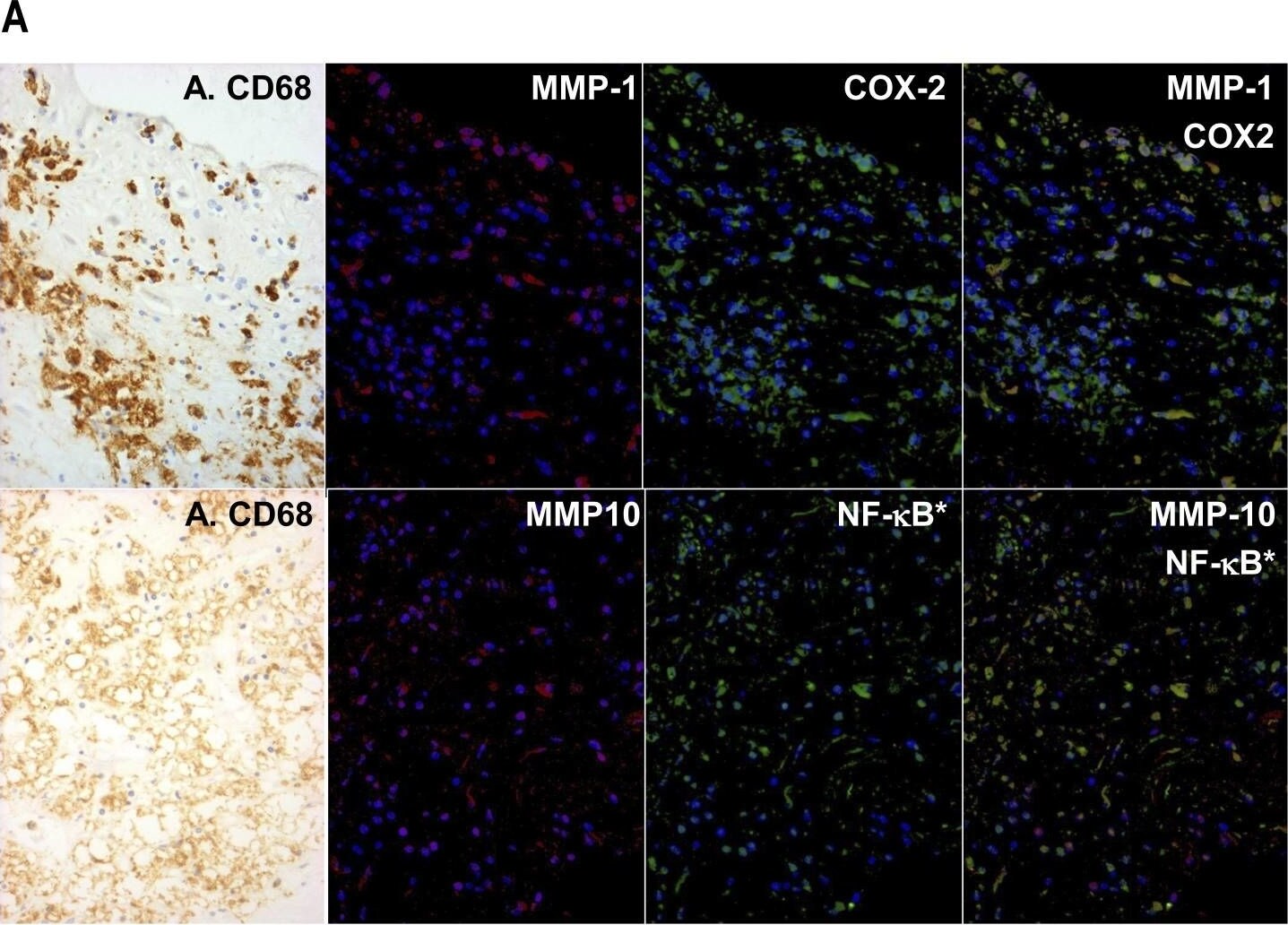
Co-localisation of MMPs with markers of classical activation in human atherosclerotic plaques.(A) Serial sections were stained by peroxidase with anti-CD68 (A. CD68) and by dual immuno-fluorescence with anti-MMP-1 or anti-MMP-10 (red) together with anti-COX-2 or p65RelA (green) as shown, and the images were superimposed digitally. Nuclear localised p65RelA (NF-kB*) was detected because of the shift in nuclear counterstain colour from dark blue (DAPI alone) to sky blue (blue plus green) in the superimposed image. (B) Areas rich in macrophages identified from the peroxidase stain were identified in the serial section. Cells in the whole field were counted and a percentage of each staining pattern calculated. Values are means ± SEM, n = 6, * p<0.05. Image collected and cropped by CiteAb from the following open publication (https://pubmed.ncbi.nlm.nih.gov/22880008), licensed under a CC-BY license. Not internally tested by R&D Systems.Applications for Human MMP-10 Antibody (117239)
Application
Recommended Usage
Immunohistochemistry
8-25 µg/mL
Sample: Immersion fixed paraffin-embedded sections of human intestine
Sample: Immersion fixed paraffin-embedded sections of human intestine
Immunoprecipitation
25 µg/mL
Sample: Conditioned cell culture medium spiked with Recombinant Human MMP‑10 (Catalog # 910‑MP), see our available Western blot detection antibodies
Sample: Conditioned cell culture medium spiked with Recombinant Human MMP‑10 (Catalog # 910‑MP), see our available Western blot detection antibodies
Western Blot
1 µg/mL
Sample: Recombinant Human MMP‑10 Western Blot Standard (Catalog # WBC026)
Sample: Recombinant Human MMP‑10 Western Blot Standard (Catalog # WBC026)
Neutralization
Measured by its ability to neutralize Recombinant Human MMP‑10 (1 µg/mL, Catalog # 910‑MP) cleavage of the fluorogenic peptide substrate Mca-RPKPVE-Nval-WRK(Dnp)-NH2 (10 µM, Catalog # ES002). The Neutralization Dose (ND50) is typically 1 µg/mL.
Reviewed Applications
Read 1 review rated 1 using MAB9101 in the following applications:
Formulation, Preparation, and Storage
Purification
Protein A or G purified from hybridoma culture supernatant
Reconstitution
Reconstitute at 0.5 mg/mL in sterile PBS. For liquid material, refer to CoA for concentration.
Loading...
Formulation
Lyophilized from a 0.2 μm filtered solution in PBS with Trehalose. *Small pack size (SP) is supplied either lyophilized or as a 0.2 µm filtered solution in PBS.
Shipping
Lyophilized product is shipped at ambient temperature. Liquid small pack size (-SP) is shipped with polar packs. Upon receipt, store immediately at the temperature recommended below.
Stability & Storage
Use a manual defrost freezer and avoid repeated freeze-thaw cycles.
- 12 months from date of receipt, -20 to -70 °C as supplied.
- 1 month, 2 to 8 °C under sterile conditions after reconstitution.
- 6 months, -20 to -70 °C under sterile conditions after reconstitution.
Calculators
Background: MMP-10
Long Name
Matrix Metalloproteinase 10
Alternate Names
MMP10, Stromelysin 2
Entrez Gene IDs
4319 (Human)
Gene Symbol
MMP10
UniProt
Additional MMP-10 Products
Product Documents for Human MMP-10 Antibody (117239)
Certificate of Analysis
To download a Certificate of Analysis, please enter a lot or batch number in the search box below.
Note: Certificate of Analysis not available for kit components.
Product Specific Notices for Human MMP-10 Antibody (117239)
For research use only
Related Research Areas
Citations for Human MMP-10 Antibody (117239)
 Powered by Bioz
Customer Reviews for Human MMP-10 Antibody (117239) (1)
1 out of 5
1 Customer Rating
Have you used Human MMP-10 Antibody (117239)?
Submit a review and receive an Amazon gift card!
$25/€18/£15/$25CAN/¥2500 for a review with an image
$10/€7/£6/$10CAN/¥1110 for a review without an image
Submit a review
Customer Images
Showing
1
-
1 的
1 review
Showing All
Filter By:
-
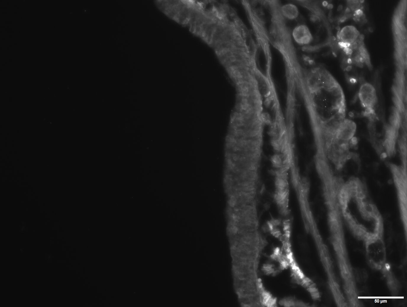
Application: Immunocytochemistry/ImmunofluorescenceSample Tested: Adult lungSpecies: HumanVerified Customer | Posted 03/13/2019Doesn't work with and without citrate retrieval, Used IgG as negative control and a different MMP10 antibody as a positive control.
 Bio-Techne ResponseThank you for reviewing our product. We are sorry to hear that this antibody did not perform as expected. We have been in touch with the customer to resolve this issue according to our Product Guarantee and to the customer’s satisfaction.
There are no reviews that match your criteria.
Protocols
Find general support by application which include: protocols, troubleshooting, illustrated assays, videos and webinars.
- Antigen Retrieval Protocol (PIER)
- Antigen Retrieval for Frozen Sections Protocol
- Appropriate Fixation of IHC/ICC Samples
- Cellular Response to Hypoxia Protocols
- Chromogenic IHC Staining of Formalin-Fixed Paraffin-Embedded (FFPE) Tissue Protocol
- Chromogenic Immunohistochemistry Staining of Frozen Tissue
- ClariTSA™ Fluorophore Kits
- Detection & Visualization of Antibody Binding
- Fluorescent IHC Staining of Frozen Tissue Protocol
- Graphic Protocol for Heat-induced Epitope Retrieval
- Graphic Protocol for the Preparation and Fluorescent IHC Staining of Frozen Tissue Sections
- Graphic Protocol for the Preparation and Fluorescent IHC Staining of Paraffin-embedded Tissue Sections
- Graphic Protocol for the Preparation of Gelatin-coated Slides for Histological Tissue Sections
- IHC Sample Preparation (Frozen sections vs Paraffin)
- Immunofluorescent IHC Staining of Formalin-Fixed Paraffin-Embedded (FFPE) Tissue Protocol
- Immunohistochemistry (IHC) and Immunocytochemistry (ICC) Protocols
- Immunohistochemistry Frozen Troubleshooting
- Immunohistochemistry Paraffin Troubleshooting
- Immunoprecipitation Protocol
- Preparing Samples for IHC/ICC Experiments
- Preventing Non-Specific Staining (Non-Specific Binding)
- Primary Antibody Selection & Optimization
- Protocol for Heat-Induced Epitope Retrieval (HIER)
- Protocol for Making a 4% Formaldehyde Solution in PBS
- Protocol for VisUCyte™ HRP Polymer Detection Reagent
- Protocol for the Preparation & Fixation of Cells on Coverslips
- Protocol for the Preparation and Chromogenic IHC Staining of Frozen Tissue Sections
- Protocol for the Preparation and Chromogenic IHC Staining of Frozen Tissue Sections - Graphic
- Protocol for the Preparation and Chromogenic IHC Staining of Paraffin-embedded Tissue Sections
- Protocol for the Preparation and Chromogenic IHC Staining of Paraffin-embedded Tissue Sections - Graphic
- Protocol for the Preparation and Fluorescent IHC Staining of Frozen Tissue Sections
- Protocol for the Preparation and Fluorescent IHC Staining of Paraffin-embedded Tissue Sections
- Protocol for the Preparation of Gelatin-coated Slides for Histological Tissue Sections
- R&D Systems Quality Control Western Blot Protocol
- TUNEL and Active Caspase-3 Detection by IHC/ICC Protocol
- The Importance of IHC/ICC Controls
- Troubleshooting Guide: Immunohistochemistry
- Troubleshooting Guide: Western Blot Figures
- Western Blot Conditions
- Western Blot Protocol
- Western Blot Protocol for Cell Lysates
- Western Blot Troubleshooting
- Western Blot Troubleshooting Guide
- View all Protocols, Troubleshooting, Illustrated assays and Webinars
Loading...