
Matrix metalloproteinases are a family of zinc and calcium dependent endopeptidases with the combined ability to degrade all the components of the extracellular matrix. MMP-13 (Collagenase-3) has been demonstrated to degrade a range of extracellular matrix proteins, including collagen types I, II, III, IV, IX, X and XIV, gelatin, aggrecan, perlecan and fibronectin. MMP-13 is distinguished from the other human collagenases by its effecient degradation of type II collagen. MMP-13 is expressed by fibroblasts, chrondrocytes and squamous epithelial cells. Structurally, MMP-13 may be divided into several distinct domains; a pro-domain which is cleaved upon activation; a catalytic domain containing the zinc binding site; a short hinge region and a carboxyl terminal (hemopexin-like) domain.

Scientific images may be resized, cropped, or adjusted for brightness or contrast for presentation purposes. These updates are limited to presentation and do not affect the underlying scientific interpretation of the data. Where available, additional source data may be provided upon request.
Key Product Details
Validated by
Species Reactivity
Validated:
Cited:
Applications
Validated:
Cited:
Label
Antibody Source
Product Specifications
Immunogen
Leu20-Cys471
Accession # P45452
Specificity
Clonality
Host
Isotype
Scientific Data Images for Human MMP-13 Antibody (87512)
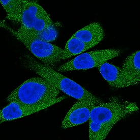
MMP‑13 in MDA‑MB‑231 Human Cell Line.
MMP-13 was detected in immersion fixed MDA-MB-231 human breast cancer cell line using Human MMP-13 Monoclonal Antibody (Catalog # MAB511) at 5 µg/mL for 3 hours at room temperature. Cells were stained using the NorthernLights™ 493-conjugated Anti-Mouse IgG Secondary Antibody (green; Catalog # NL009) and counterstained with DAPI (blue). View our protocol for Fluorescent ICC Staining of Cells on Coverslips.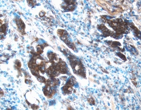
MMP‑13 in Human Ovarian Cancer Tissue.
MMP‑13 was detected in immersion fixed paraffin-embedded sections of human ovarian cancer tissue using 8 µg/mL Mouse Anti-Human MMP‑13 Monoclonal Antibody (Catalog # MAB511) overnight at 4 °C. Tissue was stained with the Anti-Mouse HRP-DAB Cell & Tissue Staining Kit (brown; Catalog # CTS002) and counterstained with hematoxylin (blue). View our protocol for Chromogenic IHC Staining of Paraffin-embedded Tissue Sections.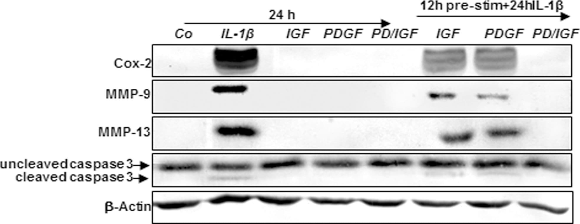
Detection of Canine MMP-13 by Western Blot
Effects of IGF-1 or/and PDGF-bb on IL-1 beta -induced NF-kappa B-dependent pro-inflammatory, pro-apoptotic and matrix degrading gene products in chondrocytes.To determine whether IGF-1 or/and PDGF-bbexert effects on IL-1 beta -induced NF-kappa B-dependent expression of pro-inflammatory, pro-apoptotic and matrix degrading gene products, primary chondrocytes were either stimulated with 10 ng/ml IL-1 beta, 10 ng/ml PDGF-bb, 10 ng/ml IGF-1 or combination of both growth factors (5 ng/ml each) or pre-stimulated for 12 h with 10 ng/ml PDGF-bb, 10 ng/ml IGF-1 or combination of both growth factors (5 ng/ml each) followed by 10 ng/ml IL-1 beta for 24. Equal amounts of total proteins were separated by SDS-PAGE and analyzed by immunoblotting using antibodies raised against COX-2, MMP-9 and MMP-13 and active caspase-3. Stimulation with IL-1 beta resulted in production of COX-2, MMP-9, MMP-13 and caspase-3 cleavage. Pre-treatment with a combination of both IGF-1 or/and PDGF-bb downregulated COX-2, MMP-9, MMP-13 and cleaved caspase-3. Image collected and cropped by CiteAb from the following publication (https://dx.plos.org/10.1371/journal.pone.0028663), licensed under a CC-BY license. Not internally tested by R&D Systems.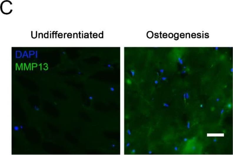
Detection of Human MMP-13 by Immunohistochemistry
Enhanced MMP13 gene expression and production due to 3D osteogenic differentiation. (A) Multivariate analysis of the sixty-six tested genes (NanoString® kit) displayed as a volcano plot (created with nSolver® software) with the 2D or 3D condition set as covariate. The plot shows each gene’s −log10 (p-value) and log2 fold change associated to the 2D or 3D condition. Highly statistically significant genes fall at the top of the plot above the indicated p-value lines, and highly differentially expressed genes fall to either side (right = upregulated, left = downregulated). The only significantly (p-value < 0.01) affected gene that was upregulated was MMP13. (B) Relative gene expression of MMP13 is shown in the four various conditions: 2D undifferentiated, 2D differentiated, 3D undifferentiated, 3D differentiated, after 5 weeks of culture. The significance levels for relative gene expression difference are indicated as ** (p-value < 0.01), *** (p-value < 0.001). (C) Immunohistochemistry of 5-week undifferentiated vs. differentiated (osteogenic) 3D Col I gels against MMP13 and nuclear DAPI. Scale bar = 50 μm. Image collected and cropped by CiteAb from the following publication (https://pubmed.ncbi.nlm.nih.gov/34948393), licensed under a CC-BY license. Not internally tested by R&D Systems.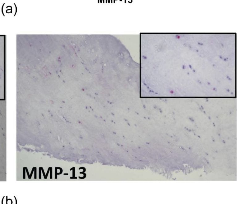
Detection of MMP-13 by Immunohistochemistry
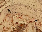
Immunohistochemistry of metalloproteinase in meniscal samples. (a) number of patients with different expression of MMP-1 and -13; (b) one representative patient for MMP-1 (grade 3) and MMP-13 (grade 1), (magnification 10×, inset 40×, respectively). Image collected and cropped by CiteAb from the following open publication (https://www.mdpi.com/1422-0067/23/7/3903), licensed under a CC-BY license. Not internally tested by R&D Systems.Applications for Human MMP-13 Antibody (87512)
Immunoaffinity Purification
Immunocytochemistry
Sample: Immersion fixed MDA-MB-231 human breast cancer cell line
Immunohistochemistry
Sample: Immersion fixed paraffin-embedded sections of human ovarian cancer tissue
Immunoprecipitation
Sample: Conditioned cell culture medium spiked with Recombinant Human MMP‑13 (Catalog # 511‑MM), see our available Western blot detection antibodies
Western Blot
Sample: Recombinant Human MMP‑13 Western Blot Standard (Catalog # WBC020)
Reviewed Applications
Read 6 reviews rated 4.7 using MAB511 in the following applications:
Formulation, Preparation, and Storage
Purification
Reconstitution
Reconstitute at 0.5 mg/mL in sterile PBS. For liquid material, refer to CoA for concentration.
Formulation
Shipping
Stability & Storage
- 12 months from date of receipt, -20 to -70 °C as supplied.
- 1 month, 2 to 8 °C under sterile conditions after reconstitution.
- 6 months, -20 to -70 °C under sterile conditions after reconstitution.
Calculators
Background: MMP-13
References
- Jeffery, J.J. (1998) in Collagenase 3. A.J. Barrett, et al. (eds): Handbook of Proteolytic Enzymes, San Diego: Academic Press, p. 1167.
Long Name
Alternate Names
Entrez Gene IDs
Gene Symbol
UniProt
Additional MMP-13 Products
Product Documents for Human MMP-13 Antibody (87512)
Certificate of Analysis
To download a Certificate of Analysis, please enter a lot or batch number in the search box below.
Note: Certificate of Analysis not available for kit components.
Product Specific Notices for Human MMP-13 Antibody (87512)
For research use only
Related Research Areas
Citations for Human MMP-13 Antibody (87512)
 Powered by Bioz
Customer Reviews for Human MMP-13 Antibody (87512) (6)
Have you used Human MMP-13 Antibody (87512)?
Submit a review and receive an Amazon gift card!
$25/€18/£15/$25CAN/¥2500 for a review with an image
$10/€7/£6/$10CAN/¥1110 for a review without an image
Submit a review
Customer Images
-
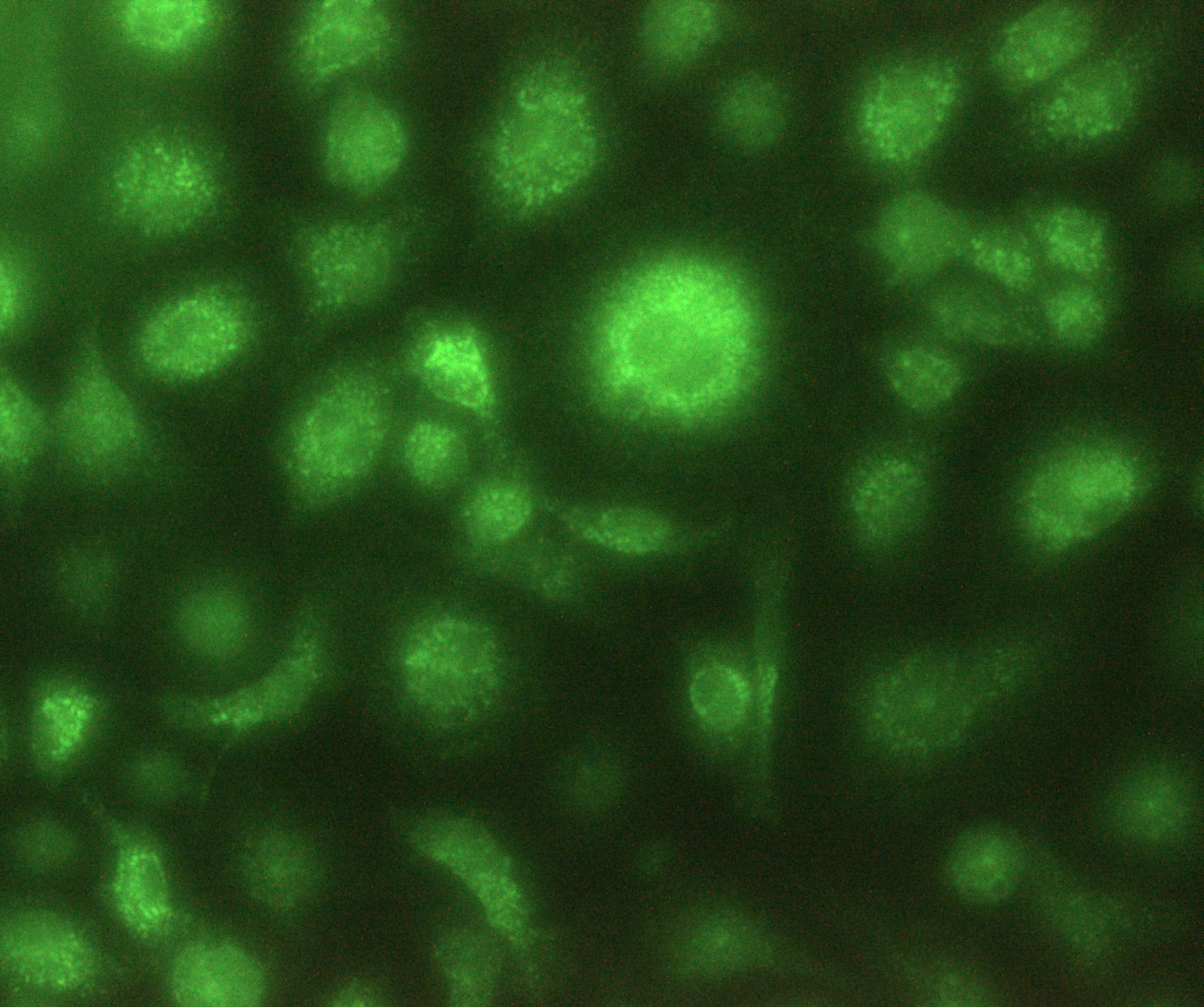
Application: ImmunocytochemistrySample Tested: Bone Marrrow Derived MacrophagesSpecies: MouseVerified Customer | Posted 02/07/2022Macrophages in vitro produce MMP131:200 Dilution Overnight incubation at RT
 Bio-Techne ResponseThis review was submitted through the legacy Novus Innovators Program, reflecting a new species or application tested on a primary antibody.
 -
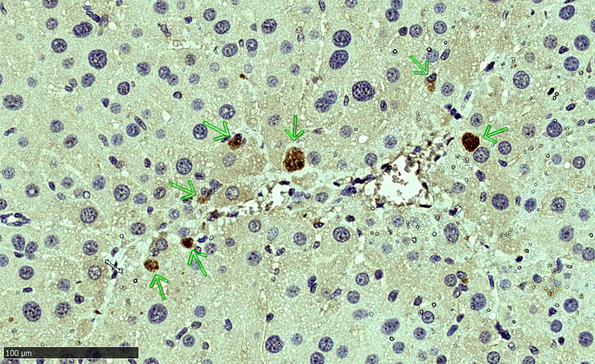
Application: Immunocytochemistry/ImmunofluorescenceSample Tested: Liver tissueSpecies: MouseVerified Customer | Posted 02/07/20221:100 dilution Citrate buffer Ag retrieval Overnight incubation of the Ab, RT
-
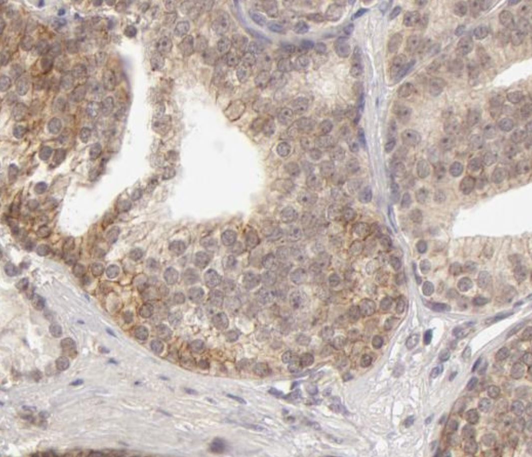
Application: ImmunohistochemistrySample Tested: Prostate cancer tissueSpecies: HumanVerified Customer | Posted 12/06/2021
-
Application: ImmunohistochemistrySample Tested: Joint tissueSpecies: HumanVerified Customer | Posted 10/14/2021
-
Application: Western BlotSample Tested: HeLa cellsSpecies: HumanVerified Customer | Posted 07/13/2018
-
Application: ELISASample Tested: Serum and PlasmaSpecies: HumanVerified Customer | Posted 11/10/2017The antibody AF511 was used as the detection antibody with this antibody as the capture in an ELISA targeting MMP-13. The immunoassay was used to measure MMP-13 levels in human plasma samples.
There are no reviews that match your criteria.
Protocols
Find general support by application which include: protocols, troubleshooting, illustrated assays, videos and webinars.
- Antigen Retrieval Protocol (PIER)
- Antigen Retrieval for Frozen Sections Protocol
- Appropriate Fixation of IHC/ICC Samples
- Cellular Response to Hypoxia Protocols
- Chromogenic IHC Staining of Formalin-Fixed Paraffin-Embedded (FFPE) Tissue Protocol
- Chromogenic Immunohistochemistry Staining of Frozen Tissue
- ClariTSA™ Fluorophore Kits
- Detection & Visualization of Antibody Binding
- Fluorescent IHC Staining of Frozen Tissue Protocol
- Graphic Protocol for Heat-induced Epitope Retrieval
- Graphic Protocol for the Preparation and Fluorescent IHC Staining of Frozen Tissue Sections
- Graphic Protocol for the Preparation and Fluorescent IHC Staining of Paraffin-embedded Tissue Sections
- Graphic Protocol for the Preparation of Gelatin-coated Slides for Histological Tissue Sections
- ICC Cell Smear Protocol for Suspension Cells
- ICC Immunocytochemistry Protocol Videos
- ICC for Adherent Cells
- IHC Sample Preparation (Frozen sections vs Paraffin)
- Immunocytochemistry (ICC) Protocol
- Immunocytochemistry Troubleshooting
- Immunofluorescence of Organoids Embedded in Cultrex Basement Membrane Extract
- Immunofluorescent IHC Staining of Formalin-Fixed Paraffin-Embedded (FFPE) Tissue Protocol
- Immunohistochemistry (IHC) and Immunocytochemistry (ICC) Protocols
- Immunohistochemistry Frozen Troubleshooting
- Immunohistochemistry Paraffin Troubleshooting
- Immunoprecipitation Protocol
- Preparing Samples for IHC/ICC Experiments
- Preventing Non-Specific Staining (Non-Specific Binding)
- Primary Antibody Selection & Optimization
- Protocol for Heat-Induced Epitope Retrieval (HIER)
- Protocol for Making a 4% Formaldehyde Solution in PBS
- Protocol for VisUCyte™ HRP Polymer Detection Reagent
- Protocol for the Fluorescent ICC Staining of Cell Smears - Graphic
- Protocol for the Fluorescent ICC Staining of Cultured Cells on Coverslips - Graphic
- Protocol for the Preparation & Fixation of Cells on Coverslips
- Protocol for the Preparation and Chromogenic IHC Staining of Frozen Tissue Sections
- Protocol for the Preparation and Chromogenic IHC Staining of Frozen Tissue Sections - Graphic
- Protocol for the Preparation and Chromogenic IHC Staining of Paraffin-embedded Tissue Sections
- Protocol for the Preparation and Chromogenic IHC Staining of Paraffin-embedded Tissue Sections - Graphic
- Protocol for the Preparation and Fluorescent ICC Staining of Cells on Coverslips
- Protocol for the Preparation and Fluorescent ICC Staining of Non-adherent Cells
- Protocol for the Preparation and Fluorescent ICC Staining of Stem Cells on Coverslips
- Protocol for the Preparation and Fluorescent IHC Staining of Frozen Tissue Sections
- Protocol for the Preparation and Fluorescent IHC Staining of Paraffin-embedded Tissue Sections
- Protocol for the Preparation of Gelatin-coated Slides for Histological Tissue Sections
- Protocol for the Preparation of a Cell Smear for Non-adherent Cell ICC - Graphic
- R&D Systems Quality Control Western Blot Protocol
- TUNEL and Active Caspase-3 Detection by IHC/ICC Protocol
- The Importance of IHC/ICC Controls
- Troubleshooting Guide: Immunohistochemistry
- Troubleshooting Guide: Western Blot Figures
- Western Blot Conditions
- Western Blot Protocol
- Western Blot Protocol for Cell Lysates
- Western Blot Troubleshooting
- Western Blot Troubleshooting Guide
- View all Protocols, Troubleshooting, Illustrated assays and Webinars