ATG5 (Autophagy-related Protein), also known as APG5L and Apoptosis-specific Protein, is a ubiquitous 32 kDa member of the ATG family of proteins. ATG5 exists as a covalent heterodimer with ATG12 through the creation of a Lys-Gly linkage. The ATG5:ATG12 heterodimer associates noncovalently with an ATG16 multimer to generate autophagosomes. Human ATG5 is 275 amino acids in length and contains N- and C-terminal ubiquitin-like domains (aa 15‑105 and 187‑275) separated by a helix-rich linker region that contains a dimerizing Lys at position 130. There are two potential alternate start sites at Met80 and Met173. Over aa 99‑193, human ATG5 is 97% aa identical to mouse ATG5.

Key Product Details
Species Reactivity
Validated:
Human, Mouse, Rat
Cited:
Human, Mouse, Rat
Applications
Validated:
Immunohistochemistry, Western Blot, Simple Western
Cited:
Immunohistochemistry, Western Blot
Label
Unconjugated
Antibody Source
Monoclonal Mouse IgG2B Clone # 603813
Loading...
Product Specifications
Immunogen
E. coli-derived recombinant human ATG5
Asn99-Thr193
Accession # Q9H1Y0
Asn99-Thr193
Accession # Q9H1Y0
Specificity
Detects human, mouse, and rat ATG5 in Western blots.
Clonality
Monoclonal
Host
Mouse
Isotype
IgG2B
Scientific Data Images for ATG5 Antibody (603813)
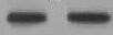
Detection of Human, Mouse, and Rat ATG5 by Western Blot.
Western blot shows lysates of HeLa human cervical epithelial carcinoma cell line, CH-1 mouse B cell lymphoma cell line, and PC-12 rat adrenal pheochromocytoma cell line. PVDF Membrane was probed with 0.5 µg/mL of Mouse Anti-Human/Mouse/Rat ATG5 Monoclonal Antibody (Catalog # MAB5294) followed by HRP-conjugated Anti-Mouse IgG Secondary Antibody (Catalog # HAF007). A specific band was detected for ATG5 at approximately 50 kDa (as indicated). This experiment was conducted under reducing conditions and using Immunoblot Buffer Group 2.
Detection of Human and Rat ATG5 by Simple WesternTM.
Simple Western lane view shows lysates of HeLa human cervical epithelial carcinoma cell line and PC‑12 rat adrenal pheochromocytoma cell line, loaded at 0.2 mg/mL. A specific band was detected for ATG5 at approximately 51 kDa (as indicated) using 25 µg/mL of Mouse Anti-Human/Mouse/Rat ATG5 Monoclonal Antibody (Catalog # MAB5294). This experiment was conducted under reducing conditions and using the 12-230 kDa separation system.Applications for ATG5 Antibody (603813)
Application
Recommended Usage
Immunohistochemistry
8-25 µg/mL
Sample: Immersion fixed paraffin-embedded human small intestine
Sample: Immersion fixed paraffin-embedded human small intestine
Simple Western
25 µg/mL
Sample: HeLa human cervical epithelial carcinoma cell line and PC‑12 rat adrenal pheochromocytoma cell line
Sample: HeLa human cervical epithelial carcinoma cell line and PC‑12 rat adrenal pheochromocytoma cell line
Western Blot
0.5 µg/mL
Sample: HeLa human cervical epithelial carcinoma cell line, CH‑1 mouse B cell lymphoma cell line, and PC‑12 rat adrenal pheochromocytoma cell line
Sample: HeLa human cervical epithelial carcinoma cell line, CH‑1 mouse B cell lymphoma cell line, and PC‑12 rat adrenal pheochromocytoma cell line
Reviewed Applications
Read 1 review rated 5 using MAB5294 in the following applications:
Formulation, Preparation, and Storage
Purification
Protein A or G purified from hybridoma culture supernatant
Reconstitution
Sterile PBS to a final concentration of 0.5 mg/mL. For liquid material, refer to CoA for concentration.
Loading...
Formulation
Lyophilized from a 0.2 μm filtered solution in PBS with Trehalose. *Small pack size (SP) is supplied either lyophilized or as a 0.2 µm filtered solution in PBS.
Shipping
Lyophilized product is shipped at ambient temperature. Liquid small pack size (-SP) is shipped with polar packs. Upon receipt, store immediately at the temperature recommended below.
Stability & Storage
Use a manual defrost freezer and avoid repeated freeze-thaw cycles.
- 12 months from date of receipt, -20 to -70 °C as supplied.
- 1 month, 2 to 8 °C under sterile conditions after reconstitution.
- 6 months, -20 to -70 °C under sterile conditions after reconstitution.
Calculators
Background: ATG5
Long Name
ATG5 Autophagy Related 5 Homolog
Alternate Names
APG5, ASP
Gene Symbol
ATG5
UniProt
Additional ATG5 Products
Product Documents for ATG5 Antibody (603813)
Certificate of Analysis
To download a Certificate of Analysis, please enter a lot or batch number in the search box below.
Note: Certificate of Analysis not available for kit components.
Product Specific Notices for ATG5 Antibody (603813)
For research use only
Related Research Areas
Citations for ATG5 Antibody (603813)
 Powered by Bioz
Customer Reviews for ATG5 Antibody (603813) (1)
5 out of 5
1 Customer Rating
Have you used ATG5 Antibody (603813)?
Submit a review and receive an Amazon gift card!
$25/€18/£15/$25CAN/¥2500 Yen for a review with an image
$10/€7/£6/$10CAN/¥1110 Yen for a review without an image
Submit a review
Customer Images
Showing
1
-
1 的
1 review
Showing All
Filter By:
-
Application: Western BlotSample Tested: CH-1 mouse B cell lymphoma cell lineSpecies: MouseVerified Customer | Posted 07/25/2022

There are no reviews that match your criteria.
Protocols
Find general support by application which include: protocols, troubleshooting, illustrated assays, videos and webinars.
- Antigen Retrieval Protocol (PIER)
- Antigen Retrieval for Frozen Sections Protocol
- Appropriate Fixation of IHC/ICC Samples
- Cellular Response to Hypoxia Protocols
- Chromogenic IHC Staining of Formalin-Fixed Paraffin-Embedded (FFPE) Tissue Protocol
- Chromogenic Immunohistochemistry Staining of Frozen Tissue
- ClariTSA™ Fluorophore Kits
- Detection & Visualization of Antibody Binding
- Fluorescent IHC Staining of Frozen Tissue Protocol
- Graphic Protocol for Heat-induced Epitope Retrieval
- Graphic Protocol for the Preparation and Fluorescent IHC Staining of Frozen Tissue Sections
- Graphic Protocol for the Preparation and Fluorescent IHC Staining of Paraffin-embedded Tissue Sections
- Graphic Protocol for the Preparation of Gelatin-coated Slides for Histological Tissue Sections
- IHC Sample Preparation (Frozen sections vs Paraffin)
- Immunofluorescent IHC Staining of Formalin-Fixed Paraffin-Embedded (FFPE) Tissue Protocol
- Immunohistochemistry (IHC) and Immunocytochemistry (ICC) Protocols
- Immunohistochemistry Frozen Troubleshooting
- Immunohistochemistry Paraffin Troubleshooting
- Preparing Samples for IHC/ICC Experiments
- Preventing Non-Specific Staining (Non-Specific Binding)
- Primary Antibody Selection & Optimization
- Protocol for Heat-Induced Epitope Retrieval (HIER)
- Protocol for Making a 4% Formaldehyde Solution in PBS
- Protocol for VisUCyte™ HRP Polymer Detection Reagent
- Protocol for the Preparation & Fixation of Cells on Coverslips
- Protocol for the Preparation and Chromogenic IHC Staining of Frozen Tissue Sections
- Protocol for the Preparation and Chromogenic IHC Staining of Frozen Tissue Sections - Graphic
- Protocol for the Preparation and Chromogenic IHC Staining of Paraffin-embedded Tissue Sections
- Protocol for the Preparation and Chromogenic IHC Staining of Paraffin-embedded Tissue Sections - Graphic
- Protocol for the Preparation and Fluorescent IHC Staining of Frozen Tissue Sections
- Protocol for the Preparation and Fluorescent IHC Staining of Paraffin-embedded Tissue Sections
- Protocol for the Preparation of Gelatin-coated Slides for Histological Tissue Sections
- R&D Systems Quality Control Western Blot Protocol
- TUNEL and Active Caspase-3 Detection by IHC/ICC Protocol
- The Importance of IHC/ICC Controls
- Troubleshooting Guide: Immunohistochemistry
- Troubleshooting Guide: Western Blot Figures
- Western Blot Conditions
- Western Blot Protocol
- Western Blot Protocol for Cell Lysates
- Western Blot Troubleshooting
- Western Blot Troubleshooting Guide
- View all Protocols, Troubleshooting, Illustrated assays and Webinars
Loading...