
Human Notch-1 is a 300 kDa type I transmembrane glycoprotein that is one of four human Notch homologues involved in developmental processes (1-3). Notch signaling is important for maintaining stem cells and inducing differentiation, especially in the nervous system and lymphoid tissues (2-4). Notch can specify binary cell fates; for example, promoting T- over B-cell development from a common precursor (2). More than 50% of human T-lineage acute lymphoblastic leukemia (T‑ALL) have activating mutations of Notch1 (1, 5). Human Notch-1 is synthesized as a 2556 amino acid (aa) precursor that contains an 18 aa signal sequence, a 1718 aa extracellular domain (ECD) with 36 EGF-like repeats and three Lin-12/Notch repeats (LNR), a 23 aa transmembrane (TM) segment and a 785 aa cytoplasmic domain containing six ankyrin repeats, a glutamine-rich domain and a PEST sequence. The 11th and 12th EGF-like repeats bind ligands including Jagged and Delta-like families in humans (6). O-fucosylation by Fringe family members at a site within this region can inhibit the interaction of Notch with Jagged ligands, thereby promoting Delta-like ligand interactions (7). Notch-1 receptor undergoes post-translational furin-type proteolytic cleavage, forming a heterodimer through interaction of a hydrophobic area C-terminal to the LNR on the 1647 aa ligand-binding extracellular region with the 891 aa transmembrane/cytoplasmic portion (8, 9). Upon ligand binding, additional sequential proteolysis by TNF-converting enzyme (ADAM17) and the presenilin-dependent gamma -secretase results in the release of the Notch intracellular domain (NICD) which translocates into the nucleus, activating transcription of Notch-responsive genes (10). Human Notch-1 ECD aa 19-526, including the first 13 EGF repeats, shows 91% aa identity with corresponding regions of mouse and rat, 89% with canine, and 79% with chicken Notch-1. This region also exhibits 60% aa identity with human Notch-2 and Notch-3.
Human Notch-1 Intracellular Domain Antibody (433802)
R&D Systems | Catalog # MAB3647

Scientific images may be resized, cropped, or adjusted for brightness or contrast for presentation purposes. These updates are limited to presentation and do not affect the underlying scientific interpretation of the data. Where available, additional source data may be provided upon request.
Key Product Details
Species Reactivity
Applications
Label
Antibody Source
Product Specifications
Immunogen
Gly2428-Lys2556
Accession # AAG33848
Specificity
Clonality
Host
Isotype
Scientific Data Images for Human Notch-1 Intracellular Domain Antibody (433802)
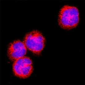
Notch‑1 in MOLT‑4 Human Cell Line.
Notch‑1 was detected in immersion fixed MOLT‑4 human acute lymphoblastic leukemia cell line using Mouse Anti-Human Notch‑1 Intracellular Domain Monoclonal Antibody (Catalog # MAB3647) at 8 µg/mL for 3 hours at room temperature. Cells were stained using the NorthernLights™ 557-conjugated Anti-Mouse IgG Secondary Antibody (red; NL007) and counterstained with DAPI (blue). Specific staining was localized to cell surface and cell nuclei. Staining was performed using our protocol for Fluorescent ICC Staining of Non-adherent Cells.Applications for Human Notch-1 Intracellular Domain Antibody (433802)
Immunocytochemistry
Sample: Immersion fixed MOLT‑4 human acute lymphoblastic leukemia cell line
Western Blot
Sample: Recombinant Human Notch‑1 Intracellular Domain
Formulation, Preparation, and Storage
Purification
Reconstitution
Reconstitute at 0.5 mg/mL in sterile PBS. For liquid material, refer to CoA for concentration.
Formulation
Shipping
Stability & Storage
- 12 months from date of receipt, -20 to -70 °C as supplied.
- 1 month, 2 to 8 °C under sterile conditions after reconstitution.
- 6 months, -20 to -70 °C under sterile conditions after reconstitution.
Calculators
Background: Notch-1
References
- Ellisen, L.W. et al. (1991) Cell 66:649.
- Dumortier, A. et al. (2005) Int. J. Hematol. 82:277.
- Yoon, K. and N. Gaiano (2005) Nat. Neurosci. 8:709.
- Androutsellis-Theotokis, A. et al. (2006) Nature 442:823.
- Weng, A.P. et al. (2004) Science 306:269.
- Hambleton, S. et al. (2004) Structure 12:2173.
- Yang, L. et al. (2005) Mol. Biol. Cell 16:927.
- Sanchez-Irizarry, C. et al. (2004) Mol. Cell. Biol. 24:9265.
- Logeat, F. et al. (1998) Proc. Natl. Acad. Sci. USA 95:8108.
- Mumm, J.S. and R. Kopan (2000) Dev. Biol. 228:151.
Alternate Names
Gene Symbol
UniProt
Additional Notch-1 Products
Product Documents for Human Notch-1 Intracellular Domain Antibody (433802)
Certificate of Analysis
To download a Certificate of Analysis, please enter a lot or batch number in the search box below.
Note: Certificate of Analysis not available for kit components.
Product Specific Notices for Human Notch-1 Intracellular Domain Antibody (433802)
For research use only
Related Research Areas
Citations for Human Notch-1 Intracellular Domain Antibody (433802)
 Powered by Bioz
Customer Reviews for Human Notch-1 Intracellular Domain Antibody (433802)
There are currently no reviews for this product. Be the first to review Human Notch-1 Intracellular Domain Antibody (433802) and earn rewards!
Have you used Human Notch-1 Intracellular Domain Antibody (433802)?
Submit a review and receive an Amazon gift card!
$25/€18/£15/$25CAN/¥2500 for a review with an image
$10/€7/£6/$10CAN/¥1110 for a review without an image
Submit a review
Protocols
Find general support by application which include: protocols, troubleshooting, illustrated assays, videos and webinars.
- Appropriate Fixation of IHC/ICC Samples
- Cellular Response to Hypoxia Protocols
- ClariTSA™ Fluorophore Kits
- Detection & Visualization of Antibody Binding
- ICC Cell Smear Protocol for Suspension Cells
- ICC Immunocytochemistry Protocol Videos
- ICC for Adherent Cells
- Immunocytochemistry (ICC) Protocol
- Immunocytochemistry Troubleshooting
- Immunofluorescence of Organoids Embedded in Cultrex Basement Membrane Extract
- Immunohistochemistry (IHC) and Immunocytochemistry (ICC) Protocols
- Preparing Samples for IHC/ICC Experiments
- Preventing Non-Specific Staining (Non-Specific Binding)
- Primary Antibody Selection & Optimization
- Protocol for VisUCyte™ HRP Polymer Detection Reagent
- Protocol for the Fluorescent ICC Staining of Cell Smears - Graphic
- Protocol for the Fluorescent ICC Staining of Cultured Cells on Coverslips - Graphic
- Protocol for the Preparation and Fluorescent ICC Staining of Cells on Coverslips
- Protocol for the Preparation and Fluorescent ICC Staining of Non-adherent Cells
- Protocol for the Preparation and Fluorescent ICC Staining of Stem Cells on Coverslips
- Protocol for the Preparation of a Cell Smear for Non-adherent Cell ICC - Graphic
- R&D Systems Quality Control Western Blot Protocol
- TUNEL and Active Caspase-3 Detection by IHC/ICC Protocol
- The Importance of IHC/ICC Controls
- Troubleshooting Guide: Western Blot Figures
- Western Blot Conditions
- Western Blot Protocol
- Western Blot Protocol for Cell Lysates
- Western Blot Troubleshooting
- Western Blot Troubleshooting Guide
- View all Protocols, Troubleshooting, Illustrated assays and Webinars
Associated Pathways


