Human phospho-Src (Y419) Antibody
R&D Systems | Catalog # AF2685
 by Western Blot.")
Key Product Details
Validated by
Biological Validation
Species Reactivity
Validated:
Human
Cited:
Human, Mouse
Applications
Validated:
Immunohistochemistry, Western Blot, Simple Western
Cited:
Immunohistochemistry-Paraffin, Immunohistochemistry-Frozen, Western Blot
Label
Unconjugated
Antibody Source
Polyclonal Rabbit IgG
Loading...
Product Specifications
Immunogen
Phosphopeptide containing human Src Y419 site
Specificity
Detects human Src family members when phosphorylated at sites corresponding to Y419 of human Src in Western blots.
Clonality
Polyclonal
Host
Rabbit
Isotype
IgG
Scientific Data Images for Human phospho-Src (Y419) Antibody
Detection of Human Phospho-Src (Y419) by Western Blot.
Western blot shows lysates of MDA-MB-468 human breast cancer cell line and HepG2 human hepatocellular carcinoma cell line untreated (-) or treated (+) with 50 µM pervanadate (PV) for 10 minutes. PVDF membrane was probed with 1 µg/mL of Rabbit Anti-Human Phospho-Src (Y419) Antigen Affinity-purified Polyclonal Antibody (Catalog # AF2685), followed by HRP-conjugated Anti-Rabbit IgG Secondary Antibody (Catalog # HAF008). A specific band was detected for Phospho-Src (Y419) at approximately 56 - 61 kDa (as indicated). This experiment was conducted under reducing conditions and using Immunoblot Buffer Group 1.
Src in Human Colon.
Src phosphorylated at Y419 was detected in immersion fixed paraffin-embedded sections of human colon using Rabbit Anti-Human Phospho-Src (Y419) Antigen Affinity-purified Polyclonal Antibody (Catalog # AF2685) at 15 µg/mL overnight at 4 °C. Tissue was stained using the Anti-Rabbit HRP-DAB Cell & Tissue Staining Kit (brown; Catalog # CTS005) and counterstained with hematoxylin (blue). Specific labeling was localized to the cytoplasm of epithelial cells. View our protocol for Chromogenic IHC Staining of Paraffin-embedded Tissue Sections.
Src in Human Liver Cancer Tissue.
Src phosphorylated at Y419 was detected in immersion fixed paraffin-embedded sections of human liver cancer tissue using Rabbit Anti-Human Phospho-Src (Y419) Antigen Affinity-purified Polyclonal Antibody (Catalog # AF2685) at 10 µg/mL overnight at 4 °C. Tissue was stained using the Anti-Rabbit HRP-DAB Cell & Tissue Staining Kit (brown; Catalog # CTS005) and counterstained with hematoxylin (blue). Lower panel shows a lack of labeling if primary antibodies are omitted and tissue is stained only with secondary antibody followed by incubation with detection reagents. View our protocol for Chromogenic IHC Staining of Paraffin-embedded Tissue Sections.
Src in Human Liver.
Src phosphorylated at Y419 was detected in immersion fixed paraffin-embedded sections of human liver array using Rabbit Anti-Human Phospho-Src (Y419) Antigen Affinity-purified Polyclonal Antibody (Catalog # AF2685) at 10 µg/mL overnight at 4 °C. Tissue was stained using the Anti-Rabbit HRP-DAB Cell & Tissue Staining Kit (brown; Catalog # CTS005) and counterstained with hematoxylin (blue). Lower panel shows a lack of labeling if primary antibodies are omitted and tissue is stained only with secondary antibody followed by incubation with detection reagents. View our protocol for Chromogenic IHC Staining of Paraffin-embedded Tissue Sections. by Simple Western<SUP>TM</SUP>.")
Detection of Human Phospho-Src (Y419) by Simple WesternTM.
Simple Western lane view shows lysates of MBA‑MB‑468 human breast cancer cell line untreated (-) or treated (+) with 50 µM Pervanadate (PV) for 10 minutes, loaded at 0.2 mg/mL. A specific band was detected for Phospho-Src (Y419) at approximately 64 kDa (as indicated) using 10 µg/mL of Rabbit Anti-Human Phospho-Src (Y419) Antigen Affinity-purified Polyclonal Antibody (Catalog # AF2685). This experiment was conducted under reducing conditions and using the 12-230 kDa separation system.
Detection of Mouse Src by Immunohistochemistry
Yap heterogeneity is unrelated to markers examinedAll panels show examples of confocal stains of lung slices from KP mice, 14 weeks after infection. (A, B) Adjacent sections stained for E-cad, ZO-1, and either Yap (A), or phospho-Yap (B). (C, D) Adjacent sections stained for E-cad, SpC, and either Yap (C), or phospho-Acc (pAcc) (D). (E, F) Adjacent sections stained for E-cad, SpC, and either Yap (E), or phospho-Src (pSrc) (F). (G, H) Adjacent sections stained for E-cad, SpC, and either Yap (E), or diphospho-Erk (F). (I) Section stained for YAP (red), Vimentin (green), and DNA (blue). (J) Section stained for YAP (red), ZO-1 (green), and E-Cad (blue). Image collected and cropped by CiteAb from the following publication (https://pubmed.ncbi.nlm.nih.gov/29340023), licensed under a CC-BY license. Not internally tested by R&D Systems.Applications for Human phospho-Src (Y419) Antibody
Application
Recommended Usage
Immunohistochemistry
5-15 µg/mL
Sample: Immersion fixed paraffin-embedded sections of human colon, human liver cancer tissue, and human liver array
Sample: Immersion fixed paraffin-embedded sections of human colon, human liver cancer tissue, and human liver array
Simple Western
10 µg/mL
Sample: MBA‑MB‑468 human breast cancer cell line treated with Pervanadate (PV)
Sample: MBA‑MB‑468 human breast cancer cell line treated with Pervanadate (PV)
Western Blot
1 µg/mL
Sample: Pervanadate-treated MDA‑MB‑468 human breast cancer cell line and HepG2 human hepatocellular carcinoma cell line
Sample: Pervanadate-treated MDA‑MB‑468 human breast cancer cell line and HepG2 human hepatocellular carcinoma cell line
Reviewed Applications
Read 1 review rated 4 using AF2685 in the following applications:
Formulation, Preparation, and Storage
Purification
Antigen Affinity-purified
Reconstitution
Reconstitute at 0.2 mg/mL in sterile PBS. For liquid material, refer to CoA for concentration.
Loading...
Formulation
Lyophilized from a 0.2 μm filtered solution in PBS with Trehalose. *Small pack size (SP) is supplied either lyophilized or as a 0.2 µm filtered solution in PBS.
Shipping
Lyophilized product is shipped at ambient temperature. Liquid small pack size (-SP) is shipped with polar packs. Upon receipt, store immediately at the temperature recommended below.
Stability & Storage
Use a manual defrost freezer and avoid repeated freeze-thaw cycles.
- 12 months from date of receipt, -20 to -70 °C as supplied.
- 1 month, 2 to 8 °C under sterile conditions after reconstitution.
- 6 months, -20 to -70 °C under sterile conditions after reconstitution.
Calculators
Background: Src
Long Name
v-src Sarcoma [Schmidt-Ruppin A-2] Viral Oncogene Homolog
Alternate Names
ASV, c-Src, RSVgp4
Gene Symbol
SRC
Additional Src Products
Product Documents for Human phospho-Src (Y419) Antibody
Certificate of Analysis
To download a Certificate of Analysis, please enter a lot or batch number in the search box below.
Note: Certificate of Analysis not available for kit components.
Product Specific Notices for Human phospho-Src (Y419) Antibody
For research use only
Citations for Human phospho-Src (Y419) Antibody
 Powered by Bioz
Customer Reviews for Human phospho-Src (Y419) Antibody (1)
4 out of 5
1 Customer Rating
Have you used Human phospho-Src (Y419) Antibody?
Submit a review and receive an Amazon gift card!
$25/€18/£15/$25CAN/¥2500 Yen for a review with an image
$10/€7/£6/$10CAN/¥1110 Yen for a review without an image
Submit a review
Customer Images
Showing
1
-
1 的
1 review
Showing All
Filter By:
-
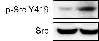
Application: Western BlotSample Tested: Breast cancer cellsSpecies: HumanVerified Customer | Posted 10/11/2018Human breast cancer cell line MDA-MB-231 was treated with carboplatin for 72 hours and the expression of p-Src at Y419 and total Src were detected by western blot.

There are no reviews that match your criteria.
Protocols
Find general support by application which include: protocols, troubleshooting, illustrated assays, videos and webinars.
- Antigen Retrieval Protocol (PIER)
- Antigen Retrieval for Frozen Sections Protocol
- Appropriate Fixation of IHC/ICC Samples
- Cellular Response to Hypoxia Protocols
- Chromogenic IHC Staining of Formalin-Fixed Paraffin-Embedded (FFPE) Tissue Protocol
- Chromogenic Immunohistochemistry Staining of Frozen Tissue
- ClariTSA™ Fluorophore Kits
- Detection & Visualization of Antibody Binding
- Fluorescent IHC Staining of Frozen Tissue Protocol
- Graphic Protocol for Heat-induced Epitope Retrieval
- Graphic Protocol for the Preparation and Fluorescent IHC Staining of Frozen Tissue Sections
- Graphic Protocol for the Preparation and Fluorescent IHC Staining of Paraffin-embedded Tissue Sections
- Graphic Protocol for the Preparation of Gelatin-coated Slides for Histological Tissue Sections
- IHC Sample Preparation (Frozen sections vs Paraffin)
- Immunofluorescent IHC Staining of Formalin-Fixed Paraffin-Embedded (FFPE) Tissue Protocol
- Immunohistochemistry (IHC) and Immunocytochemistry (ICC) Protocols
- Immunohistochemistry Frozen Troubleshooting
- Immunohistochemistry Paraffin Troubleshooting
- Preparing Samples for IHC/ICC Experiments
- Preventing Non-Specific Staining (Non-Specific Binding)
- Primary Antibody Selection & Optimization
- Protocol for Heat-Induced Epitope Retrieval (HIER)
- Protocol for Making a 4% Formaldehyde Solution in PBS
- Protocol for VisUCyte™ HRP Polymer Detection Reagent
- Protocol for the Preparation & Fixation of Cells on Coverslips
- Protocol for the Preparation and Chromogenic IHC Staining of Frozen Tissue Sections
- Protocol for the Preparation and Chromogenic IHC Staining of Frozen Tissue Sections - Graphic
- Protocol for the Preparation and Chromogenic IHC Staining of Paraffin-embedded Tissue Sections
- Protocol for the Preparation and Chromogenic IHC Staining of Paraffin-embedded Tissue Sections - Graphic
- Protocol for the Preparation and Fluorescent IHC Staining of Frozen Tissue Sections
- Protocol for the Preparation and Fluorescent IHC Staining of Paraffin-embedded Tissue Sections
- Protocol for the Preparation of Gelatin-coated Slides for Histological Tissue Sections
- R&D Systems Quality Control Western Blot Protocol
- TUNEL and Active Caspase-3 Detection by IHC/ICC Protocol
- The Importance of IHC/ICC Controls
- Troubleshooting Guide: Immunohistochemistry
- Troubleshooting Guide: Western Blot Figures
- Western Blot Conditions
- Western Blot Protocol
- Western Blot Protocol for Cell Lysates
- Western Blot Troubleshooting
- Western Blot Troubleshooting Guide
- View all Protocols, Troubleshooting, Illustrated assays and Webinars
Loading...
Associated Pathways


MAPK Signaling: Oxidative Stress Pathway
 MAPK Signaling Pathway: Mitogen Stimulation Pathway
 Pathogen or Damage-activated C-Type Lectin Receptor Signaling Pathways
 VEGF - VEGF R2 Signaling Pathways
 Wnt Signaling Pathways: beta-Catenin-dependent Wnt Signaling
