
Key Product Details
Validated by
Knockout/Knockdown
Species Reactivity
Validated:
Human
Cited:
Human
Applications
Validated:
Knockout Validated, Western Blot, Immunocytochemistry
Cited:
Western Blot
Label
Unconjugated
Antibody Source
Monoclonal Mouse IgG2A Clone # 863028
Loading...
Product Specifications
Immunogen
E. coli-derived recombinant human Rab13
Lys94-Thr191
Accession # P51153
Lys94-Thr191
Accession # P51153
Specificity
Detects human Rab13 in direct ELISA and Western Blot.
Clonality
Monoclonal
Host
Mouse
Isotype
IgG2A
Scientific Data Images for Human Rab13 Antibody (863028)
Detection of Human Rab13 by Western Blot.
Western blot shows lysates of human lung tissue and human aorta tissue. PVDF membrane was probed with 2 µg/mL of Mouse Anti-Human Rab13 Monoclonal Antibody (Catalog # MAB8305) followed by HRP-conjugated Anti-Mouse IgG Secondary Antibody (HAF018). A specific band was detected for Rab13 at approximately 23 kDa (as indicated). This experiment was conducted under reducing conditions and using Immunoblot Buffer Group 1.
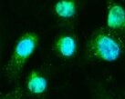
Rab13 in Caco‑2 Human Cell Line.
Rab13 was detected in immersion fixed Caco-2 human colorectal adenocarcinoma cell line using Mouse Anti-Human Rab13 Monoclonal Antibody (Catalog # MAB8305) at 25 µg/mL for 3 hours at room temperature. Cells were stained using the NorthernLights™ 557-conjugated Anti-Mouse IgG Secondary Antibody (red; NL007) and counterstained with DAPI (blue). Specific staining was localized to plasma membrane. View our protocol for Fluorescent ICC Staining of Cells on Coverslips.
Western Blot Shows Rab13 Specificity Using Knockout Cell Line.
Western blot shows lysates of HEK-293T parental cell line and Rab13 knockout HEK-293T cell line (KO). Nitrocellulose membrane was probed with Mouse Anti-Human Rab13 Monoclonal Antibody (Catalog # MAB8305) followed by HRP-conjugated secondary antibody. A specific band was detected for Rab13 at approximately 23 kDa (as indicated) in the parental HEK-293T cell line, but is not detectable in knockout HEK-293T cell line. Primary antibody dilution used: 2.5 µg/mL. The Ponceau stained transfer of the blot is shown. This experiment was conducted under reducing conditions. Image, protocol, and testing courtesy of YCharOS Inc. See ycharos.com for additional details.
Western Blot Shows Rab13 Specificity Using Knockdown Cell Line.
Western blot shows lysates of U87 MG parental cell line and Rab13 knockout U87 MG cell line (KD). Nitrocellulose membrane was probed with Mouse Anti-Human Rab13 Monoclonal Antibody (Catalog # MAB8305) followed by HRP-conjugated secondary antibody. A specific band was detected for Rab13 at approximately 23 kDa (as indicated) in the parental U87 MG cell line, but is significantly reduced in the U87 MG cell line. Primary antibody dilution used: 2.5 µg/mL. The Ponceau stained transfer of the blot is shown. This experiment was conducted under reducing conditions. Image, protocol, and testing courtesy of YCharOS Inc. See ycharos.com for additional details.
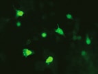
Rab13 Specificity is Shown by Immunocytochemistry in Knockdown Cell Line.
U‑87 MG human glioblastoma/astrocytoma parental cell line WT and Rab13 U-87 MG KD cells were labelled with a green or a far-red fluorescent dye, respectively. Cells were stained with Mouse Anti-Human Rab13 Monoclonal Antibody (Catalog # MAB8305) followed by incubation with an Alexa-fluor 555 conjugated secondary antibody (upper panel). DAPI-only counterstained cells shown on a lower panel. Acquisition of the blue (nucleus-DAPI), green (identification of WT cells), red (antibody staining) and far-red (identification of KD cells) channels was performed. Representative images of the blue and red (grayscale) channels are shown. WT and KD cells are outlined with green and magenta dashed line, respectively. Primary antibody dilution used: 1 µg/mL. Image, protocol and testing courtesy of YCharOS Inc. (ycharos.com).Applications for Human Rab13 Antibody (863028)
Application
Recommended Usage
Immunocytochemistry
8-25 µg/mL
Sample: Immersion fixed Caco‑2 human colorectal adenocarcinoma cell line
Sample: Immersion fixed Caco‑2 human colorectal adenocarcinoma cell line
Knockout Validated
Rab13 is specifically detected in HEK-293T parental cell line but is not detectable in knockout HEK-293T cell line.
Western Blot
2 µg/mL
Sample: Human lung tissue and human aorta tissue
Sample: Human lung tissue and human aorta tissue
Reviewed Applications
Read 2 reviews rated 5 using MAB8305 in the following applications:
Formulation, Preparation, and Storage
Purification
Protein A or G purified from hybridoma culture supernatant
Reconstitution
Reconstitute at 0.5 mg/mL in sterile PBS. For liquid material, refer to CoA for concentration.
Loading...
Formulation
Lyophilized from a 0.2 μm filtered solution in PBS with Trehalose. See Certificate of Analysis for details.
*Small pack size (-SP) is supplied either lyophilized or as a 0.2 µm filtered solution in PBS.
*Small pack size (-SP) is supplied either lyophilized or as a 0.2 µm filtered solution in PBS.
Shipping
Lyophilized product is shipped at ambient temperature. Liquid small pack size (-SP) is shipped with polar packs. Upon receipt, store immediately at the temperature recommended below.
Stability & Storage
Use a manual defrost freezer and avoid repeated freeze-thaw cycles.
- 12 months from date of receipt, -20 to -70 °C as supplied.
- 1 month, 2 to 8 °C under sterile conditions after reconstitution.
- 6 months, -20 to -70 °C under sterile conditions after reconstitution.
Calculators
Background: Rab13
Long Name
RAs Genes from Brain Protein 13
Alternate Names
Cell growth-inhibiting gene 4 protein, growth-inhibiting gene 4 protein, RAB13, member RAS oncogene family, RAS-associated protein RAB13, ras-related protein Rab-13
Gene Symbol
RAB13
UniProt
Additional Rab13 Products
Product Documents for Human Rab13 Antibody (863028)
Certificate of Analysis
To download a Certificate of Analysis, please enter a lot or batch number in the search box below.
Note: Certificate of Analysis not available for kit components.
Product Specific Notices for Human Rab13 Antibody (863028)
For research use only
Related Research Areas
Citations for Human Rab13 Antibody (863028)
 Powered by Bioz
Customer Reviews for Human Rab13 Antibody (863028) (2)
5 out of 5
2 Customer Ratings
Have you used Human Rab13 Antibody (863028)?
Submit a review and receive an Amazon gift card!
$25/€18/£15/$25CAN/¥2500 Yen for a review with an image
$10/€7/£6/$10CAN/¥1110 Yen for a review without an image
Submit a review
Customer Images
Showing
1
-
2 的
2 reviews
Showing All
Filter By:
-
Application: Immunocytochemistry/ImmunofluorescenceSample Tested: Aortic endothelial cellsSpecies: HumanVerified Customer | Posted 07/08/2022

-
Application: Immunocytochemistry/ImmunofluorescenceSample Tested: NeuroblastsSpecies: HumanVerified Customer | Posted 02/17/2022
There are no reviews that match your criteria.
Protocols
Find general support by application which include: protocols, troubleshooting, illustrated assays, videos and webinars.
- Appropriate Fixation of IHC/ICC Samples
- Cellular Response to Hypoxia Protocols
- ClariTSA™ Fluorophore Kits
- Detection & Visualization of Antibody Binding
- ICC Cell Smear Protocol for Suspension Cells
- ICC Immunocytochemistry Protocol Videos
- ICC for Adherent Cells
- Immunocytochemistry (ICC) Protocol
- Immunocytochemistry Troubleshooting
- Immunofluorescence of Organoids Embedded in Cultrex Basement Membrane Extract
- Immunohistochemistry (IHC) and Immunocytochemistry (ICC) Protocols
- Preparing Samples for IHC/ICC Experiments
- Preventing Non-Specific Staining (Non-Specific Binding)
- Primary Antibody Selection & Optimization
- Protocol for VisUCyte™ HRP Polymer Detection Reagent
- Protocol for the Fluorescent ICC Staining of Cell Smears - Graphic
- Protocol for the Fluorescent ICC Staining of Cultured Cells on Coverslips - Graphic
- Protocol for the Preparation and Fluorescent ICC Staining of Cells on Coverslips
- Protocol for the Preparation and Fluorescent ICC Staining of Non-adherent Cells
- Protocol for the Preparation and Fluorescent ICC Staining of Stem Cells on Coverslips
- Protocol for the Preparation of a Cell Smear for Non-adherent Cell ICC - Graphic
- R&D Systems Quality Control Western Blot Protocol
- TUNEL and Active Caspase-3 Detection by IHC/ICC Protocol
- The Importance of IHC/ICC Controls
- Troubleshooting Guide: Western Blot Figures
- Western Blot Conditions
- Western Blot Protocol
- Western Blot Protocol for Cell Lysates
- Western Blot Troubleshooting
- Western Blot Troubleshooting Guide
- View all Protocols, Troubleshooting, Illustrated assays and Webinars
Loading...