S100A8 (also known as MRP8 and calgranulin A) is a 10 kDa member of the S100 family, EF-hand superfamily of Ca-binding proteins. It is produced by neutrophils and monocytes, and forms Ca‑dependent heterodimer/heterotetramer complexes (termed calprotectin) with S100A9. It functions both intracellularly and extracellularly, where it binds to RAGE and CD36. Human S100A8 is 93 amino acids (aa) in length. It contains two EF-hand motifs (aa 12-47 and 46-81) and one high-affinity Ca‑binding site (aa 59-70). There may be one splice form that shows a 15 aa substitution for the C-terminal 14 amino acids. Although mouse S100A8 is cleaved by MMP‑2 after Asn21, it is unclear if human S100A8 is susceptible. Full-length human S100A8 is 57% and 74% aa identical to mouse and canine S100A8, respectively.

Key Product Details
Species Reactivity
Validated:
Human
Cited:
Human
Applications
Validated:
Western Blot, Intracellular Staining by Flow Cytometry, Immunocytochemistry, Simple Western, CyTOF-ready
Cited:
Western Blot, ELISA
Label
Unconjugated
Antibody Source
Polyclonal Sheep IgG
Loading...
Product Specifications
Immunogen
E. coli-derived recombinant human S100A8
Met1-Glu93
Accession # P05109
Met1-Glu93
Accession # P05109
Specificity
Detects human S100A8 in direct ELISAs and Western blots. In these formats, less than 1% cross-reactivity with recombinant mouse S100A8, recombinant human (rh) S100A1, and rhS100P is observed.
Clonality
Polyclonal
Host
Sheep
Isotype
IgG
Scientific Data Images for Human S100A8 Antibody
Detection of Human S100A8 by Western Blot.
Western blot shows lysates of human brain tissue, K562 human chronic myelogenous leukemia cell line, and HeLa human cervical epithelial carcinoma cell line. PVDF membrane was probed with 1 µg/mL of Sheep Anti-Human S100A8 Antigen Affinity-purified Polyclonal Antibody (Catalog # AF4570) followed by HRP-conjugated Anti-Sheep IgG Secondary Antibody (Catalog # HAF016). A specific band was detected for S100A8 at approximately 10 kDa (as indicated). This experiment was conducted under reducing conditions and using Immunoblot Buffer Group 8.
S100A8 in HeLa Human Cell Line.
S100A8 was detected in immersion fixed HeLa human cervical epithelial carcinoma cell line using Sheep Anti-Human S100A8 Antigen Affinity-purified Polyclonal Antibody (Catalog # AF4570) at 10 µg/mL for 3 hours at room temperature. Cells were stained using the NorthernLights™ 557-conjugated Anti-Sheep IgG Secondary Antibody (red; Catalog # NL010) and counterstained with DAPI (blue). Specific staining was localized to cytoplasm. View our protocol for Fluorescent ICC Staining of Cells on Coverslips.
Detection of S100A8 in HeLa Human Cell Line by Flow Cytometry.
HeLa human cervical epithelial carcinoma cell line was stained with Sheep Anti-Human S100A8 Antigen Affinity-purified Polyclonal Antibody (Catalog # AF4570, filled histogram) or control antibody (Catalog # 5-001-A, open histogram), followed by NorthernLights™ 637-conjugated Anti-Sheep IgG Secondary Antibody (Catalog # NL011). To facilitate intracellular staining, cells were fixed with paraformaldehyde and permeabilized with saponin.
Detection of Human S100A8 by Simple WesternTM.
Simple Western lane view shows lysates of normal adjacent tissue and Crohns tissue, loaded at 0.2 mg/mL. A specific band was detected for S100A8 at approximately 10 kDa (as indicated) using 20 µg/mL of Sheep Anti-Human S100A8 Antigen Affinity-purified Polyclonal Antibody (Catalog # AF4570) followed by 1:50 dilution of HRP-conjugated Anti-Sheep IgG Secondary Antibody (Catalog # HAF016). This experiment was conducted under reducing conditions and using the 12-230 kDa separation system.Applications for Human S100A8 Antibody
Application
Recommended Usage
CyTOF-ready
Ready to be labeled using established conjugation methods. No BSA or other carrier proteins that could interfere with conjugation.
Immunocytochemistry
5-15 µg/mL
Sample: Immersion fixed HeLa human cervical epithelial carcinoma cell line
Sample: Immersion fixed HeLa human cervical epithelial carcinoma cell line
Intracellular Staining by Flow Cytometry
2.5 µg/106 cells
Sample: HeLa human cervical epithelial carcinoma cell line, fixed with paraformaldehyde and permeabilized with saponin
Sample: HeLa human cervical epithelial carcinoma cell line, fixed with paraformaldehyde and permeabilized with saponin
Simple Western
10 µg/mL
Sample: Normal adjacent tissue and Crohns tissue
Sample: Normal adjacent tissue and Crohns tissue
Western Blot
1 µg/mL
Sample: Human brain tissue, K562 human chronic myelogenous leukemia cell line, and HeLa human cervical epithelial carcinoma cell line
Sample: Human brain tissue, K562 human chronic myelogenous leukemia cell line, and HeLa human cervical epithelial carcinoma cell line
Reviewed Applications
Read 2 reviews rated 4 using AF4570 in the following applications:
Flow Cytometry Panel Builder
Bio-Techne Knows Flow Cytometry
Save time and reduce costly mistakes by quickly finding compatible reagents using the Panel Builder Tool.
Advanced Features
- Spectra Viewer - Custom analysis of spectra from multiple fluorochromes
- Spillover Popups - Visualize the spectra of individual fluorochromes
- Antigen Density Selector - Match fluorochrome brightness with antigen density

Formulation, Preparation, and Storage
Purification
Antigen Affinity-purified
Reconstitution
Reconstitute at 0.2 mg/mL in sterile PBS. For liquid material, refer to CoA for concentration.
Loading...
Formulation
Lyophilized from a 0.2 μm filtered solution in PBS with Trehalose. *Small pack size (SP) is supplied either lyophilized or as a 0.2 µm filtered solution in PBS.
Shipping
Lyophilized product is shipped at ambient temperature. Liquid small pack size (-SP) is shipped with polar packs. Upon receipt, store immediately at the temperature recommended below.
Stability & Storage
Use a manual defrost freezer and avoid repeated freeze-thaw cycles.
- 12 months from date of receipt, -20 to -70 °C as supplied.
- 1 month, 2 to 8 °C under sterile conditions after reconstitution.
- 6 months, -20 to -70 °C under sterile conditions after reconstitution.
Calculators
Background: S100A8
Long Name
S100 Calcium Binding Protein A8
Alternate Names
Calgranulin A, CFAG, MRP8
Gene Symbol
S100A8
UniProt
Additional S100A8 Products
Product Documents for Human S100A8 Antibody
Certificate of Analysis
To download a Certificate of Analysis, please enter a lot or batch number in the search box below.
Note: Certificate of Analysis not available for kit components.
Product Specific Notices for Human S100A8 Antibody
For research use only
Citations for Human S100A8 Antibody
 Powered by Bioz
Customer Reviews for Human S100A8 Antibody (2)
4 out of 5
2 Customer Ratings
Have you used Human S100A8 Antibody?
Submit a review and receive an Amazon gift card!
$25/€18/£15/$25CAN/¥2500 Yen for a review with an image
$10/€7/£6/$10CAN/¥1110 Yen for a review without an image
Submit a review
Customer Images
Showing
1
-
2 的
2 reviews
Showing All
Filter By:
-
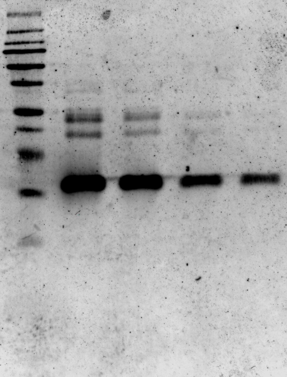
Application: Western BlotSample Tested: Recombinant proteinSpecies: HumanVerified Customer | Posted 07/01/2016S100A8 Antibody tested against different concentrations of human recombinant S100A8/A9.

-
Application: Western BlotSample Tested: See PMID 20806971Species: HumanVerified Customer | Posted 01/08/2015
There are no reviews that match your criteria.
Protocols
Find general support by application which include: protocols, troubleshooting, illustrated assays, videos and webinars.
- 7-Amino Actinomycin D (7-AAD) Cell Viability Flow Cytometry Protocol
- Appropriate Fixation of IHC/ICC Samples
- Cellular Response to Hypoxia Protocols
- ClariTSA™ Fluorophore Kits
- Detection & Visualization of Antibody Binding
- Extracellular Membrane Flow Cytometry Protocol
- Flow Cytometry Protocol for Cell Surface Markers
- Flow Cytometry Protocol for Staining Membrane Associated Proteins
- Flow Cytometry Staining Protocols
- Flow Cytometry Troubleshooting Guide
- ICC Cell Smear Protocol for Suspension Cells
- ICC Immunocytochemistry Protocol Videos
- ICC for Adherent Cells
- Immunocytochemistry (ICC) Protocol
- Immunocytochemistry Troubleshooting
- Immunofluorescence of Organoids Embedded in Cultrex Basement Membrane Extract
- Immunohistochemistry (IHC) and Immunocytochemistry (ICC) Protocols
- Intracellular Flow Cytometry Protocol Using Alcohol (Methanol)
- Intracellular Flow Cytometry Protocol Using Detergents
- Intracellular Nuclear Staining Flow Cytometry Protocol Using Detergents
- Intracellular Staining Flow Cytometry Protocol Using Alcohol Permeabilization
- Intracellular Staining Flow Cytometry Protocol Using Detergents to Permeabilize Cells
- Preparing Samples for IHC/ICC Experiments
- Preventing Non-Specific Staining (Non-Specific Binding)
- Primary Antibody Selection & Optimization
- Propidium Iodide Cell Viability Flow Cytometry Protocol
- Protocol for Liperfluo
- Protocol for VisUCyte™ HRP Polymer Detection Reagent
- Protocol for the Characterization of Human Th22 Cells
- Protocol for the Characterization of Human Th9 Cells
- Protocol for the Fluorescent ICC Staining of Cell Smears - Graphic
- Protocol for the Fluorescent ICC Staining of Cultured Cells on Coverslips - Graphic
- Protocol for the Preparation and Fluorescent ICC Staining of Cells on Coverslips
- Protocol for the Preparation and Fluorescent ICC Staining of Non-adherent Cells
- Protocol for the Preparation and Fluorescent ICC Staining of Stem Cells on Coverslips
- Protocol for the Preparation of a Cell Smear for Non-adherent Cell ICC - Graphic
- Protocol: Annexin V and PI Staining by Flow Cytometry
- Protocol: Annexin V and PI Staining for Apoptosis by Flow Cytometry
- R&D Systems Quality Control Western Blot Protocol
- TUNEL and Active Caspase-3 Detection by IHC/ICC Protocol
- The Importance of IHC/ICC Controls
- Troubleshooting Guide: Fluorokine Flow Cytometry Kits
- Troubleshooting Guide: Western Blot Figures
- Western Blot Conditions
- Western Blot Protocol
- Western Blot Protocol for Cell Lysates
- Western Blot Troubleshooting
- Western Blot Troubleshooting Guide
- View all Protocols, Troubleshooting, Illustrated assays and Webinars
Loading...