Human TRAIL R1, also called DR4, is a type 1, TNF R family, membrane protein which is a receptor for TRAIL (APO2 ligand). In the TNF superfamily nomenclature, TRAIL RI is referred to as TNFRSF10A. TRAIL R1 cDNA encodes a 468 amino acid residue precursor protein containing extracellular cysteine-rich domains, a transmembrane domain and a cytoplasmic death domain. Among the TNF receptor family proteins, TRAIL R1 is most closely related to TRAIL R2/DR5, sharing 55% amino acid sequence identity. Binding of trimeric TRAIL to TRAIL R1 induces apoptosis. The induction of apoptosis likely requires oligomerization of the receptor. The human TRAIL R1/Fc chimera neutralizes the ability of TRAIL to induce apoptosis. Besides TRAIL R1, an additional TRAIL R2/DR5, which tranduces apoptosis signal, and two TRAIL decoy receptors, which antagonize TRAIL-induced apoptosis, have been reported.

Key Product Details
Validated by
Biological Validation
Species Reactivity
Validated:
Human
Cited:
Human
Applications
Validated:
Immunohistochemistry, Western Blot, Neutralization
Cited:
Immunohistochemistry-Paraffin, Immunohistochemistry-Frozen, Western Blot, Neutralization, Flow Cytometry, Immunocytochemistry, Immunoprecipitation, Bioassay
Label
Unconjugated
Antibody Source
Polyclonal Goat IgG
Loading...
Product Specifications
Immunogen
S. frugiperda insect ovarian cell line Sf 21-derived recombinant human TRAIL R1/TNFRSF10A
Ala24-Asn239
Accession # AAC51226
Ala24-Asn239
Accession # AAC51226
Specificity
Detects human TRAIL R1/TNFRSF10A in direct ELISAs and Western blots. In Western blots, approximately 10% cross-reactivity with recombinant human (rh) TRAIL R2 is observed and less than 1% cross-reactivity with rhTRAIL R3 and rhTRAIL R4 is observed.
Clonality
Polyclonal
Host
Goat
Isotype
IgG
Endotoxin Level
<0.10 EU per 1 μg of the antibody by the LAL method.
Scientific Data Images for Human TRAILR1/TNFRSF10A Antibody
Detection of Human TRAIL R1/TNFRSF10A by Western Blot.
Western blot shows lysates of TF-1 human erythroleukemic cell line. PVDF membrane was probed with 1 µg/mL of Goat Anti-Human TRAIL R1/TNFRSF10A Antigen Affinity-purified Polyclonal Antibody (Catalog # AF347) followed by HRP-conjugated Anti-Goat IgG Secondary Antibody (Catalog # HAF019). A specific band was detected for TRAIL R1/TNFRSF10A at approximately 50 kDa (as indicated). This experiment was conducted under reducing conditions.
TRAIL R1/TNFRSF10A Inhibition of TRAIL/TNFSF10-induced Cytotoxicity and Neutralization by Human TRAIL R1/TNFRSF10A Antibody.
In the presence of a cross-linking antibody, Mouse polyHistidine Monoclonal Antibody (Catalog # MAB050) and the metabolic inhibitor actinomycin D (1 µg/mL), Recombinant Human TRAIL R1/TNFRSF10A Fc Chimera (Catalog # 347-DR) inhibits Recombinant Human TRAIL/TNFSF10 (Catalog # 375-TL) induced cytotoxicity in the L-929 mouse fibroblast cell line in a dose-dependent manner (orange line), as measured by crystal violet staining. Under these conditions, inhibition of Recombinant Human TRAIL/TNFSF10 (12 ng/mL) activity elicited by Recombinant Human TRAIL R1/TNFRSF10A Fc Chimera (10 ng/mL) is neutralized (green line) by increasing concentrations of Goat Anti-Human TRAIL R1/ TNFRSF10A Antigen Affinity-purified Polyclonal Antibody (Catalog # AF347). The ND50 is typically 0.02-0.055 µg/mL.
TRAIL R1/TNFRSF10A in Human Brain.
TRAIL R1/TNFRSF10A was detected in immersion fixed paraffin-embedded sections of human brain using Goat Anti-Human TRAIL R1/TNFRSF10A Antigen Affinity-purified Polyclonal Antibody (Catalog # AF347) at 10 µg/mL overnight at 4 °C. Tissue was stained using the Anti-Goat HRP-DAB Cell & Tissue Staining Kit (brown; Catalog # CTS008) and counterstained with hematoxylin (blue). Specific staining was localized to cytoplasm in neurons. View our protocol for Chromogenic IHC Staining of Paraffin-embedded Tissue Sections.
Detection of Human TRAILR1/TNFRSF10A by Western Blot
B3GNT2 disrupts ligand–receptor interactions between tumor and T cells.a Cell survival against T cell cytotoxicity (top) and T cell IFN gamma secretion (bottom) in A375 cells overexpressing B3GNT2 or GFP that have been treated with different concentrations of kifunensine. Kifunensine was used to pretreat A375 cells and was present during co-culture with T cells at E:T ratio of 3. Kifunensine-treated cells that were co-cultured with ESO T cells were compared to kifunensine-treated cells cultured in parallel without T cells to determine percent survival. N = 6. Two-tailed t tests were performed. b Tomato lectin IP of A375 cells overexpressing GFP or B3GNT2 followed by western blot for different B3GNT2 target proteins. 2% of the input and no lectin IP controls are shown. Data are representative of two independent experiments. c Western blots of A375 cells overexpressing B3GNT2 or GFP that were treated with kifunensine (KIF) or benzyl-2-acetamido-2-deoxy-alpha -D-galactopyranoside (BAG) to remove N- or O-glycosylation respectively. Data are representative of two independent experiments. d Histograms (top) and corresponding median fluorescence intensity (MFI; bottom) showing binding of recombinant T cell proteins to A375 cells measured by flow cytometry. A375 cells were overexpressing GFP or B3GNT2 and treated with KIF or BAG. N = 3. Two-tailed t tests were performed. e Schematic showing the tumor cell surface ligands and receptors modified by B3GNT2 to disrupt interactions with T cells that mediate cytotoxicity. All values are mean ± s.e.m. ns not significant. Source data are provided in Source Data 4. Image collected and cropped by CiteAb from the following open publication (https://pubmed.ncbi.nlm.nih.gov/35338135), licensed under a CC-BY license. Not internally tested by R&D Systems.Applications for Human TRAILR1/TNFRSF10A Antibody
Application
Recommended Usage
Immunohistochemistry
5-15 µg/mL
Sample: Immersion fixed paraffin-embedded sections of human brain
Sample: Immersion fixed paraffin-embedded sections of human brain
Western Blot
1 µg/mL
Sample: TF‑1 human erythroleukemic cell line
Sample: TF‑1 human erythroleukemic cell line
Neutralization
Measured by its ability to neutralize TRAIL R1/TNFRSF10A-mediated inhibition of cytotoxicity in the L‑929 mouse fibroblast cell line. The Neutralization Dose (ND50) is typically 0.02-0.055 µg/mL in the presence of 10 ng/mL Recombinant Human TRAIL R1/TNFRSF10A Fc Chimera, 12 ng/mL Recombinant Human TRAIL/TNFSF10, a cross-linking antibody, Mouse polyHistidine Monoclonal Antibody, and 1 µg/mL actinomycin D.
Reviewed Applications
Read 1 review rated 4 using AF347 in the following applications:
Formulation, Preparation, and Storage
Purification
Antigen Affinity-purified
Reconstitution
Reconstitute at 0.2 mg/mL in sterile PBS. For liquid material, refer to CoA for concentration.
Loading...
Formulation
Lyophilized from a 0.2 μm filtered solution in PBS with Trehalose. *Small pack size (SP) is supplied either lyophilized or as a 0.2 µm filtered solution in PBS.
Shipping
Lyophilized product is shipped at ambient temperature. Liquid small pack size (-SP) is shipped with polar packs. Upon receipt, store immediately at the temperature recommended below.
Stability & Storage
Use a manual defrost freezer and avoid repeated freeze-thaw cycles.
- 12 months from date of receipt, -20 to -70 °C as supplied.
- 1 month, 2 to 8 °C under sterile conditions after reconstitution.
- 6 months, -20 to -70 °C under sterile conditions after reconstitution.
Calculators
Background: TRAILR1/TNFRSF10A
References
- Pan, G. et al. (1997) Science 276:111.
- Golstein, P. (1997) Curr. Biol. 7:R750.
Long Name
TRAIL Receptor 1
Alternate Names
CD261, DR4, TNFRSF10A, TRAIL R1
Entrez Gene IDs
8797 (Human)
Gene Symbol
TNFRSF10A
UniProt
Additional TRAILR1/TNFRSF10A Products
Product Documents for Human TRAILR1/TNFRSF10A Antibody
Certificate of Analysis
To download a Certificate of Analysis, please enter a lot or batch number in the search box below.
Note: Certificate of Analysis not available for kit components.
Product Specific Notices for Human TRAILR1/TNFRSF10A Antibody
For research use only
Citations for Human TRAILR1/TNFRSF10A Antibody
 Powered by Bioz
Customer Reviews for Human TRAILR1/TNFRSF10A Antibody (1)
4 out of 5
1 Customer Rating
Have you used Human TRAILR1/TNFRSF10A Antibody?
Submit a review and receive an Amazon gift card!
$25/€18/£15/$25CAN/¥2500 Yen for a review with an image
$10/€7/£6/$10CAN/¥1110 Yen for a review without an image
Submit a review
Customer Images
Showing
1
-
1 的
1 review
Showing All
Filter By:
-
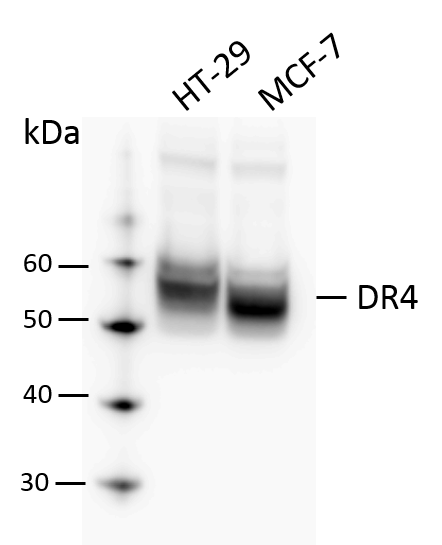
Application: Western BlotSample Tested: HT-29 human colon adenocarcinoma cell line and MCF-7 human breast cancer cell lineSpecies: HumanVerified Customer | Posted 07/23/2018Total cell lysates from HT29 and MCF-7 were subjected to western blot. PVDF membrane were probed with 1 mm/ml Human DR4 Antibody (AF347). A specific band was detected for DR4 at approximately 55 kDa. This experiment was conducted under reducing conditions.

There are no reviews that match your criteria.
Protocols
Find general support by application which include: protocols, troubleshooting, illustrated assays, videos and webinars.
- Antigen Retrieval Protocol (PIER)
- Antigen Retrieval for Frozen Sections Protocol
- Appropriate Fixation of IHC/ICC Samples
- Cellular Response to Hypoxia Protocols
- Chromogenic IHC Staining of Formalin-Fixed Paraffin-Embedded (FFPE) Tissue Protocol
- Chromogenic Immunohistochemistry Staining of Frozen Tissue
- ClariTSA™ Fluorophore Kits
- Detection & Visualization of Antibody Binding
- Fluorescent IHC Staining of Frozen Tissue Protocol
- Graphic Protocol for Heat-induced Epitope Retrieval
- Graphic Protocol for the Preparation and Fluorescent IHC Staining of Frozen Tissue Sections
- Graphic Protocol for the Preparation and Fluorescent IHC Staining of Paraffin-embedded Tissue Sections
- Graphic Protocol for the Preparation of Gelatin-coated Slides for Histological Tissue Sections
- IHC Sample Preparation (Frozen sections vs Paraffin)
- Immunofluorescent IHC Staining of Formalin-Fixed Paraffin-Embedded (FFPE) Tissue Protocol
- Immunohistochemistry (IHC) and Immunocytochemistry (ICC) Protocols
- Immunohistochemistry Frozen Troubleshooting
- Immunohistochemistry Paraffin Troubleshooting
- Preparing Samples for IHC/ICC Experiments
- Preventing Non-Specific Staining (Non-Specific Binding)
- Primary Antibody Selection & Optimization
- Protocol for Heat-Induced Epitope Retrieval (HIER)
- Protocol for Making a 4% Formaldehyde Solution in PBS
- Protocol for VisUCyte™ HRP Polymer Detection Reagent
- Protocol for the Preparation & Fixation of Cells on Coverslips
- Protocol for the Preparation and Chromogenic IHC Staining of Frozen Tissue Sections
- Protocol for the Preparation and Chromogenic IHC Staining of Frozen Tissue Sections - Graphic
- Protocol for the Preparation and Chromogenic IHC Staining of Paraffin-embedded Tissue Sections
- Protocol for the Preparation and Chromogenic IHC Staining of Paraffin-embedded Tissue Sections - Graphic
- Protocol for the Preparation and Fluorescent IHC Staining of Frozen Tissue Sections
- Protocol for the Preparation and Fluorescent IHC Staining of Paraffin-embedded Tissue Sections
- Protocol for the Preparation of Gelatin-coated Slides for Histological Tissue Sections
- R&D Systems Quality Control Western Blot Protocol
- TUNEL and Active Caspase-3 Detection by IHC/ICC Protocol
- The Importance of IHC/ICC Controls
- Troubleshooting Guide: Immunohistochemistry
- Troubleshooting Guide: Western Blot Figures
- Western Blot Conditions
- Western Blot Protocol
- Western Blot Protocol for Cell Lysates
- Western Blot Troubleshooting
- Western Blot Troubleshooting Guide
- View all Protocols, Troubleshooting, Illustrated assays and Webinars
Loading...
Associated Pathways

