mCherry Antibody - BSA Free
Novus Biologicals | Catalog # NBP2-25158
![Immunocytochemistry/ Immunofluorescence: mCherry Antibody [NBP2-25158]](https://resources.rndsystems.com/images/products/mCherry-Antibody-Immunocytochemistry-Immunofluorescence-NBP2-25158-img0005.jpg "Immunocytochemistry/ Immunofluorescence: mCherry Antibody [NBP2-25158]")
Loading...
Key Product Details
Validated by
Biological Validation
Species Reactivity
Validated:
Non-species specific
Cited:
Human, Mouse, Avian - Chicken, Donkey, Insect, Primate - Macaca mulatta (Rhesus Macaque), Rabbit
Applications
Validated:
Immunohistochemistry, Immunohistochemistry-Paraffin, Immunohistochemistry-Frozen, Immunohistochemistry Free-Floating, Immunohistochemistry Whole-Mount, Western Blot, ELISA, Immunocytochemistry/ Immunofluorescence
Cited:
Immunohistochemistry, Immunohistochemistry-Paraffin, Immunohistochemistry-Frozen, Immunohistochemistry Free-Floating, Immunohistochemistry Whole-Mount, Western Blot, ELISA, Immunocytochemistry/ Immunofluorescence, IF/IHC
Label
Unconjugated
Antibody Source
Polyclonal Chicken IgY
Format
BSA Free
Loading...
Product Specifications
Immunogen
This mCherry Antibody was developed against full length recombinant protein expressed in and purified from E. coli.
Clonality
Polyclonal
Host
Chicken
Isotype
IgY
Theoretical MW
27 kDa.
Disclaimer note: The observed molecular weight of the protein may vary from the listed predicted molecular weight due to post translational modifications, post translation cleavages, relative charges, and other experimental factors.
Disclaimer note: The observed molecular weight of the protein may vary from the listed predicted molecular weight due to post translational modifications, post translation cleavages, relative charges, and other experimental factors.
Scientific Data Images for mCherry Antibody - BSA Free
Immunocytochemistry/ Immunofluorescence: mCherry Antibody [NBP2-25158]
Immunocytochemistry/Immunofluorescence: mCherry Antibody [NBP2-25158] - HEK293 cells transfected with mCherry, stained with mCherry antibody and viewed in a confocal microscope. Most HEK293 cells are not transfected so only the nucleus of these cells can be visualized with a blue DNA stain. Cells which are transfected with mCherry are bright red, and staining with the mCherry antibody is shown in Green. The green antibody staining is only seen cells which express mCherry, and the superimposition of the green and red results in an orange signal. Interestingly stronger mCherry staining is seen in the nucleus, possibly due to degradation of some mCherry molecules releasing the low molecular weight mCherry fluorochrome.![Western Blot: mCherry Antibody [NBP2-25158]](https://resources.rndsystems.com/images/products/mCherry-Antibody-NBP2-25158-img0004.jpg "Western Blot: mCherry Antibody [NBP2-25158]")
Western Blot: mCherry Antibody [NBP2-25158]
Western Blot: mCherry Antibody [NBP2-25158] - Analysis of NBP2-25158. Lane 1. Lysate of HEK293 cells transfected with pFin-EF1-mCherry vector. There is a strong clean band at about 29kDa corresponding to mCherry (predicted molecular weight ~27kDa). Lane 2, lysate of untransfected HEK293 cells show no protein bands. The NBP2-25158 antibody was used at 1:2,000 dilution.![Western Blot: mCherry Antibody [NBP2-25158]](https://resources.rndsystems.com/images/products/mCherry-Antibody-Western-Blot-NBP2-25158-img0006.jpg "Western Blot: mCherry Antibody [NBP2-25158]")
Western Blot: mCherry Antibody [NBP2-25158]
Western Blot: mCherry Antibody [NBP2-25158] - Analysis of HEK293 cell lysates, and recombinant protein solutions using chicken mCherry pAb, dilution 1:2000 (Green). [1] protein standard, [2] HEK293, [3] HEK293 cells transfected with an mCherry-HA constract, [4] pure recombinant mCherry protein (predicted molecular weight ~ 27kDa), [5] pure recombinant GFP protein and [6] HEK293 transfected with a GFP construct. The major band at about 30kDa corresponds to mCherry-HA protein and the slightly larger recombinant form runs at about 33kDa due to presence of a His tag and other vector derived sequence. The mCherry antibody reacts strongly with both mCherry constructs and does not react with GFP protein. The blot was simultaneously probed with mouse HSP60 mAb, dilution 1:10000 (Red), which reveals a band at 60kDa only in the cell lysates.![Immunocytochemistry/ Immunofluorescence: mCherry Antibody [NBP2-25158]](https://resources.rndsystems.com/images/products/mCherry-Antibody-Immunocytochemistry-Immunofluorescence-NBP2-25158-img0007.jpg "Immunocytochemistry/ Immunofluorescence: mCherry Antibody [NBP2-25158]")

Western Blot: mCherry Antibody [NBP2-25158]-
Western blot analysis of HEK293 cell lysates using chicken pAb to mCherry, NBP2-25158, dilution 1:2,000, in green, and rabbit pAb to GAPDH, RPCA-GAPDH, dilution 1:5,000, in red: [1] protein molecular weight standard, [2] untransfected HEK293 control cells, [3] HEK293 cells transfected with pCI-Neo-mod vector expressing two tdTomato protein domains, [4] HEK293 cells transfected with pCI-Neo-mod vector expressing one mCherry-HA protein domain, and [5] HEK293 cells transfected with pCI-Neo-mod vector expressing one GFP domain. NBP2-25158 recognizes tdTomato and mCherry proteins revealing major bands at about 60kDa and 30kDa, but does not recognize GFP. The red band at 37kDa corresponds to GAPDH protein here used as a loading control.
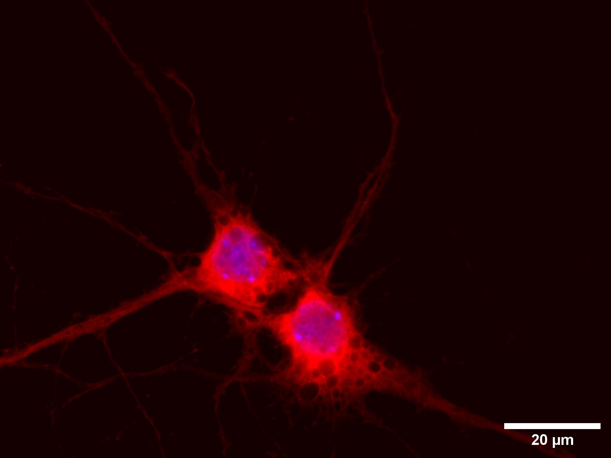
Immunocytochemistry/Immunofluorescence: Chicken Polyclonal mCherry Antibody [NBP2-25158]
DIV9 primary mouse cortical neurons infected with a shRNA construct carrying an mCherry tag, stained with mCherry (NBP2-25158) and Alexa Fluor 647 secondary (red); nuclei counterstained with DAPI (blue). Image from a verified customer review.
Immunocytochemistry/ Immunofluorescence: mCherry Antibody - BSA Free [NBP2-25158] -
Inducible Cx43 (over-)expression in stably transfected murine G4 ES cells (clone 31), in vitro differentiation into cardiomyocytes. (a) In vitro differentiation scheme of murine ES cells into spontaneously beating EBs (adapted from Boheler et al. [33]). (b) The mCherry+ EBs derived from the transgenic Cre+ G4 ES cells of clone 31 (upper picture) and transgenic mCherry− control EBs (lower picture). Scale bars 100 um. (c) Western blot analysis of EBs from Cre+ and Cre− G4 ES cells confirmed inducible Cx43 overexpression, as underscored by P2A-tagged Cx43 and mCherry expression. (d) Immunostainings of EBs illustrated clusters of cardiac alpha -actinin+ (white) cardiomyocytes in both Cre+ (upper left picture) and Cre− control EBs (lower left picture). Immunostainings yielded strong Cx43 (green, upper left picture), P2A (white), and mCherry (red) (over-)expression (upper middle picture) in Cre+ EBs. Similar apoptotic areas preferentially in the center of Cre+ and Cre− control EBs (right pictures) were found. Scale bars 10 um. Image collected and cropped by CiteAb from the following open publication (https://pubmed.ncbi.nlm.nih.gov/35203340), licensed under a CC-BY license. Not internally tested by Novus Biologicals.
Immunocytochemistry/ Immunofluorescence: mCherry Antibody - BSA Free [NBP2-25158] -
Antibody validation in MCHR1KO and Mchr1mCherry fusion allele animals. A, MCHR1 knock-out mice show ACIII-positive cilia but show no MCHR1-positive cilia. B, MCHR1 mCherry-tagged mice show colocalization of MCHR1 and mCherry tag-positive cilia. Scale bars, 10 um. Hoechst nuclei blue stain was used. Arrows indicate example cilia. N = 3 animals/genotype. Image collected and cropped by CiteAb from the following open publication (https://pubmed.ncbi.nlm.nih.gov/36849261), licensed under a CC-BY license. Not internally tested by Novus Biologicals.Applications for mCherry Antibody - BSA Free
Application
Recommended Usage
Immunocytochemistry/ Immunofluorescence
1:1000
Immunohistochemistry
1:1000
Western Blot
1:2000-1:5000
Application Notes
This mCherry antibody is useful for ICC/IF and Western Blot, where a band can be seen at ~28 kDa. Use in IHC-P, IHC-WhMt, and ELISA reported in scientific literature (PMID: 24924516, 29889212, and 29791133 respectively). Use in Immunohistochemistry-Frozen reported in scientific literature (PMID: 31591156). Use in Immunohistochemistry free floating reported in scientific literature (PMID: 31320449).
Reviewed Applications
Read 2 reviews rated 4 using NBP2-25158 in the following applications:
Formulation, Preparation, and Storage
Purification
IgY purified
Formulation
Supplied as a concentrated total IgY preparation from egg yolk, dialyzed against PBS with added preservative.
Format
BSA Free
Preservative
0.02% Sodium Azide
Concentration
Please see the vial label for concentration. If unlisted please contact technical services.
Shipping
The product is shipped with polar packs. Upon receipt, store it immediately at the temperature recommended below.
Stability & Storage
Store at 4C short term. Aliquot and store at -20C long term. Avoid freeze-thaw cycles.
Background: mCherry
mCherry can be used as a long-wavelength hetero-FRET (fluorescence resonance energy transfer) acceptor and probe for homoFRET experiments given its high peak molar absorptivity, folding efficiency, and superior spectral properties (4). Additionally, because mCherry does not interfere with other plasmids or alter the growth of Legionella species during intracellular growth, it can be used for constitutive gene expression in a variety of gram-negative bacterial species (5). For example, a plasmid developed to constitutively express mCherry under the Ptac promoter has been used in several Legionella species including L. pneumophila, the causative agent of Legionnaires' disease (5).
References
1. Shaner, N. C., Steinbach, P. A., & Tsien, R. Y. (2005). A guide to choosing fluorescent proteins. Nature Methods, 2(12), 905-909. doi:10.1038/nmeth819
2. Bevis, B. J., & Glick, B. S. (2002). Rapidly maturing variants of the Discosoma red fluorescent protein (DsRed). Nature Biotechnology, 20(1), 83-87. https://doi.org/10.1038/nbt0102-83
3. Wall, M. A., Socolich, M., & Ranganathan, R. (2000). The structural basis for red fluorescence in the tetrameric GFP homolog DsRed. Nature Structural Biology, 7(12), 1133-1138. https://doi.org/10.1038/81992
4. Akrap, N., Seidel, T., & Barisas, B. G. (2010). Forster distances for fluorescence resonant energy transfer between mCherry and other visible fluorescent proteins. Analytical Biochemistry, 402(1), 105-106. https://doi.org/10.1016/j.ab.2010.03.026
5. Gebhardt, M. J., Jacobson, R. K., & Shuman, H. A. (2017). Seeing red; the development of pON.mCherry, a broad-host range constitutive expression plasmid for Gram-negative bacteria. Plos One, 12(3), e0173116. https://doi.org/10.1371/journal.pone.0173116
Long Name
mCherry
Alternate Names
DSRED, red fluorescent protein mCherry, Red Fluoroscent Protein
Additional mCherry Products
Product Documents for mCherry Antibody - BSA Free
Certificate of Analysis
To download a Certificate of Analysis, please enter a lot or batch number in the search box below.
Product Specific Notices for mCherry Antibody - BSA Free
Chicken products cannot be exported to Canada.
This product is for research use only and is not approved for use in humans or in clinical diagnosis. Primary Antibodies are guaranteed for 1 year from date of receipt.
Citations for mCherry Antibody - BSA Free
 Powered by Bioz
Customer Reviews for mCherry Antibody - BSA Free (2)
4 out of 5
2 Customer Ratings
Have you used mCherry Antibody - BSA Free?
Submit a review and receive an Amazon gift card!
$25/€18/£15/$25CAN/¥2500 Yen for a review with an image
$10/€7/£6/$10CAN/¥1110 Yen for a review without an image
Submit a review
Customer Images
Showing
1
-
2 的
2 reviews
Showing All
Filter By:
-
Application: ImmunocytochemistrySample Tested: mouse primary neural cultures and mouse brain lysatesSpecies: MouseVerified Customer | Posted 10/31/2025DIV9 primary mouse cortical neurons infected with a shRNA construct carrying an mCherry tag, stained with mCherry (NBP2-25158) and Alexa Fluor 647 secondary (red); nuclei counterstained with DAPI (blue).

-
Application: Immunohistochemistry (whole mount)Sample Tested: Wing imaginal discSpecies: DrosophilaVerified Customer | Posted 12/08/2016The expression of mCherry under the control of the endogenous Sulf1 regulatory sequence in the wing disc.1:1000 dilution
There are no reviews that match your criteria.
Protocols
Find general support by application which include: protocols, troubleshooting, illustrated assays, videos and webinars.
- Antigen Retrieval Protocol (PIER)
- Antigen Retrieval for Frozen Sections Protocol
- Appropriate Fixation of IHC/ICC Samples
- Cellular Response to Hypoxia Protocols
- Chromogenic IHC Staining of Formalin-Fixed Paraffin-Embedded (FFPE) Tissue Protocol
- Chromogenic Immunohistochemistry Staining of Frozen Tissue
- ClariTSA™ Fluorophore Kits
- Detection & Visualization of Antibody Binding
- ELISA Sample Preparation & Collection Guide
- ELISA Troubleshooting Guide
- Fluorescent IHC Staining of Frozen Tissue Protocol
- Graphic Protocol for Heat-induced Epitope Retrieval
- Graphic Protocol for the Preparation and Fluorescent IHC Staining of Frozen Tissue Sections
- Graphic Protocol for the Preparation and Fluorescent IHC Staining of Paraffin-embedded Tissue Sections
- Graphic Protocol for the Preparation of Gelatin-coated Slides for Histological Tissue Sections
- How to Run an R&D Systems DuoSet ELISA
- How to Run an R&D Systems Quantikine ELISA
- How to Run an R&D Systems Quantikine™ QuicKit™ ELISA
- ICC Cell Smear Protocol for Suspension Cells
- ICC Immunocytochemistry Protocol Videos
- ICC for Adherent Cells
- IHC Sample Preparation (Frozen sections vs Paraffin)
- Immunocytochemistry (ICC) Protocol
- Immunocytochemistry Troubleshooting
- Immunofluorescence of Organoids Embedded in Cultrex Basement Membrane Extract
- Immunofluorescent IHC Staining of Formalin-Fixed Paraffin-Embedded (FFPE) Tissue Protocol
- Immunohistochemistry (IHC) and Immunocytochemistry (ICC) Protocols
- Immunohistochemistry Frozen Troubleshooting
- Immunohistochemistry Paraffin Troubleshooting
- Preparing Samples for IHC/ICC Experiments
- Preventing Non-Specific Staining (Non-Specific Binding)
- Primary Antibody Selection & Optimization
- Protocol for Heat-Induced Epitope Retrieval (HIER)
- Protocol for Making a 4% Formaldehyde Solution in PBS
- Protocol for VisUCyte™ HRP Polymer Detection Reagent
- Protocol for the Fluorescent ICC Staining of Cell Smears - Graphic
- Protocol for the Fluorescent ICC Staining of Cultured Cells on Coverslips - Graphic
- Protocol for the Preparation & Fixation of Cells on Coverslips
- Protocol for the Preparation and Chromogenic IHC Staining of Frozen Tissue Sections
- Protocol for the Preparation and Chromogenic IHC Staining of Frozen Tissue Sections - Graphic
- Protocol for the Preparation and Chromogenic IHC Staining of Paraffin-embedded Tissue Sections
- Protocol for the Preparation and Chromogenic IHC Staining of Paraffin-embedded Tissue Sections - Graphic
- Protocol for the Preparation and Fluorescent ICC Staining of Cells on Coverslips
- Protocol for the Preparation and Fluorescent ICC Staining of Non-adherent Cells
- Protocol for the Preparation and Fluorescent ICC Staining of Stem Cells on Coverslips
- Protocol for the Preparation and Fluorescent IHC Staining of Frozen Tissue Sections
- Protocol for the Preparation and Fluorescent IHC Staining of Paraffin-embedded Tissue Sections
- Protocol for the Preparation of Gelatin-coated Slides for Histological Tissue Sections
- Protocol for the Preparation of a Cell Smear for Non-adherent Cell ICC - Graphic
- Quantikine HS ELISA Kit Assay Principle, Alkaline Phosphatase
- Quantikine HS ELISA Kit Principle, Streptavidin-HRP Polymer
- R&D Systems Quality Control Western Blot Protocol
- Sandwich ELISA (Colorimetric) – Biotin/Streptavidin Detection Protocol
- Sandwich ELISA (Colorimetric) – Direct Detection Protocol
- TUNEL and Active Caspase-3 Detection by IHC/ICC Protocol
- The Importance of IHC/ICC Controls
- Troubleshooting Guide: ELISA
- Troubleshooting Guide: Immunohistochemistry
- Troubleshooting Guide: Western Blot Figures
- Western Blot Conditions
- Western Blot Protocol
- Western Blot Protocol for Cell Lysates
- Western Blot Troubleshooting
- Western Blot Troubleshooting Guide
- View all Protocols, Troubleshooting, Illustrated assays and Webinars
FAQs for mCherry Antibody - BSA Free
Showing
1
-
3 的
3 FAQs
Showing All
-
Q: Does this antibody cross-react with GFP epitopes? As I would like to use both GFP and mCherry antibodies during histochemistry I would not like them to cross-react.
A: mCherry and GFP share just 29% sequence similarity, so this antibody is not predicted to cross-react to GFP and has never shown any ability to detect GFP in testing.
-
Q: Hi, I would like to know the concentration of anti-mcherry antibody NBP2-25158
A: Since this is an Ammonium sulfate precipitation and not a purified antibody we cannot quantitate the antibody.
-
Q: What isotype is this anti mcherry antibody NBP2-25158?
A: IGY
-
Q: Does this antibody cross-react with GFP epitopes? As I would like to use both GFP and mCherry antibodies during histochemistry I would not like them to cross-react.
A: mCherry and GFP share just 29% sequence similarity, so this antibody is not predicted to cross-react to GFP and has never shown any ability to detect GFP in testing.
-
Q: Hi, I would like to know the concentration of anti-mcherry antibody NBP2-25158
A: Since this is an Ammonium sulfate precipitation and not a purified antibody we cannot quantitate the antibody.
-
Q: What isotype is this anti mcherry antibody NBP2-25158?
A: IGY
Loading...