MMP-2 Antibody (8B4) - (Pro and Active) - BSA Free
Novus Biologicals | Catalog # NB200-114

![Western Blot: MMP-2 Antibody (8B4)(Pro and Active)BSA Free [NB200-114]](https://resources.rndsystems.com/images/products/MMP-2-Antibody-8B4-Pro-and-Active-Western-Blot-NB200-114-img0005.jpg "Western Blot: MMP-2 Antibody (8B4)(Pro and Active)BSA Free [NB200-114]")
Scientific images may be resized, cropped, or adjusted for brightness or contrast for presentation purposes. These updates are limited to presentation and do not affect the underlying scientific interpretation of the data. Where available, additional source data may be provided upon request.
Key Product Details
Species Reactivity
Validated:
Cited:
Applications
Validated:
Cited:
Label
Antibody Source
Format
Product Specifications
Immunogen
Specificity
Clonality
Host
Isotype
Scientific Data Images for MMP-2 Antibody (8B4) - (Pro and Active) - BSA Free
Western Blot: MMP-2 Antibody (8B4)(Pro and Active)BSA Free [NB200-114]
Western Blot: MMP-2 Antibody (8B4) - (Pro and Active) [NB200-114] - Analysis of MMP2 expression in ProMMP2 (1), Active MMP2 (2) and human pressure ulcer biopsy (3).![Immunohistochemistry-Paraffin: MMP-2 Antibody (8B4) - (Pro and Active) - BSA Free [NB200-114]](https://resources.rndsystems.com/images/products/MMP-2-Antibody-8B4-Pro-and-Active-Immunohistochemistry-Paraffin-NB200-114-img0006.jpg "Immunohistochemistry-Paraffin: MMP-2 Antibody (8B4) - (Pro and Active) - BSA Free [NB200-114]")
Immunohistochemistry-Paraffin: MMP-2 Antibody (8B4) - (Pro and Active) - BSA Free [NB200-114]
Immunohistochemistry-Paraffin: MMP-2 Antibody (8B4) - (Pro and Active) [NB200-114] - Detection of MMP-2 staining in FFPE human ovary tissue. Primary antibody was used at 2.5 ug/ml with the avidin-biotin-peroxidase detection method.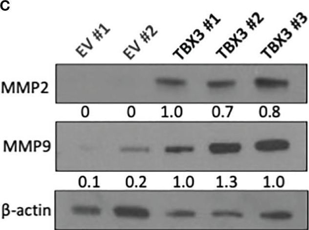
Western Blot: MMP-2 Antibody (8B4) - (Pro and Active) - BSA Free [NB200-114] -
TBX3 promotes 3D spheroid invasion. (A) 3D spheroid invasion assay of EV and TBX3 hMSCs. Representative images of the 3D invading spheroids at 0, 24 and 48 h. (40X; EVOS M5000 Imaging System; scale bars = 600 um). (B) Quantification of the invasive area. Student’s t-test was used to compare between groups, ***p < 0.001; error bars represent mean +/- SEM (n=3). (C) Western blot analysis of MMP2 and MMP9 expression in 3D hMSC spheroids (n=2). For western blotting beta -actin was used as a loading control. Densitometry readings were obtained using Fiji and protein expression levels are represented as a ratio of protein of interest/ beta -actin normalized to TBX3 #1. Image collected and cropped by CiteAb from the following open publication (https://pubmed.ncbi.nlm.nih.gov/35145908), licensed under a CC-BY license. Not internally tested by Novus Biologicals.Applications for MMP-2 Antibody (8B4) - (Pro and Active) - BSA Free
ELISA
Immunocytochemistry/ Immunofluorescence
Immunohistochemistry
Immunohistochemistry-Frozen
Immunohistochemistry-Paraffin
Knockdown Validated
Simple Western
Western Blot
Formulation, Preparation, and Storage
Purification
Formulation
Format
Preservative
Concentration
Shipping
Stability & Storage
Background: MMP-2
Long Name
Alternate Names
Gene Symbol
Additional MMP-2 Products
Product Documents for MMP-2 Antibody (8B4) - (Pro and Active) - BSA Free
Certificate of Analysis
To download a Certificate of Analysis, please enter a lot or batch number in the search box below.
Product Specific Notices for MMP-2 Antibody (8B4) - (Pro and Active) - BSA Free
This product is for research use only and is not approved for use in humans or in clinical diagnosis. Primary Antibodies are guaranteed for 1 year from date of receipt.
Citations for MMP-2 Antibody (8B4) - (Pro and Active) - BSA Free
 Powered by Bioz
Customer Reviews for MMP-2 Antibody (8B4) - (Pro and Active) - BSA Free
There are currently no reviews for this product. Be the first to review MMP-2 Antibody (8B4) - (Pro and Active) - BSA Free and earn rewards!
Have you used MMP-2 Antibody (8B4) - (Pro and Active) - BSA Free?
Submit a review and receive an Amazon gift card!
$25/€18/£15/$25CAN/¥2500 for a review with an image
$10/€7/£6/$10CAN/¥1110 for a review without an image
Submit a review
Protocols
View specific protocols for MMP-2 Antibody (8B4) - (Pro and Active) - BSA Free (NB200-114):
For IHC on Paraffin-Embedded Tissue Sections:
1. Deparaffinize in xylene. 3 changes at 10 min each.
2. Hydrate in 100% EtOH for 5 min (2X).
3. Quench endogenous peroxidase. 30 min in 1% H2O2 in methanol. (Make fresh each time: 6 ml 30% H2O2+ 194 ml methanol).
4. Hydrate to H2O in graded alcohols.
a. 5 min in 95%.
b. 5 min in 70%
c. 5 min in distilled H2O.
5. Perform antigen retrieval (Antigen Retrieval; Dako #S1700) with heating as described by manufacturer. Fill plastic Coplin jar with 50 ml Antigen Retrieval buffer. Add slides to jar when temperature reaches 95-99C start timing. Heat for 30 min. After heating, let slides cool in jar for 15 min.
6. Wash slides in PBS for 5 min (2x).
7. Infiltrate sections with PBS + 0.5% Triton X100 for 10 min, then rinse twice in PBS.
8. Block for 1 hr. at room temp in Blocking buffer- (PBS + 10% goat serum).
9. Aspirate blocking buffer. Apply MMP2/8B4 IgG diluted to 5 ug/ml in Blocking buffer. Incubate overnight at 4C (or >2 hr at 37C).
10. Rinse 3X in PBS for 10 min each.
11. Apply 2 degrees antibody. Dilute biotin conjugated goat anti-mouse 1:500 (Dako # E0433) in Blocking buffer.
12. Incubate for 2 hr. at room temperature.
13. Rinse 3X in PBS for 10 min each.
14. Prepare Avidin-biotin-peroxidase by combining each component at 1:50 in PBS + 0.1% Triton X100. Gently mix and react for 30 min. Prior to use, dilute the complexed mixture 1:5 with PBS + 0.1% BSA. Apply to sections and incubate for 2 hr at room temp.
15. Rinse in 3X in PBS for 15 min each.
16. Develop with DAB. Dissolved 1.5 mg diaminobenzidine-(HCl)4 in 3 ml of PBS, then add 2 ul of 30% hydrogen peroxide. Filter through a 0.2 um filter immediately before use. Develop for 15 min.
17. Rinse in water for 5 min. Counterstain for nuclei (e.g., hematoxylin) as desired.
18. Dehydrate through graded ethanol, clear in 100% ethanol and xylene (1:1 solution) and then 100% xylene. Coverslip with Permount.
Find general support by application which include: protocols, troubleshooting, illustrated assays, videos and webinars.
- Antigen Retrieval Protocol (PIER)
- Antigen Retrieval for Frozen Sections Protocol
- Appropriate Fixation of IHC/ICC Samples
- Cellular Response to Hypoxia Protocols
- Chromogenic IHC Staining of Formalin-Fixed Paraffin-Embedded (FFPE) Tissue Protocol
- Chromogenic Immunohistochemistry Staining of Frozen Tissue
- ClariTSA™ Fluorophore Kits
- Detection & Visualization of Antibody Binding
- ELISA Sample Preparation & Collection Guide
- ELISA Troubleshooting Guide
- Fluorescent IHC Staining of Frozen Tissue Protocol
- Graphic Protocol for Heat-induced Epitope Retrieval
- Graphic Protocol for the Preparation and Fluorescent IHC Staining of Frozen Tissue Sections
- Graphic Protocol for the Preparation and Fluorescent IHC Staining of Paraffin-embedded Tissue Sections
- Graphic Protocol for the Preparation of Gelatin-coated Slides for Histological Tissue Sections
- How to Run an R&D Systems DuoSet ELISA
- How to Run an R&D Systems Quantikine ELISA
- How to Run an R&D Systems Quantikine™ QuicKit™ ELISA
- ICC Cell Smear Protocol for Suspension Cells
- ICC Immunocytochemistry Protocol Videos
- ICC for Adherent Cells
- IHC Sample Preparation (Frozen sections vs Paraffin)
- Immunocytochemistry (ICC) Protocol
- Immunocytochemistry Troubleshooting
- Immunofluorescence of Organoids Embedded in Cultrex Basement Membrane Extract
- Immunofluorescent IHC Staining of Formalin-Fixed Paraffin-Embedded (FFPE) Tissue Protocol
- Immunohistochemistry (IHC) and Immunocytochemistry (ICC) Protocols
- Immunohistochemistry Frozen Troubleshooting
- Immunohistochemistry Paraffin Troubleshooting
- Immunoprecipitation Protocol
- Preparing Samples for IHC/ICC Experiments
- Preventing Non-Specific Staining (Non-Specific Binding)
- Primary Antibody Selection & Optimization
- Protocol for Heat-Induced Epitope Retrieval (HIER)
- Protocol for Making a 4% Formaldehyde Solution in PBS
- Protocol for VisUCyte™ HRP Polymer Detection Reagent
- Protocol for the Fluorescent ICC Staining of Cell Smears - Graphic
- Protocol for the Fluorescent ICC Staining of Cultured Cells on Coverslips - Graphic
- Protocol for the Preparation & Fixation of Cells on Coverslips
- Protocol for the Preparation and Chromogenic IHC Staining of Frozen Tissue Sections
- Protocol for the Preparation and Chromogenic IHC Staining of Frozen Tissue Sections - Graphic
- Protocol for the Preparation and Chromogenic IHC Staining of Paraffin-embedded Tissue Sections
- Protocol for the Preparation and Chromogenic IHC Staining of Paraffin-embedded Tissue Sections - Graphic
- Protocol for the Preparation and Fluorescent ICC Staining of Cells on Coverslips
- Protocol for the Preparation and Fluorescent ICC Staining of Non-adherent Cells
- Protocol for the Preparation and Fluorescent ICC Staining of Stem Cells on Coverslips
- Protocol for the Preparation and Fluorescent IHC Staining of Frozen Tissue Sections
- Protocol for the Preparation and Fluorescent IHC Staining of Paraffin-embedded Tissue Sections
- Protocol for the Preparation of Gelatin-coated Slides for Histological Tissue Sections
- Protocol for the Preparation of a Cell Smear for Non-adherent Cell ICC - Graphic
- Quantikine HS ELISA Kit Assay Principle, Alkaline Phosphatase
- Quantikine HS ELISA Kit Principle, Streptavidin-HRP Polymer
- R&D Systems Quality Control Western Blot Protocol
- Sandwich ELISA (Colorimetric) – Biotin/Streptavidin Detection Protocol
- Sandwich ELISA (Colorimetric) – Direct Detection Protocol
- TUNEL and Active Caspase-3 Detection by IHC/ICC Protocol
- The Importance of IHC/ICC Controls
- Troubleshooting Guide: ELISA
- Troubleshooting Guide: Immunohistochemistry
- Troubleshooting Guide: Western Blot Figures
- Western Blot Conditions
- Western Blot Protocol
- Western Blot Protocol for Cell Lysates
- Western Blot Troubleshooting
- Western Blot Troubleshooting Guide
- View all Protocols, Troubleshooting, Illustrated assays and Webinars
FAQs for MMP-2 Antibody (8B4) - (Pro and Active) - BSA Free
-
Q: Could you guarantee NB200-113 and NB200-114 both detect human pro-form and active form MMP2 ?
A: Yes, both of your mentioned antibodies (i.e. NB200-113 and NB200-114) detects the pro (approximately 70kD) as well as the activated forms (approximately 62kD) of MMP-2 in Western Blot. In fact, the validation image on the datasheet of #NB200-114 shows a very nice blot where these two forms are very evident and this antibody has been cited in 8 peer reviewed publications.
-
Q: I ordered the MMP-2 ms monoclonal ab 8B4, and after several tries, I cannot get it to work. I have tried in my cell lysates, concentrated conditioned media and a uterine tissue, and I am still not able to see it. Would it be possible for your company to send me a lysate that I can use as a positive control so that I can test it in something where it should be expressed? Also, are there any already known conditions for western blotting?
A:
Our lab does not have any of the positive control biopsy lysate available to send you as a control. We do offer some transfected lysates that may be of interest to you. I also have some additional information about expected samples that would express the protein. You may also find this information on UniProt useful.
-
Q: Could you guarantee NB200-113 and NB200-114 both detect human pro-form and active form MMP2 ?
A: Yes, both of your mentioned antibodies (i.e. NB200-113 and NB200-114) detects the pro (approximately 70kD) as well as the activated forms (approximately 62kD) of MMP-2 in Western Blot. In fact, the validation image on the datasheet of #NB200-114 shows a very nice blot where these two forms are very evident and this antibody has been cited in 8 peer reviewed publications.
-
Q: I ordered the MMP-2 ms monoclonal ab 8B4, and after several tries, I cannot get it to work. I have tried in my cell lysates, concentrated conditioned media and a uterine tissue, and I am still not able to see it. Would it be possible for your company to send me a lysate that I can use as a positive control so that I can test it in something where it should be expressed? Also, are there any already known conditions for western blotting?
A:
Our lab does not have any of the positive control biopsy lysate available to send you as a control. We do offer some transfected lysates that may be of interest to you. I also have some additional information about expected samples that would express the protein. You may also find this information on UniProt useful.