
Key Product Details
Species Reactivity
Validated:
Mouse
Cited:
Mouse
Applications
Validated:
Immunohistochemistry, Western Blot
Cited:
Immunohistochemistry, Western Blot
Label
Unconjugated
Antibody Source
Polyclonal Sheep IgG
Loading...
Product Specifications
Immunogen
E. coli-derived recombinant mouse FGF-10
Ser62-Thr209
Accession # NP_032028
Ser62-Thr209
Accession # NP_032028
Specificity
Detects mouse and human FGF-10 in Western blots. In direct ELISAs, approximately 100% cross-reactivity with recombinant rat FGF‑10 is observed and less than 10% cross-reactivity with recombinant mouse FGF-6 and recombinant mouse FGF-7 is observed.
Clonality
Polyclonal
Host
Sheep
Isotype
IgG
Scientific Data Images for Mouse FGF-10 Antibody
Detection of Human FGF‑10 by Western Blot.
Western blot shows lysates of A549 human lung carcinoma cell line. PVDF Membrane was probed with 1 µg/mL of Sheep Anti-Mouse FGF-10 Antigen Affinity-purified Polyclonal Antibody (Catalog # AF6224) followed by HRP-conjugated Anti-Sheep IgG Secondary Antibody (Catalog # HAF016). A specific band was detected for FGF-10 at approximately 20 kDa (as indicated). This experiment was conducted under reducing conditions and using Immunoblot Buffer Group 8.
FGF‑10 in Mouse Embryo.
FGF-10 was detected in immersion fixed frozen sections of mouse embryo (E13) using Sheep Anti-Mouse FGF-10 Antigen Affinity-purified Polyclonal Antibody (Catalog # AF6224) at 5 µg/mL overnight at 4 °C. Tissue was stained using the Anti-Sheep HRP-DAB Cell & Tissue Staining Kit (brown; Catalog # CTS019) and counterstained with hematoxylin (blue). Specific staining was localized to developing spinal cord. View our protocol for Chromogenic IHC Staining of Frozen Tissue Sections.Applications for Mouse FGF-10 Antibody
Application
Recommended Usage
Immunohistochemistry
5-15 µg/mL
Sample: Immersion fixed frozen sections of mouse embryo (E13)
Sample: Immersion fixed frozen sections of mouse embryo (E13)
Western Blot
1 µg/mL
Sample: A549 human lung carcinoma cell line
Sample: A549 human lung carcinoma cell line
Reviewed Applications
Read 1 review rated 4 using AF6224 in the following applications:
Formulation, Preparation, and Storage
Purification
Antigen Affinity-purified
Reconstitution
Sterile PBS to a final concentration of 0.2 mg/mL. For liquid material, refer to CoA for concentration.
Loading...
Formulation
Lyophilized from a 0.2 μm filtered solution in PBS with Trehalose. *Small pack size (SP) is supplied either lyophilized or as a 0.2 µm filtered solution in PBS.
Shipping
Lyophilized product is shipped at ambient temperature. Liquid small pack size (-SP) is shipped with polar packs. Upon receipt, store immediately at the temperature recommended below.
Stability & Storage
Use a manual defrost freezer and avoid repeated freeze-thaw cycles.
- 12 months from date of receipt, -20 to -70 °C as supplied.
- 1 month, 2 to 8 °C under sterile conditions after reconstitution.
- 6 months, -20 to -70 °C under sterile conditions after reconstitution.
Calculators
Background: FGF-10
References
- Beenken, A. and M. Mohammadi (2009) Nat. Rev. Drug Discov. 8:235.
- Bellusci, S. et al. (1997) Development 124:4867.
- Tagashira, S. et al. (1997) Gene 197:399.
- Beer, H.-D. et al. (2005) Oncogene 24:5269.
- Zhang, X. et al. (2006) J. Biol. Chem. 281:15694.
- Min, H. et al. (1998) Genes Dev. 12:3156.
- Rice, R. et al. (2004) J. Clin. Invest. 113:1692.
- Weaver, M. et al. (2000) Development 127:2695.
- Pirvola, U. et al. (2000) J. Neurosci. 20:6125.
- Sakaue, H. et al. (2002) Genes Dev. 16:908.
- Donjacour, A.A. et al. (2003) Dev. Biol. 261:39.
- Mailleux, A.A. et al. (2002) Development 129:53.
- Makarenkova, H.P. et al. (2000) Development 127:2563.
- Jaskoll, T. et al. (2005) BMC Dev. Biol. 5:11.
- Nomura, S. et al. (2008) Br. J. Cancer 99:305.
Long Name
Fibroblast Growth Factor 10
Alternate Names
FGF10, KGF-2, KGF2
Gene Symbol
FGF10
UniProt
Additional FGF-10 Products
Product Documents for Mouse FGF-10 Antibody
Certificate of Analysis
To download a Certificate of Analysis, please enter a lot or batch number in the search box below.
Note: Certificate of Analysis not available for kit components.
Product Specific Notices for Mouse FGF-10 Antibody
For research use only
Related Research Areas
Citations for Mouse FGF-10 Antibody
 Powered by Bioz
Customer Reviews for Mouse FGF-10 Antibody (1)
4 out of 5
1 Customer Rating
Have you used Mouse FGF-10 Antibody?
Submit a review and receive an Amazon gift card!
$25/€18/£15/$25CAN/¥2500 Yen for a review with an image
$10/€7/£6/$10CAN/¥1110 Yen for a review without an image
Submit a review
Customer Images
Showing
1
-
1 的
1 review
Showing All
Filter By:
-
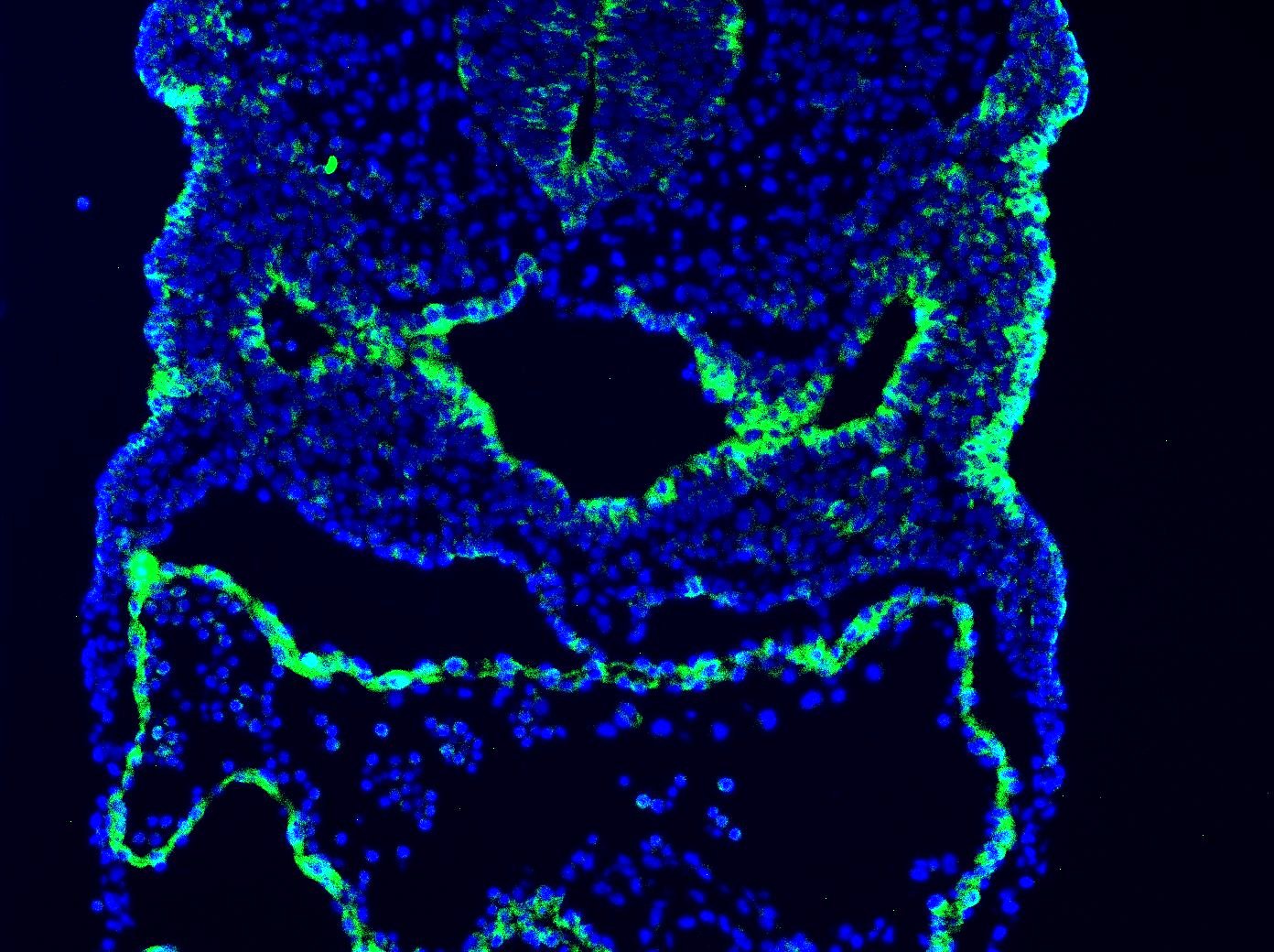
Application: Immunocytochemistry/ImmunofluorescenceSample Tested: E9.5 mouse embryo fixed in 4% PFASpecies: MouseVerified Customer | Posted 12/02/2020Antibody was used on E9.5 mouse sections. Concentration used 10ug/mL.

There are no reviews that match your criteria.
Protocols
Find general support by application which include: protocols, troubleshooting, illustrated assays, videos and webinars.
- Antigen Retrieval Protocol (PIER)
- Antigen Retrieval for Frozen Sections Protocol
- Appropriate Fixation of IHC/ICC Samples
- Cellular Response to Hypoxia Protocols
- Chromogenic IHC Staining of Formalin-Fixed Paraffin-Embedded (FFPE) Tissue Protocol
- Chromogenic Immunohistochemistry Staining of Frozen Tissue
- ClariTSA™ Fluorophore Kits
- Detection & Visualization of Antibody Binding
- Fluorescent IHC Staining of Frozen Tissue Protocol
- Graphic Protocol for Heat-induced Epitope Retrieval
- Graphic Protocol for the Preparation and Fluorescent IHC Staining of Frozen Tissue Sections
- Graphic Protocol for the Preparation and Fluorescent IHC Staining of Paraffin-embedded Tissue Sections
- Graphic Protocol for the Preparation of Gelatin-coated Slides for Histological Tissue Sections
- IHC Sample Preparation (Frozen sections vs Paraffin)
- Immunofluorescent IHC Staining of Formalin-Fixed Paraffin-Embedded (FFPE) Tissue Protocol
- Immunohistochemistry (IHC) and Immunocytochemistry (ICC) Protocols
- Immunohistochemistry Frozen Troubleshooting
- Immunohistochemistry Paraffin Troubleshooting
- Preparing Samples for IHC/ICC Experiments
- Preventing Non-Specific Staining (Non-Specific Binding)
- Primary Antibody Selection & Optimization
- Protocol for Heat-Induced Epitope Retrieval (HIER)
- Protocol for Making a 4% Formaldehyde Solution in PBS
- Protocol for VisUCyte™ HRP Polymer Detection Reagent
- Protocol for the Preparation & Fixation of Cells on Coverslips
- Protocol for the Preparation and Chromogenic IHC Staining of Frozen Tissue Sections
- Protocol for the Preparation and Chromogenic IHC Staining of Frozen Tissue Sections - Graphic
- Protocol for the Preparation and Chromogenic IHC Staining of Paraffin-embedded Tissue Sections
- Protocol for the Preparation and Chromogenic IHC Staining of Paraffin-embedded Tissue Sections - Graphic
- Protocol for the Preparation and Fluorescent IHC Staining of Frozen Tissue Sections
- Protocol for the Preparation and Fluorescent IHC Staining of Paraffin-embedded Tissue Sections
- Protocol for the Preparation of Gelatin-coated Slides for Histological Tissue Sections
- R&D Systems Quality Control Western Blot Protocol
- TUNEL and Active Caspase-3 Detection by IHC/ICC Protocol
- The Importance of IHC/ICC Controls
- Troubleshooting Guide: Immunohistochemistry
- Troubleshooting Guide: Western Blot Figures
- Western Blot Conditions
- Western Blot Protocol
- Western Blot Protocol for Cell Lysates
- Western Blot Troubleshooting
- Western Blot Troubleshooting Guide
- View all Protocols, Troubleshooting, Illustrated assays and Webinars


