
Interleukin 10, also known as cytokine synthesis inhibitory factor (CSIF), is the charter member of the IL-10 family of alpha -helical cytokines that also includes IL-19, IL‑20, IL‑22, and IL-24 (1, 2). IL-10 is secreted by many activated hematopoietic cell types as well as hepatic stellate cells, keratinocytes, and placental cytotrophoblasts (2‑5). Mature rat IL‑10 shares 85% amino acid sequence identity with mouse and 71%‑79% amino acid sequence identity with bovine, canine, equine, feline, human, ovine, and porcine IL-10. Whereas human IL-10 is active on mouse cells, mouse IL-10 does not act on human cells (6, 7). IL-10 is a 178 amino acid molecule that contains two intrachain disulfide bridges and is expressed as a 36 kDa noncovalently associated homodimer (8‑10). The IL-10 dimer binds to two IL‑10 R alpha /IL‑10 R1 chains, resulting in recruitment of two IL-10 R beta /IL-10R2 chains and activation of a signaling cascade involving JAK1, TYK2, and STAT3 (11). IL‑10 R beta does not bind IL-10 by itself but is required for signal transduction (1). IL-10 R beta also associates with IL‑20 R alpha, IL-22 R alpha, or IL-28 R alpha to form the receptor complexes for IL-22, IL-26, IL-28, and IL-29 (12‑14). IL-10 is a critical molecule in the control of viral infections and allergic and autoimmune inflammation (15‑17). It promotes phagocytic uptake and Th2 responses but suppresses antigen presentation and Th1 proinflammatory responses (2).

Scientific images may be resized, cropped, or adjusted for brightness or contrast for presentation purposes. These updates are limited to presentation and do not affect the underlying scientific interpretation of the data. Where available, additional source data may be provided upon request.
Key Product Details
Species Reactivity
Validated:
Cited:
Applications
Validated:
Cited:
Label
Antibody Source
Product Specifications
Immunogen
Ser19-Asn178
Accession # P29456
Specificity
Clonality
Host
Isotype
Endotoxin Level
Scientific Data Images for IL-10 Antibody
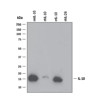
Detection of Recombinant Mouse and Rat IL‑10 by Western Blot.
Western blot shows 25 ng of Recombinant Mouse IL-10 (Catalog # 417-ML), Recombinant Human IL-10 (Catalog # 217-IL), Recombinant Rat IL-10 (Catalog # 522-RLB), and Recombinant Human IL-26/AK155 Monomer (Catalog # 1375-IL). PVDF Membrane was probed with 0.1 µg/mL of Goat Anti-Mouse/Rat IL-10 Antigen Affinity-purified Polyclonal Antibody (Catalog # AF519) followed by HRP-conjugated Anti-Goat IgG Secondary Antibody (Catalog # HAF109). A specific band was detected for IL-10 at approximately 16 kDa (as indicated). This experiment was conducted under reducing conditions and using Immunoblot Buffer Group 3.
Cell Proliferation Induced by IL‑10 and Neutralization by Rat IL‑10 Antibody.
Recombinant Rat IL-10 (Catalog # 522-RL) stimulates proliferation in the MC/9-2 mouse mast cell line in a dose-dependent manner (orange line). Proliferation elicited by Recombinant Rat IL-10 (40 ng/mL) is neutralized (green line) by increasing concentrations of Goat Anti-Rat IL-10 Antigen Affinity-purified Polyclonal Antibody (Catalog # AF519). The ND50 is typically 0.3-1.0 µg/mL.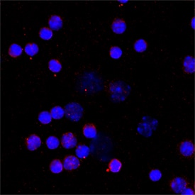
IL‑10 in Mouse Splenocytes.
IL‑10 was detected in immersion fixed mouse splenocytes using Goat Anti-Mouse/Rat IL‑10 Antigen Affinity-purified Polyclonal Antibody (Catalog # AF519) at 15 µg/mL for 3 hours at room temperature. Cells were stained using the NorthernLights™ 557-conjugated Anti-Goat IgG Secondary Antibody (red; NL001) and counterstained with DAPI (blue). Specific staining was localized to cytoplasm. Staining was performed using our protocol for Fluorescent ICC Staining of Non-adherent Cells.
Rat IL-10 ELISA Standard Curve
Mouse Chordin was serially diluted and captured by Mouse Anti-Rat IL‑10 Monoclonal Antibody (Catalog # MAB519) coated on a Clear Polystyrene Microplate (Catalog # DY990). Goat Anti-Mouse/Rat IL‑10 Antigen Affinity-purified Polyclonal Antibody (Catalog # AF519) was biotinylated and incubated with the protein captured on the plate. Detection of the standard curve was achieved by incubating Streptavidin-HRP (Catalog # DY998)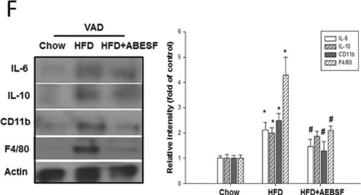
Detection of IL-10 by Western Blot
4-(2-Aminoethyl) benzenesulfonyl fluoride hydrochloride (AEBSF) recovered high-fat diet (HFD)-impaired insulin signaling and inflammation in visceral adipose (VAD) tissue of LDLR−/− mice. (A) The peripheral-tissue insulin signaling pathway [insulin receptor, pyruvate dehydrogenase kinase isozyme 1 (PDK1), Akt, glycogen synthase kinase 3 beta (GSK3 beta )] was analyzed by western immunoblots. (B) Quantification of insulin receptor-alpha and -beta expression, measured by imageQuant. Amounts of phosphorylated and total PDK1 (C), Akt (D), and GSK3 beta (E). (F) VAD tissue inflammatory cytokines and macrophage markers [interleukin (IL)-6, IL-10, CD11b, F4/80] were analyzed by western immunoblots, with protein levels quantified by imageQuant. (G) Tumor necrosis factor-alpha (TNF-alpha ) expression in VAD tissue was analyzed by ELISA. (H) Serine protease activity in VAD tissue of mice receiving the chow, HFD, and HFD + AEBSF treatments (n = 6/group). *P < 0.05, chow vs. HFD; ♯p < 0.05, HFD vs. HFD + AEBSF. Values are means ± standard deviations. Full-length blots were presented in Supplementary Figs. 3 and 4. Image collected and cropped by CiteAb from the following open publication (https://pubmed.ncbi.nlm.nih.gov/32015418), licensed under a CC-BY license. Not internally tested by R&D Systems.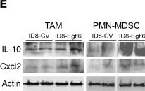
Detection of IL-10 by Western Blot
IL-10 and Cxcl2 mediate Egfl6 antitumor immunosuppression.(A) Volcano plot showing differentially expressed genes (DEGs) between CD11b+ cells infiltrating 2F8c-Egfl6 versus 2F8c tumors. Negative Log10P values determined via t test are plotted on the y axis. (B) IPA protein analysis of Egfl6 treatment associated DEG pathways identified as significantly (P < 0.05) upregulated (left panel) or downregulated (right panel). (C and D) Summary of PD-L1 expression determined by flow cytometry in infiltrating TAMs (C) and by qPCR in BM-derived macrophages polarized with different stimuli as indicated D. (E) Western blotting analysis of IL-10 and Cxcl2 in TAMs and PMN-MDSCs isolated from ID8+/–Egfl6 ascites. Actin was used as loading control. (F) ELISA of IFN-gamma in CD8+ T cells cultured with the Ly6G+ cells isolated from ID8+/–Egfl6 ascites in the absence/presence of IL-10 or Cxcl2 NAbs. (G) Western blotting showing the indicated protein expression in BM-isolated CD11b+ cells treated with GM-CSF and rEgfl6 for 0, 7.5, and 15 minutes. beta -Actin was used as loading control. Results are representative of at least 3 independent experiments. (H and I) ELISA showing IL-10 and Cxcl2 protein secretion in GM-CSF-treated BM CD11b+ cells +/– rEgfl6 and/or Syk inhibitor (R406) (H), and GM-CSF-treated BM CD11b+ cells +/– rEgfl6 and/or the integrin inhibitor Cyclo-RGD (c-RGD) (I). (J) Graph represents a ChIP assay performed with anti-Jun Ab followed by qPCR to measure IL-10 promoter in ID8+/–Egfl6 ascites. Data are presented as mean ± SEM. P values were calculated using unpaired 2-tailed t test or 1-way ANOVA with Tukey’s post test for multiple comparisons. *P < 0.05, **P < 0.01, ***P < 0.001, and ****P < 0.0001. All results are representative of 3 independent experiments. Image collected and cropped by CiteAb from the following open publication (https://pubmed.ncbi.nlm.nih.gov/39312740), licensed under a CC-BY license. Not internally tested by R&D Systems.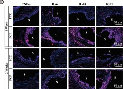
Detection of IL-10 by Immunohistochemistry
BP regulates immune signaling in bone regeneration. A) A KEGG enrichment analysis of differentially expressed genes between PCP and PCC groups. B) A summary of the expression and role of immune molecules associated with bone regeneration. C) A PPI analysis of the interactions of immune molecules associated with bone regeneration. D) IF staining images of local pro‐inflammatory factors, TNF‐ alpha and IL‐6, and anti‐inflammatory factors, IL‐10 and IGF1, at 1 and 2 weeks after PCC and PCP scaffold implantation. S, scaffold. E) Analysis of mean grays value of IF staining, n = 5. Image collected and cropped by CiteAb from the following open publication (https://pubmed.ncbi.nlm.nih.gov/37953457), licensed under a CC-BY license. Not internally tested by R&D Systems.Applications for IL-10 Antibody
Immunocytochemistry
Sample: Immersion fixed mouse splenocytes
Western Blot
Sample: Recombinant Mouse IL‑10 (Catalog # 417-ML) and Recombinant Rat IL‑10 (Catalog # 522-RLB)
Neutralization
Reviewed Applications
Read 5 reviews rated 4.6 using AF519 in the following applications:
Formulation, Preparation, and Storage
Purification
Reconstitution
Reconstitute at 0.2 mg/mL in sterile PBS. For liquid material, refer to CoA for concentration.
Formulation
Shipping
Stability & Storage
- 12 months from date of receipt, -20 to -70 °C as supplied.
- 1 month, 2 to 8 °C under sterile conditions after reconstitution.
- 6 months, -20 to -70 °C under sterile conditions after reconstitution.
Calculators
Background: IL-10
References
- Pestka, S. et al. (2004) Annu. Rev. Immunol. 22:929.
- O’Garra, A. and P. Vieira (2007) Nat. Rev. Immunol. 7:425.
- Mathurin, P. et al. (2002) Am. J. Physiol. Gastrointest. Liver Physiol. 282:G981.
- Grewe, M. et al. (1995) J. Invest. Dermatol. 104:3.
- Szony, B.J. et al. (1999) Mol. Hum. Reprod. 5:1059.
- Vieira, P. et al. (1991) Proc. Natl. Acad. Sci. 88:1172.
- Hsu, D.-H. et al. (1990) Science 250:830.
- Goodman, R. et al. (1992) Biochem. Biophys. Res. Commun. 189:1.
- Windsor, W.T. et al. (1993) Biochemistry 32:8807.
- Syto, R. et al. (1998) Biochemistry 37:16943.
- Kotenko, S.V. et al. (1997) EMBO J. 16:5894.
- Kotenko, S.V. et al. (2000) J. Biol. Chem. 276:2725.
- Hor, S. et al. (2004) J. Biol. Chem. 279:33343.
- Sheppard, P. et al. (2003) Nat. Immunol. 4:63.
- Fitzgerald, D.C. et al. (2007) Nat. Immunol. 8:1372.
- Wu, K. et al. (2007) Cell. Mol. Immunol. 4:269.
- Blackburn, S.D. and E.J. Wherry (2007) Trends Microbiol. 15:143.
Long Name
Alternate Names
Entrez Gene IDs
Gene Symbol
UniProt
Additional IL-10 Products
Product Documents for IL-10 Antibody
Certificate of Analysis
To download a Certificate of Analysis, please enter a lot or batch number in the search box below.
Note: Certificate of Analysis not available for kit components.
Product Specific Notices for IL-10 Antibody
For research use only
Related Research Areas
Citations for IL-10 Antibody
 Powered by Bioz
Customer Reviews for IL-10 Antibody (5)
Have you used IL-10 Antibody?
Submit a review and receive an Amazon gift card!
$25/€18/£15/$25CAN/¥2500 for a review with an image
$10/€7/£6/$10CAN/¥1110 for a review without an image
Submit a review
Customer Images
-
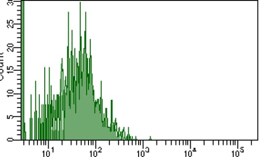
Application: Flow CytometrySample Tested: Myeloid cellsSpecies: MouseVerified Customer | Posted 08/28/2022Try this for flow, the signal is not too strong, may need optimize.

-
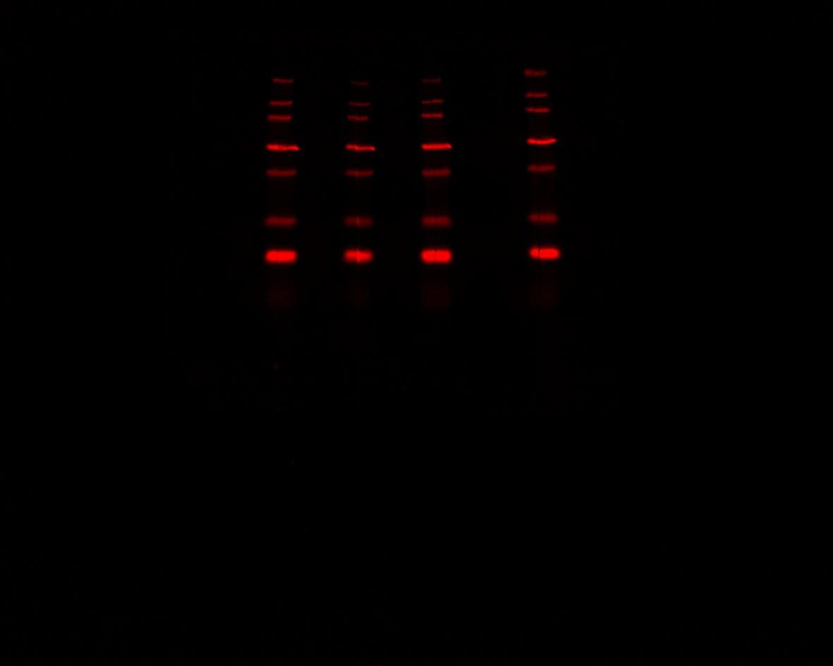
Application: Western BlotSample Tested: Bronchoalveolar lavageSpecies: RatVerified Customer | Posted 11/30/2020We had an issue with shipping due to COVID, that caused the antibody to sit at room temperature for too long and become unusable. Fortunately we placed another order of the exact same product number, and it performed perfectly. I've included the photo of the gel after the shipping mishap
-
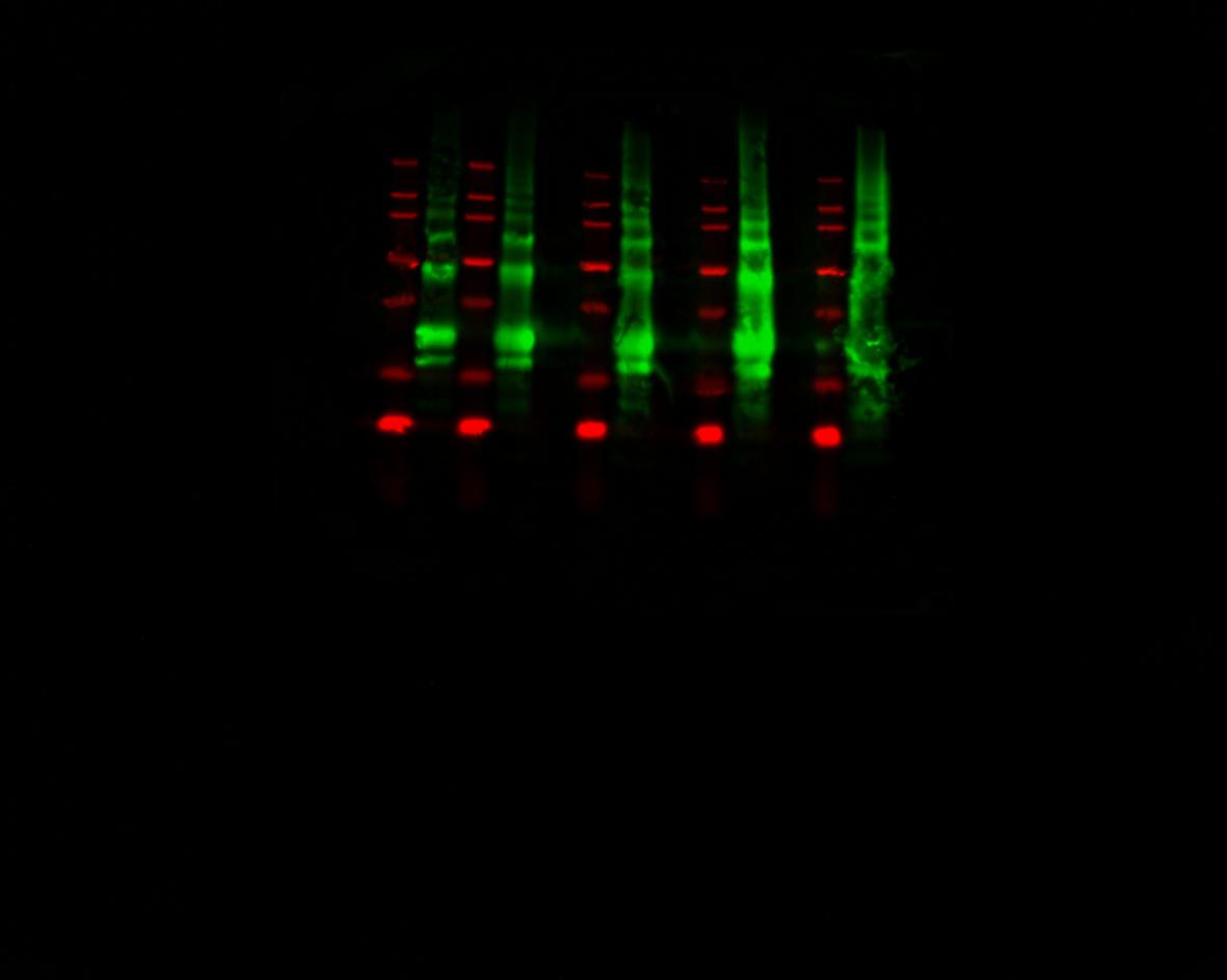
Application: Western BlotSample Tested: ControlSpecies: RatVerified Customer | Posted 11/30/2020This image was purely a test with an IL-10 control protein at different concentrations. Initially we were unable to detect the IL-10 protein in our samples, so we were testing to ensure that this antibody was functional. Turns out it was!
-
Application: ImmunohistochemistrySample Tested: Spleen tissueSpecies: MouseVerified Customer | Posted 04/24/2018
-
Application: Block/NeutralizeSample Tested: Splenic B cellsSpecies: MouseVerified Customer | Posted 07/27/2016
There are no reviews that match your criteria.
Protocols
Find general support by application which include: protocols, troubleshooting, illustrated assays, videos and webinars.
- Appropriate Fixation of IHC/ICC Samples
- Cellular Response to Hypoxia Protocols
- ClariTSA™ Fluorophore Kits
- Detection & Visualization of Antibody Binding
- ICC Cell Smear Protocol for Suspension Cells
- ICC Immunocytochemistry Protocol Videos
- ICC for Adherent Cells
- Immunocytochemistry (ICC) Protocol
- Immunocytochemistry Troubleshooting
- Immunofluorescence of Organoids Embedded in Cultrex Basement Membrane Extract
- Immunohistochemistry (IHC) and Immunocytochemistry (ICC) Protocols
- Preparing Samples for IHC/ICC Experiments
- Preventing Non-Specific Staining (Non-Specific Binding)
- Primary Antibody Selection & Optimization
- Protocol for VisUCyte™ HRP Polymer Detection Reagent
- Protocol for the Fluorescent ICC Staining of Cell Smears - Graphic
- Protocol for the Fluorescent ICC Staining of Cultured Cells on Coverslips - Graphic
- Protocol for the Preparation and Fluorescent ICC Staining of Cells on Coverslips
- Protocol for the Preparation and Fluorescent ICC Staining of Non-adherent Cells
- Protocol for the Preparation and Fluorescent ICC Staining of Stem Cells on Coverslips
- Protocol for the Preparation of a Cell Smear for Non-adherent Cell ICC - Graphic
- R&D Systems Quality Control Western Blot Protocol
- TUNEL and Active Caspase-3 Detection by IHC/ICC Protocol
- The Importance of IHC/ICC Controls
- Troubleshooting Guide: Western Blot Figures
- Western Blot Conditions
- Western Blot Protocol
- Western Blot Protocol for Cell Lysates
- Western Blot Troubleshooting
- Western Blot Troubleshooting Guide
- View all Protocols, Troubleshooting, Illustrated assays and Webinars
Associated Pathways



