
Interleukin 10 (IL-10) is a pleiotropic cytokine that can exert either immunostimulatory or immunosuppressive effects on a variety of cell types. It is produced predominantly by Th2 lymphocytes and other activated lymphoid cells.

Scientific images may be resized, cropped, or adjusted for brightness or contrast for presentation purposes. These updates are limited to presentation and do not affect the underlying scientific interpretation of the data. Where available, additional source data may be provided upon request.
Key Product Details
Validated by
Biological Validation
Species Reactivity
Validated:
Rat
Cited:
Rat
Applications
Validated:
Western Blot, ELISA Capture (Matched Antibody Pair)
Cited:
ELISA Development
Label
Unconjugated
Antibody Source
Monoclonal Mouse IgG2B Clone # 67232
Loading...
Product Specifications
Immunogen
E. coli-derived recombinant rat IL‑10
Ser19-Asn178
Accession # P29456
Ser19-Asn178
Accession # P29456
Specificity
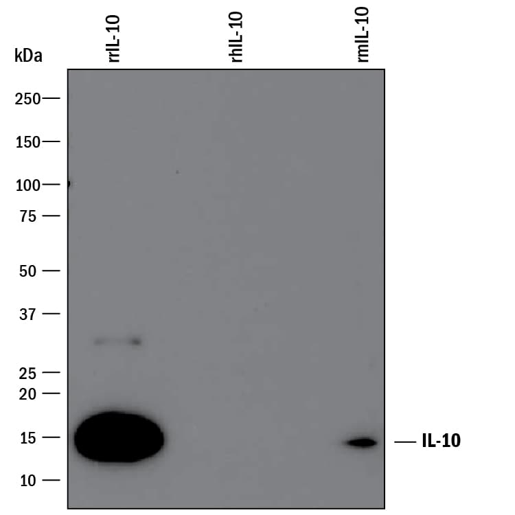
Detects rat IL-10 in ELISAs and Western blots. In Western blots, shows approximately 15% cross-reactivity with recombinant mouse IL‑10 and less than 2% cross-reactivity with recombinant human IL-10.
Clonality
Monoclonal
Host
Mouse
Isotype
IgG2B
Scientific Data Images for Rat IL-10 Antibody (67232)
Detection of Recombinant Mouse and Rat IL‑10 by Western Blot.
Western blot shows 25 ng of Recombinant Rat IL-10 (Catalog # 522-RLB), Recombinant Human IL-10 (Catalog # 217-IL) and Recombinant Mouse IL-10 (Catalog # 417-ML). PVDF Membrane was probed with 1 µg/mL of Mouse Anti-Rat IL-10 Monoclonal Antibody (Catalog # MAB519) followed by HRP-conjugated Anti-Mouse IgG Secondary Antibody (Catalog # HAF007). A specific band was detected for IL-10 at approximately 15 kDa (as indicated). This experiment was conducted under reducing conditions and using Immunoblot Buffer Group 3.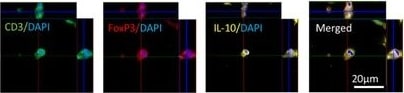
Detection of Rat IL-10 by Immunohistochemistry
Nrg-1 promotes Treg cell response in the injured spinal cord. a, b Representative images of the gating strategy for flow cytometry after singlet selection are provided for vehicle- and Nrg-1-treated groups. c At 3-day post-injury, the number of CD3+CD4+ T cells in the spinal cord was significantly higher in vehicle-treated animals compared to uninjured control group. There was no significant difference in the population of infiltrated helper T cells and the number of FoxP3+ Treg cells (CD3+CD4+FoxP3+) between vehicle- and Nrg-1-treated groups. However, the population of IL-10 producing CD4+ T cells (CD3+CD4+IL-10+) was significantly increased in Nrg-1-treated animals in comparison to vehicle-treated group. d At 7 days post-SCI, despite no significant difference in the total helper and regulatory T cell populations between vehicle- and Nrg-1-treated groups, a significant reduction (1.9-fold) was observed in IFN gamma producing effector T cell population in Nrg-1-treated SCI rats compared to their vehicle-treated counterparts. e At 14 days post-SCI, infiltrated helper T cells reached their lowest level among all examined time-points, and Nrg-1 treatment had no significant effect on the total helper T cell population. IL-10 expressing Treg cells were significantly higher in both vehicle and Nrg-1 injured rats compared to uninjured animals. Nrg-1-treated animals showed a significantly higher number of Treg cells in their spinal cord at 14-day time-point compared to vehicle-treated rats. f However, at chronic (42-day) time-point, the number of T helper cells reached a maximum, with Nrg-1-treated animals harboring a significantly decreased population of CD4+ T-cells in their spinal cord compared to the vehicle-treated group. Most importantly, CD3+CD4+FoxP3+ and CD3+CD4+IL-10+ regulatory T cell populations were significantly increased in Nrg-1-treated groups. g Immunohistochemical images show the presence of Treg cells in the perilesional area (N = 5/group/time-point, *p < 0.05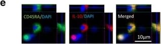
Detection of Rat IL-10 by Immunohistochemistry
Nrg-1 treatment promotes Breg cell population following SCI. a Representative images of the gating strategy for flow cytometry of spinal cord are provided. b At 7 days post-injury, the number of CD45RA+ B cells was significantly increased in the spinal cord without any significant difference in the total and regulatory B cell populations between vehicle- and Nrg-1-treated groups. c At 14-day post-SCI, a significant increase in the number of Breg cells was observed in Nrg-1-treated animals compared to vehicle-treated group. d Chronically at 42 days post-SCI, the number of B cells in the spinal cord reached the highest level compared to all earlier time-points. Nrg-1 treatment resulted in a significant increase in the number of infiltrated B cells in the spinal cord and promoted IL-10 expressing Breg cells compared to vehicle treatment. e Immunohistochemical analysis verified the presence of Breg cells in the perilesional area of the injured spinal cord. f Representative images of the gating strategy for flow cytometry of blood are provided. g Analysis of the blood revealed a significant decline in B cell population at 7 days post-SCI without any significant change in the Breg cell population at this time-point. h, i No significant change in total and regulatory B cell populations were observed in the blood at 14- and 42-day time-points. (N = 5/group/time-point, *p < 0.05, **p < 0.01, ***p < 0.001, one-way ANOVA followed by Holm-Sidak post hoc test) Image collected and cropped by CiteAb from the following publication (https://jneuroinflammation.biomedcentral.com/articles/10.1186/s12974-01…), licensed under a CC-BY license. Not internally tested by R&D Systems.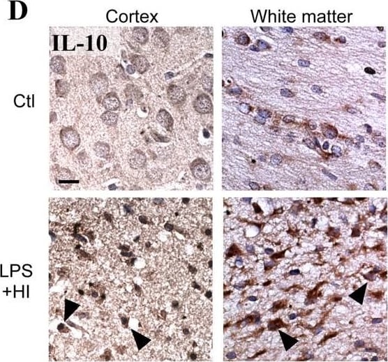
Detection of Rat IL-10 by Immunohistochemistry
Comparison between P1 and P12 immunostaining intensities of IL-6 and IL-10 in brain. IL-6 and IL-10 expressions were increased only at P12 in brains of rat pups exposed to HI+/- LPS. Data are presented (A, C) as fold increase of IL-6 and IL-10 expressions compared to control (set at 1). (B) Increased IL-6 staining (arrowheads) in P12 spongiotic frontal cortex (pyknotic neurons with abnormal reduced contrast between nucleus and cytoplasm), and spongiotic underlying external capsule following LPS+HI exposure compared to control. (D) Increased IL-10 staining (arrowheads) in P12 lesioned frontal cortex and underlying external capsule exposed to LPS+HI compared to control. IHC was performed at 48 h post-HI, in 3 to 4 brains under each experimental condition. *p < 0.05, **p < 0.01, ***p < 0.001, one-way ANOVA with Newman-Keuls post test. Scale bars = 15 μm (B, D) Image collected and cropped by CiteAb from the following open publication (https://pubmed.ncbi.nlm.nih.gov/21599903), licensed under a CC-BY license. Not internally tested by R&D Systems.
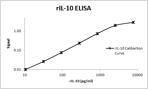
Rat IL-10 ELISA Standard Curve
Rat IL-10 was serially diluted and captured by Mouse Anti-Rat IL‑10 Monoclonal Antibody (Catalog # MAB519) coated on a Clear Polystyrene Microplate (Catalog # DY990). Goat Anti-Mouse/Rat IL‑10 Antigen Affinity-purified Polyclonal Antibody (Catalog # AF519) was biotinylated and incubated with the protein captured on the plate. Detection of the standard curve was achieved by incubating Streptavidin-HRP (Catalog # DY998)Applications for Rat IL-10 Antibody (67232)
Application
Recommended Usage
Western Blot
1 µg/mL
Sample: Recombinant Rat IL‑10 (Catalog # 522-RLB)
Sample: Recombinant Rat IL‑10 (Catalog # 522-RLB)
Rat IL-10 Sandwich Immunoassay
Please Note: Optimal dilutions of this antibody should be experimentally determined.
Reviewed Applications
Read 1 review rated 5 using MAB519 in the following applications:
Formulation, Preparation, and Storage
Purification
Protein A or G purified from hybridoma culture supernatant
Reconstitution
Reconstitute at 0.5 mg/mL in sterile PBS. For liquid material, refer to CoA for concentration.
Loading...
Formulation
Lyophilized from a 0.2 μm filtered solution in PBS with Trehalose. *Small pack size (SP) is supplied either lyophilized or as a 0.2 µm filtered solution in PBS.
Shipping
Lyophilized product is shipped at ambient temperature. Liquid small pack size (-SP) is shipped with polar packs. Upon receipt, store immediately at the temperature recommended below.
Stability & Storage
Use a manual defrost freezer and avoid repeated freeze-thaw cycles.
- 12 months from date of receipt, -20 to -70 °C as supplied.
- 1 month, 2 to 8 °C under sterile conditions after reconstitution.
- 6 months, -20 to -70 °C under sterile conditions after reconstitution.
Calculators
Background: IL-10
Long Name
Interleukin 10
Alternate Names
CSIF, GVHDS, IL10, IL10A, TGIF
Entrez Gene IDs
Gene Symbol
IL10
UniProt
Additional IL-10 Products
Product Documents for Rat IL-10 Antibody (67232)
Certificate of Analysis
To download a Certificate of Analysis, please enter a lot or batch number in the search box below.
Note: Certificate of Analysis not available for kit components.
Product Specific Notices for Rat IL-10 Antibody (67232)
For research use only
Related Research Areas
Citations for Rat IL-10 Antibody (67232)
 Powered by Bioz
Customer Reviews for Rat IL-10 Antibody (67232) (1)
5 out of 5
1 Customer Rating
Have you used Rat IL-10 Antibody (67232)?
Submit a review and receive an Amazon gift card!
$25/€18/£15/$25CAN/¥2500 for a review with an image
$10/€7/£6/$10CAN/¥1110 for a review without an image
Submit a review
Customer Images
Showing
1
-
1 的
1 review
Showing All
Filter By:
-
Application: ELISASample Tested: SerumSpecies: RatVerified Customer | Posted 11/08/2017This antibody was used to build an ELISA to measure rat IL-10. This antibody worked well as a capture antibody and BAF519 as the detection antibody.

There are no reviews that match your criteria.
Protocols
Find general support by application which include: protocols, troubleshooting, illustrated assays, videos and webinars.
- Cellular Response to Hypoxia Protocols
- R&D Systems Quality Control Western Blot Protocol
- Troubleshooting Guide: Western Blot Figures
- Western Blot Conditions
- Western Blot Protocol
- Western Blot Protocol for Cell Lysates
- Western Blot Troubleshooting
- Western Blot Troubleshooting Guide
- View all Protocols, Troubleshooting, Illustrated assays and Webinars
Loading...
Associated Pathways



