
NDRG2 Antibody (6A5) - Azide and BSA Free
Novus Biologicals | Catalog # H00057447-M03

![Knockdown Validated: NDRG2 Antibody (6A5) [H00057447-M03]](https://resources.rndsystems.com/images/products/NDRG2-Antibody-6A5-Western-Blot-H00057447-M03-img0013.jpg "Western Blot: NDRG2 Antibody (6A5) [H00057447-M03]")
Scientific images may be resized, cropped, or adjusted for brightness or contrast for presentation purposes. These updates are limited to presentation and do not affect the underlying scientific interpretation of the data. Where available, additional source data may be provided upon request.
Key Product Details
Validated by
Species Reactivity
Applications
Validated:
Cited:
Label
Antibody Source
Format
Product Specifications
Immunogen
Specificity
Clonality
Host
Isotype
Scientific Data Images for NDRG2 Antibody (6A5) - Azide and BSA Free
![Immunocytochemistry/ Immunofluorescence: NDRG2 Antibody (6A5) [H00057447-M03]](https://resources.rndsystems.com/images/products/NDRG2-Antibody-6A5-Immunocytochemistry-Immunofluorescence-H00057447-M03-img0005.jpg "Immunocytochemistry/ Immunofluorescence: NDRG2 Antibody (6A5) [H00057447-M03]")
Immunocytochemistry/ Immunofluorescence: NDRG2 Antibody (6A5) [H00057447-M03]
NDRG2-Antibody-6A5-Immunocytochemistry-Immunofluorescence-H00057447-M03-img0005.jpg![Immunohistochemistry: NDRG2 Antibody (6A5) [H00057447-M03]](https://resources.rndsystems.com/images/products/NDRG2-Antibody-6A5-Immunohistochemistry-H00057447-M03-img0007.jpg "Immunohistochemistry: NDRG2 Antibody (6A5) [H00057447-M03]")
Immunohistochemistry: NDRG2 Antibody (6A5) [H00057447-M03]
NDRG2-Antibody-6A5-Immunohistochemistry-H00057447-M03-img0007.jpg![Western Blot: NDRG2 Antibody (6A5) [H00057447-M03]](https://resources.rndsystems.com/images/products/NDRG2-Antibody-6A5-Western-Blot-H00057447-M03-img0008.jpg "Western Blot: NDRG2 Antibody (6A5) [H00057447-M03]")
Western Blot: NDRG2 Antibody (6A5) [H00057447-M03]
NDRG2-Antibody-6A5-Western-Blot-H00057447-M03-img0008.jpg![Western Blot: NDRG2 Antibody (6A5) [H00057447-M03]](https://resources.rndsystems.com/images/products/NDRG2-Antibody-6A5-Western-Blot-H00057447-M03-img0009.jpg "Western Blot: NDRG2 Antibody (6A5) [H00057447-M03]")
Western Blot: NDRG2 Antibody (6A5) [H00057447-M03]
NDRG2-Antibody-6A5-Western-Blot-H00057447-M03-img0009.jpg![Western Blot: NDRG2 Antibody (6A5) [H00057447-M03]](https://resources.rndsystems.com/images/products/NDRG2-Antibody-6A5-Western-Blot-H00057447-M03-img0011.jpg "Western Blot: NDRG2 Antibody (6A5) [H00057447-M03]")
![Western Blot: NDRG2 Antibody (6A5) [H00057447-M03]](https://resources.rndsystems.com/images/products/NDRG2-Antibody-6A5-Western-Blot-H00057447-M03-img0010.jpg "Western Blot: NDRG2 Antibody (6A5) [H00057447-M03]")
Western Blot: NDRG2 Antibody (6A5) [H00057447-M03]
NDRG2-Antibody-6A5-Western-Blot-H00057447-M03-img0010.jpg![Western Blot: NDRG2 Antibody (6A5) [H00057447-M03]](https://resources.rndsystems.com/images/products/NDRG2-Antibody-6A5-Western-Blot-H00057447-M03-img0014.jpg "Western Blot: NDRG2 Antibody (6A5) [H00057447-M03]")
![Immunocytochemistry/ Immunofluorescence: NDRG2 Antibody (6A5) [H00057447-M03]](https://resources.rndsystems.com/images/products/NDRG2-Antibody-6A5-Immunocytochemistry-Immunofluorescence-H00057447-M03-img0006.jpg "Immunocytochemistry/ Immunofluorescence: NDRG2 Antibody (6A5) [H00057447-M03]")
Immunocytochemistry/ Immunofluorescence: NDRG2 Antibody (6A5) [H00057447-M03]
NDRG2-Antibody-6A5-Immunocytochemistry-Immunofluorescence-H00057447-M03-img0006.jpg![Western Blot: NDRG2 Antibody (6A5) [H00057447-M03]](https://resources.rndsystems.com/images/products/NDRG2-Antibody-6A5-Western-Blot-H00057447-M03-img0003.jpg "Western Blot: NDRG2 Antibody (6A5) [H00057447-M03]")
Western Blot: NDRG2 Antibody (6A5) [H00057447-M03]
Western Blot: NDRG2 Antibody (6A5) [H00057447-M03] - Analysis of NDRG2 expression in Hela S3 NE.![Western Blot: NDRG2 Antibody (6A5) [H00057447-M03]](https://resources.rndsystems.com/images/products/NDRG2-Antibody-6A5-Western-Blot-H00057447-M03-img0012.jpg "Western Blot: NDRG2 Antibody (6A5) [H00057447-M03]")
Western Blot: NDRG2 Antibody (6A5) [H00057447-M03]
NDRG2-Antibody-6A5-Western-Blot-H00057447-M03-img0012.jpg![Western Blot: NDRG2 Antibody (6A5) [H00057447-M03]](https://resources.rndsystems.com/images/products/NDRG2-Antibody-6A5-Western-Blot-H00057447-M03-img0015.jpg "Western Blot: NDRG2 Antibody (6A5) [H00057447-M03]")
Western Blot: NDRG2 Antibody (6A5) [H00057447-M03]
NDRG2-Antibody-6A5-Western-Blot-H00057447-M03-img0015.jpg![ELISA: NDRG2 Antibody (6A5) [H00057447-M03]](https://resources.rndsystems.com/images/products/NDRG2-Antibody-6A5-ELISA-H00057447-M03-img0002.jpg "ELISA: NDRG2 Antibody (6A5) [H00057447-M03]")
ELISA: NDRG2 Antibody (6A5) [H00057447-M03]
ELISA: NDRG2 Antibody (6A5) [H00057447-M03] - Detection limit for recombinant GST tagged NDRG2 is approximately 0.03ng/ml as a capture antibody.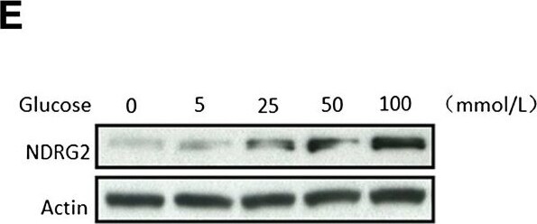
Western Blot: NDRG2 Antibody (6A5) [H00057447-M03] -
Western Blot: NDRG2 Antibody (6A5) [H00057447-M03] - NDRG2 inhibits cell proliferation & reduces intracellular glucose levels of breast cancer cells. (E) & (F) SK-BR-3 cells cultured to glucose medium at concentrations of 0, 5, 25, 50 & 100 mM for 24 hrs, & then the protein or mRNA was extracted for analysis by immunoblotting (E) or real-time PCR (F). beta -actin used as a loading control. The data presented means ± SD; error bars represented SD from 3 replicative wells. *P < 0.05 & **P < 0.01 versus control group. Image collected & cropped by CiteAb from the following publication (http://breast-cancer-research.biomedcentral.com/articles/10.1186/bcr3628), licensed under a CC-BY license. Not internally tested by Novus Biologicals.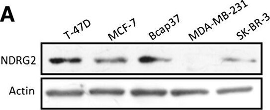
Western Blot: NDRG2 Antibody (6A5) [H00057447-M03] -
Western Blot: NDRG2 Antibody (6A5) [H00057447-M03] - NDRG2 inhibits cell proliferation & reduces intracellular glucose levels of breast cancer cells. (A) T-47D, MCF-7, Bcap37, MDA-MB-231 & SK-BR-3 cells collected for the extraction of proteins & analysed for N-myc downstream-regulated gene 2 (NDRG2) expression by immunoblotting. Image collected & cropped by CiteAb from the following publication (http://breast-cancer-research.biomedcentral.com/articles/10.1186/bcr3628), licensed under a CC-BY license. Not internally tested by Novus Biologicals.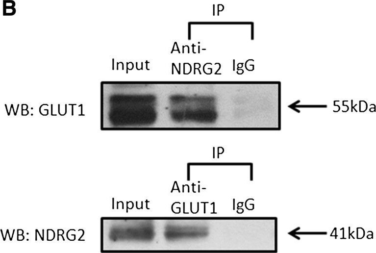
Western Blot: NDRG2 Antibody (6A5) [H00057447-M03] -
Western Blot: NDRG2 Antibody (6A5) [H00057447-M03] - NDRG2 interacts with GLUT1. (A) SK-BR-3 cells were fixed & incubated with primary antibodies against N-myc downstream-regulated gene 2 (NDRG2) or glucose transporter 1 (GLUT1) & with fluorescein isothiocyanate or a cyanine 3 secondary antibody. Green fluorescence indicates NDRG2 expression, red fluorescence indicates GLUT1 expression & blue fluorescence indicates nuclear staining. The results of the merged images reveal that NDRG2 & GLUT1 were colocalised in the cytoplasm. (B) Immunoprecipitation (IP) assays were performed with whole-cell lysates of SK-BR-3 cells pretreated with protein A–conjugated sepharose beads. Whole-cell lysates were probed for input. The antibodies for immunoprecipitation & Western blot (WB) analyses were carried out as indicated. The locations of various proteins are indicated by arrowheads. IgG, Immunoglobulin G. Image collected & cropped by CiteAb from the following publication (http://breast-cancer-research.biomedcentral.com/articles/10.1186/bcr3628), licensed under a CC-BY license. Not internally tested by Novus Biologicals.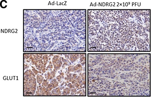
Immunocytochemistry/ Immunofluorescence: NDRG2 Antibody (6A5) [H00057447-M03] -
Immunocytochemistry/ Immunofluorescence: NDRG2 Antibody (6A5) [H00057447-M03] - NDRG2 decreases the glucose uptake & GLUT1 protein levels in SK-BR-3-based subcutaneously xenograft tumours. The experiments illustrated are described in the Methods section. (A) Tumour growth was assessed every 3 days until day 21 treatment by measuring two perpendicular diameters & calculating the volume in cubic centimetres. Ad-LacZ, adenovirus expressing LacZ; Ad-NDRG2, adenovirus expressing NDRG2; PFU, Plaque-forming units. The data presented are means ± SD; error bars represent SD from 6 mice. *P < 0.05 & **P < 0.01 versus phosphate-buffered saline (PBS) or Ad-LacZ. (B) Tumour cells were dissociated from xenograft tumours & suspended in PBS after the number of cells was counted. Next, the glucose uptake of cells in each group was detected. The data presented are means ± SD of three independent experiments; error bars represent SD from 6 mice. *P < 0.05 & **P < 0.01 versus PBS or Ad-LacZ. (C) Intratumoural protein expression was assessed by N-myc downstream-regulated gene 2 (NDRG2) & glucose transporter 1 (GLUT1) IHC staining. Representative images are shown. Original magnification: 400 x; Scale bars = 50 μm. (D) Proteins of the xenograft tumours from each group were extracted & analysed by immunoblotting to quantify NDRG2 & GLUT1 protein changes. Image collected & cropped by CiteAb from the following publication (http://breast-cancer-research.biomedcentral.com/articles/10.1186/bcr3628), licensed under a CC-BY license. Not internally tested by Novus Biologicals.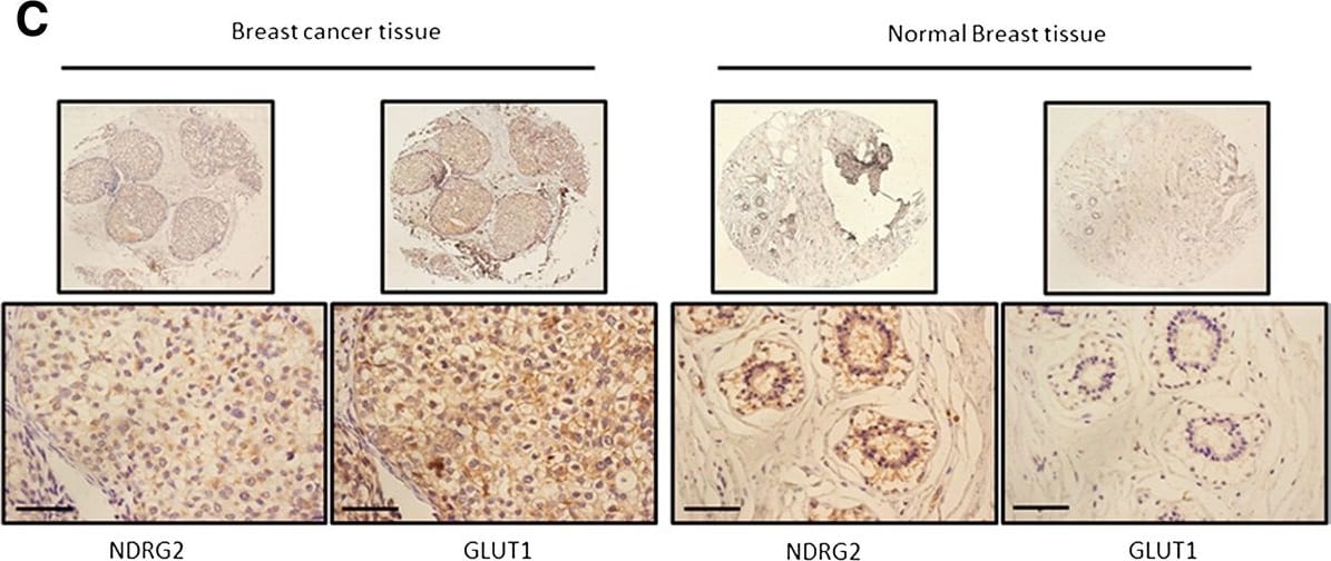
Immunohistochemistry: NDRG2 Antibody (6A5) [H00057447-M03] -
Immunohistochemistry: NDRG2 Antibody (6A5) [H00057447-M03] - NDRG2 is correlated with increased survival & negatively correlated with GLUT1 in breast carcinoma. Kaplan–Meier analysis was carried out according to N-myc downstream-regulated gene 2 (NDRG2) expression levels of disease-free survival (A) & overall survival (B). (C) Serial immunostained sections for NDRG2 & glucose transporter 1 (GLUT1) in breast cancer & normal tissues were analysed. Original magnification, 40× (top) & 400× (bottom); scale bars = 50 μm. (D) Protein was extracted from matched breast tumour tissue (T) & adjacent normal tissue (N) & subjected to immunoblot analysis to examine NDRG2 & GLUT1 expression. beta -actin served as a loading control. P: patient. Relative expression levels of NDRG2 (E) & GLUT1 (F) in human breast cancer & adjacent normal tissue are shown. immunoreactivity score distribution of cancer & adjacent normal tissue were represented with black & brown closed circles, respectively. The horizontal lines presented are means; error bars represented SD from 30 samples. P < 0.01 was considered a statistically significant difference. Image collected & cropped by CiteAb from the following publication (http://breast-cancer-research.biomedcentral.com/articles/10.1186/bcr3628), licensed under a CC-BY license. Not internally tested by Novus Biologicals.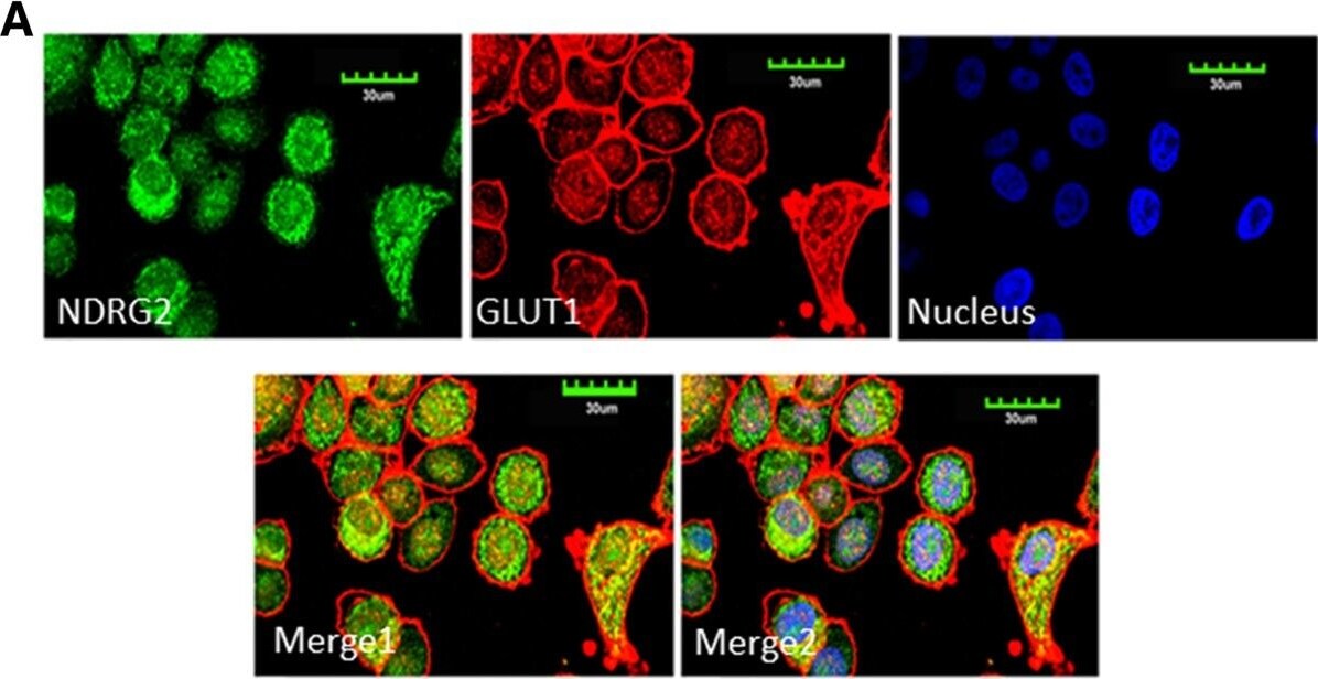
Immunocytochemistry/ Immunofluorescence: NDRG2 Antibody (6A5) [H00057447-M03] -
Immunocytochemistry/ Immunofluorescence: NDRG2 Antibody (6A5) [H00057447-M03] - NDRG2 interacts with GLUT1. (A) SK-BR-3 cells were fixed & incubated with primary antibodies against N-myc downstream-regulated gene 2 (NDRG2) or glucose transporter 1 (GLUT1) & with fluorescein isothiocyanate or a cyanine 3 secondary antibody. Green fluorescence indicates NDRG2 expression, red fluorescence indicates GLUT1 expression & blue fluorescence indicates nuclear staining. The results of the merged images reveal that NDRG2 & GLUT1 were colocalised in the cytoplasm. (B) Immunoprecipitation (IP) assays were performed with whole-cell lysates of SK-BR-3 cells pretreated with protein A–conjugated sepharose beads. Whole-cell lysates were probed for input. The antibodies for immunoprecipitation & Western blot (WB) analyses were carried out as indicated. The locations of various proteins are indicated by arrowheads. IgG, Immunoglobulin G. Image collected & cropped by CiteAb from the following publication (http://breast-cancer-research.biomedcentral.com/articles/10.1186/bcr3628), licensed under a CC-BY license. Not internally tested by Novus Biologicals.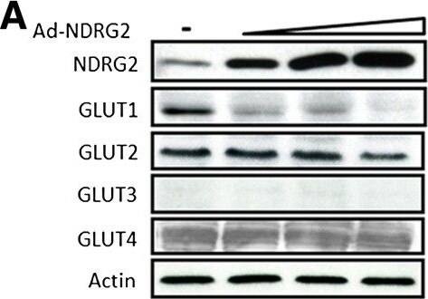
Western Blot: NDRG2 Antibody (6A5) [H00057447-M03] -
Western Blot: NDRG2 Antibody (6A5) [H00057447-M03] - NDRG2 downregulates GLUT1 by promoting its ubiquitination. (A), (C) & (E) SK-BR-3 cells were infected with an adenovirus carrying N-myc downstream-regulated gene 2 (Ad-NDRG2) at 1, 5 & 10 multiplicity of infection (MOI) or Ad-LacZ for 48 hours. (B), (D) & (F) T-47D cells were transfected with NDRG2 small interfering RNA (siRNA) 10, 25 & 100 pmol or control siRNA for 48 hours. Next, cell proteins or mRNA were extracted & analysed by immunoblotting (A) & (B) or by real-time PCR (C) to (F). beta -actin was used as a loading control. (C) – (F) The data presented are the means ± SD of three independent experiments; error bars represent SD from 3 replicative wells. *P < 0.05 & **P < 0.01 versus control group. (G) SK-BR-3 cells were infected with 10 MOI Ad-NDRG2 or Ad-LacZ for 48 hours & then treated with 2 μM, 6 μM or 8 μM MG-132 for 4 hours. Next, the protein was extracted & analysed by immunoblotting. (H) Cell fractions were prepared from the SK-BR-3 cells infected with 10 MOI Ad-NDRG2 or Ad-LacZ for 48 hours, & the membrane & cytosolic fractions of endogenous glucose transporter 1 (GLUT1) protein were detected. Tubulin & beta -actin served as loading controls. (I) SK-BR-3 cells were transfected with hemagglutinin (HA)-ubiquitin plasmid for 6 hours & infected with Ad-NDRG2 or Ad-LacZ for another 48 hours. Subsequently, the cell lysates were collected & analysed by immunoprecipitation (IP) & immunoblotting with GLUT1 & HA antibodies. WB, Western blot. Image collected & cropped by CiteAb from the following publication (http://breast-cancer-research.biomedcentral.com/articles/10.1186/bcr3628), licensed under a CC-BY license. Not internally tested by Novus Biologicals.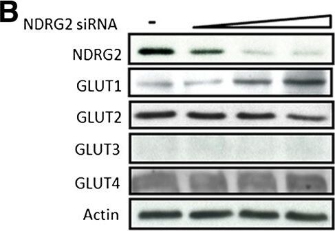
Western Blot: NDRG2 Antibody (6A5) [H00057447-M03] -
Western Blot: NDRG2 Antibody (6A5) [H00057447-M03] - NDRG2 downregulates GLUT1 by promoting its ubiquitination. (A), (C) & (E) SK-BR-3 cells were infected with an adenovirus carrying N-myc downstream-regulated gene 2 (Ad-NDRG2) at 1, 5 & 10 multiplicity of infection (MOI) or Ad-LacZ for 48 hours. (B), (D) & (F) T-47D cells were transfected with NDRG2 small interfering RNA (siRNA) 10, 25 & 100 pmol or control siRNA for 48 hours. Next, cell proteins or mRNA were extracted & analysed by immunoblotting (A) & (B) or by real-time PCR (C) to (F). beta -actin was used as a loading control. (C) – (F) The data presented are the means ± SD of three independent experiments; error bars represent SD from 3 replicative wells. *P < 0.05 & **P < 0.01 versus control group. (G) SK-BR-3 cells were infected with 10 MOI Ad-NDRG2 or Ad-LacZ for 48 hours & then treated with 2 μM, 6 μM or 8 μM MG-132 for 4 hours. Next, the protein was extracted & analysed by immunoblotting. (H) Cell fractions were prepared from the SK-BR-3 cells infected with 10 MOI Ad-NDRG2 or Ad-LacZ for 48 hours, & the membrane & cytosolic fractions of endogenous glucose transporter 1 (GLUT1) protein were detected. Tubulin & beta -actin served as loading controls. (I) SK-BR-3 cells were transfected with hemagglutinin (HA)-ubiquitin plasmid for 6 hours & infected with Ad-NDRG2 or Ad-LacZ for another 48 hours. Subsequently, the cell lysates were collected & analysed by immunoprecipitation (IP) & immunoblotting with GLUT1 & HA antibodies. WB, Western blot. Image collected & cropped by CiteAb from the following publication (http://breast-cancer-research.biomedcentral.com/articles/10.1186/bcr3628), licensed under a CC-BY license. Not internally tested by Novus Biologicals.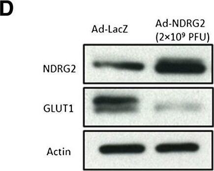
Western Blot: NDRG2 Antibody (6A5) [H00057447-M03] -
Western Blot: NDRG2 Antibody (6A5) [H00057447-M03] - NDRG2 decreases the glucose uptake & GLUT1 protein levels in SK-BR-3-based subcutaneously xenograft tumours. The experiments illustrated are described in the Methods section. (A) Tumour growth was assessed every 3 days until day 21 treatment by measuring two perpendicular diameters & calculating the volume in cubic centimetres. Ad-LacZ, adenovirus expressing LacZ; Ad-NDRG2, adenovirus expressing NDRG2; PFU, Plaque-forming units. The data presented are means ± SD; error bars represent SD from 6 mice. *P < 0.05 & **P < 0.01 versus phosphate-buffered saline (PBS) or Ad-LacZ. (B) Tumour cells were dissociated from xenograft tumours & suspended in PBS after the number of cells was counted. Next, the glucose uptake of cells in each group was detected. The data presented are means ± SD of three independent experiments; error bars represent SD from 6 mice. *P < 0.05 & **P < 0.01 versus PBS or Ad-LacZ. (C) Intratumoural protein expression was assessed by N-myc downstream-regulated gene 2 (NDRG2) & glucose transporter 1 (GLUT1) IHC staining. Representative images are shown. Original magnification: 400 x; Scale bars = 50 μm. (D) Proteins of the xenograft tumours from each group were extracted & analysed by immunoblotting to quantify NDRG2 & GLUT1 protein changes. Image collected & cropped by CiteAb from the following publication (http://breast-cancer-research.biomedcentral.com/articles/10.1186/bcr3628), licensed under a CC-BY license. Not internally tested by Novus Biologicals.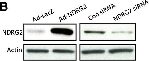
Western Blot: NDRG2 Antibody (6A5) [H00057447-M03] -
Western Blot: NDRG2 Antibody (6A5) [H00057447-M03] - NDRG2 inhibits cell proliferation & reduces intracellular glucose levels of breast cancer cells. (B) SK-BR-3 cells with low NDRG2 expression infected by an adenovirus carrying NDRG2 (Ad-NDRG2) or negative control LacZ (Ad-LacZ), & T-47D cells with high NDRG2 transfected with small interfering RNA targeting NDRG2 (NDRG2 siRNA) or negative control siRNA (Con siRNA). Thereafter proteins extracted from these cells & analysed by immunoblotting. beta -actin used as a loading control. Before being cultured in 25 mM high-glucose (H.G.) or 5.5 mM low-glucose (L.G.) medium, SK-BR-3 cells infected by Ad-NDRG2 (C) & T-47D cells transfected by NDRG2 siRNA (D). Cell proliferation was detected by 3-(4,5-dimethylthiazol-2-yl)-2,5-diphenyltetrazolium bromide assay for 1 to 5 days.Image collected & cropped by CiteAb from the following publication (http://breast-cancer-research.biomedcentral.com/articles/10.1186/bcr3628), licensed under a CC-BY license. Not internally tested by Novus Biologicals.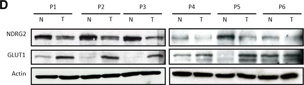
Western Blot: NDRG2 Antibody (6A5) [H00057447-M03] -
Western Blot: NDRG2 Antibody (6A5) [H00057447-M03] - NDRG2 is correlated with increased survival & negatively correlated with GLUT1 in breast carcinoma. Kaplan–Meier analysis was carried out according to N-myc downstream-regulated gene 2 (NDRG2) expression levels of disease-free survival (A) & overall survival (B). (C) Serial immunostained sections for NDRG2 & glucose transporter 1 (GLUT1) in breast cancer & normal tissues were analysed. Original magnification, 40× (top) & 400× (bottom); scale bars = 50 μm. (D) Protein was extracted from matched breast tumour tissue (T) & adjacent normal tissue (N) & subjected to immunoblot analysis to examine NDRG2 & GLUT1 expression. beta -actin served as a loading control. P: patient. Relative expression levels of NDRG2 (E) & GLUT1 (F) in human breast cancer & adjacent normal tissue are shown. immunoreactivity score distribution of cancer & adjacent normal tissue were represented with black & brown closed circles, respectively. The horizontal lines presented are means; error bars represented SD from 30 samples. P < 0.01 was considered a statistically significant difference. Image collected & cropped by CiteAb from the following publication (http://breast-cancer-research.biomedcentral.com/articles/10.1186/bcr3628), licensed under a CC-BY license. Not internally tested by Novus Biologicals.Applications for NDRG2 Antibody (6A5) - Azide and BSA Free
Western Blot
Formulation, Preparation, and Storage
Purification
Formulation
Format
Preservative
Concentration
Shipping
Stability & Storage
Background: NDRG2
Alternate Names
Gene Symbol
OMIM
UniProt
Additional NDRG2 Products
Product Documents for NDRG2 Antibody (6A5) - Azide and BSA Free
Certificate of Analysis
To download a Certificate of Analysis, please enter a lot or batch number in the search box below.
Product Specific Notices for NDRG2 Antibody (6A5) - Azide and BSA Free
This product is produced by and distributed for Abnova, a company based in Taiwan.
This product is for research use only and is not approved for use in humans or in clinical diagnosis. Primary Antibodies are guaranteed for 1 year from date of receipt.
Citations for NDRG2 Antibody (6A5) - Azide and BSA Free
 Powered by Bioz
Customer Reviews for NDRG2 Antibody (6A5) - Azide and BSA Free
There are currently no reviews for this product. Be the first to review NDRG2 Antibody (6A5) - Azide and BSA Free and earn rewards!
Have you used NDRG2 Antibody (6A5) - Azide and BSA Free?
Submit a review and receive an Amazon gift card!
$25/€18/£15/$25CAN/¥2500 for a review with an image
$10/€7/£6/$10CAN/¥1110 for a review without an image
Submit a review
Protocols
Find general support by application which include: protocols, troubleshooting, illustrated assays, videos and webinars.
- Antigen Retrieval Protocol (PIER)
- Antigen Retrieval for Frozen Sections Protocol
- Appropriate Fixation of IHC/ICC Samples
- Cellular Response to Hypoxia Protocols
- Chromogenic IHC Staining of Formalin-Fixed Paraffin-Embedded (FFPE) Tissue Protocol
- Chromogenic Immunohistochemistry Staining of Frozen Tissue
- ClariTSA™ Fluorophore Kits
- Detection & Visualization of Antibody Binding
- ELISA Sample Preparation & Collection Guide
- ELISA Troubleshooting Guide
- Fluorescent IHC Staining of Frozen Tissue Protocol
- Graphic Protocol for Heat-induced Epitope Retrieval
- Graphic Protocol for the Preparation and Fluorescent IHC Staining of Frozen Tissue Sections
- Graphic Protocol for the Preparation and Fluorescent IHC Staining of Paraffin-embedded Tissue Sections
- Graphic Protocol for the Preparation of Gelatin-coated Slides for Histological Tissue Sections
- How to Run an R&D Systems DuoSet ELISA
- How to Run an R&D Systems Quantikine ELISA
- How to Run an R&D Systems Quantikine™ QuicKit™ ELISA
- ICC Cell Smear Protocol for Suspension Cells
- ICC Immunocytochemistry Protocol Videos
- ICC for Adherent Cells
- IHC Sample Preparation (Frozen sections vs Paraffin)
- Immunocytochemistry (ICC) Protocol
- Immunocytochemistry Troubleshooting
- Immunofluorescence of Organoids Embedded in Cultrex Basement Membrane Extract
- Immunofluorescent IHC Staining of Formalin-Fixed Paraffin-Embedded (FFPE) Tissue Protocol
- Immunohistochemistry (IHC) and Immunocytochemistry (ICC) Protocols
- Immunohistochemistry Frozen Troubleshooting
- Immunohistochemistry Paraffin Troubleshooting
- Preparing Samples for IHC/ICC Experiments
- Preventing Non-Specific Staining (Non-Specific Binding)
- Primary Antibody Selection & Optimization
- Protocol for Heat-Induced Epitope Retrieval (HIER)
- Protocol for Making a 4% Formaldehyde Solution in PBS
- Protocol for VisUCyte™ HRP Polymer Detection Reagent
- Protocol for the Fluorescent ICC Staining of Cell Smears - Graphic
- Protocol for the Fluorescent ICC Staining of Cultured Cells on Coverslips - Graphic
- Protocol for the Preparation & Fixation of Cells on Coverslips
- Protocol for the Preparation and Chromogenic IHC Staining of Frozen Tissue Sections
- Protocol for the Preparation and Chromogenic IHC Staining of Frozen Tissue Sections - Graphic
- Protocol for the Preparation and Chromogenic IHC Staining of Paraffin-embedded Tissue Sections
- Protocol for the Preparation and Chromogenic IHC Staining of Paraffin-embedded Tissue Sections - Graphic
- Protocol for the Preparation and Fluorescent ICC Staining of Cells on Coverslips
- Protocol for the Preparation and Fluorescent ICC Staining of Non-adherent Cells
- Protocol for the Preparation and Fluorescent ICC Staining of Stem Cells on Coverslips
- Protocol for the Preparation and Fluorescent IHC Staining of Frozen Tissue Sections
- Protocol for the Preparation and Fluorescent IHC Staining of Paraffin-embedded Tissue Sections
- Protocol for the Preparation of Gelatin-coated Slides for Histological Tissue Sections
- Protocol for the Preparation of a Cell Smear for Non-adherent Cell ICC - Graphic
- Quantikine HS ELISA Kit Assay Principle, Alkaline Phosphatase
- Quantikine HS ELISA Kit Principle, Streptavidin-HRP Polymer
- R&D Systems Quality Control Western Blot Protocol
- Sandwich ELISA (Colorimetric) – Biotin/Streptavidin Detection Protocol
- Sandwich ELISA (Colorimetric) – Direct Detection Protocol
- TUNEL and Active Caspase-3 Detection by IHC/ICC Protocol
- The Importance of IHC/ICC Controls
- Troubleshooting Guide: ELISA
- Troubleshooting Guide: Immunohistochemistry
- Troubleshooting Guide: Western Blot Figures
- Western Blot Conditions
- Western Blot Protocol
- Western Blot Protocol for Cell Lysates
- Western Blot Troubleshooting
- Western Blot Troubleshooting Guide
- View all Protocols, Troubleshooting, Illustrated assays and Webinars