NKX3.1 Antibody (0361) - BSA Free
Novus Biologicals | Catalog # NB100-1828
Key Product Details
Validated by
Species Reactivity
Validated:
Cited:
Applications
Validated:
Cited:
Label
Antibody Source
Format
Product Specifications
Immunogen
Reactivity Notes
Localization
Clonality
Host
Isotype
Scientific Data Images for NKX3.1 Antibody (0361) - BSA Free
![Western Blot: NKX3.1 Antibody (0361)BSA Free [NB100-1828]](https://resources.rndsystems.com/images/products/NKX3-1-Antibody-0361-Western-Blot-NB100-1828-img0006.jpg "Western Blot: NKX3.1 Antibody (0361)BSA Free [NB100-1828]")
Western Blot: NKX3.1 Antibody (0361)BSA Free [NB100-1828]
Western Blot: NKX3.1 Antibody (0361) [NB100-1828] - Analysis of NKX3.1 in HeLa cell lysate using anti-NKX3.1 antibody. WB image submitted by a verified customer review.![Immunohistochemistry-Paraffin: NKX3.1 Antibody (0361) - BSA Free [NB100-1828]](https://resources.rndsystems.com/images/products/NKX3-1-Antibody-0361-Immunohistochemistry-Paraffin-NB100-1828-img0008.jpg "Immunohistochemistry-Paraffin: NKX3.1 Antibody (0361) - BSA Free [NB100-1828]")
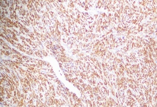
Immunohistochemistry-Paraffin: NKX3.1 Antibody (0361) - BSA Free [NB100-1828]
Immunohistochemistry-Paraffin: NKX3.1 Antibody (0361) [NB100-1828] - Staining of human chondrosarcoma tissue with NKX3.1 monoclonal antibody. IHC-P image submitted by a verified customer review.![Western Blot: NKX3.1 Antibody (0361)BSA Free [NB100-1828]](https://resources.rndsystems.com/images/products/Nkx3-1-Antibody-0361-Western-Blot-NB100-1828-img0003.jpg "Western Blot: NKX3.1 Antibody (0361)BSA Free [NB100-1828]")
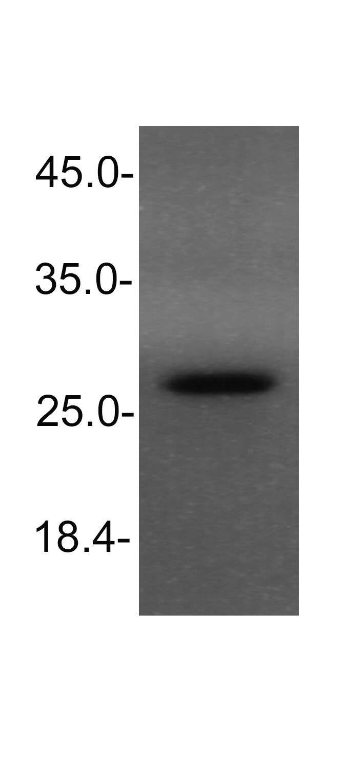
Western Blot: NKX3.1 Antibody (0361)BSA Free [NB100-1828]
Western Blot: Nkx3.1 Antibody (0361) [NB100-1828] - Detection of NKX3.1 in mouse testis lysate using NB 100-1828. ECL detection 1 minute.![Immunohistochemistry-Paraffin: NKX3.1 Antibody (0361) - BSA Free [NB100-1828]](https://resources.rndsystems.com/images/products/NKX3-1-Antibody-0361-Immunohistochemistry-Paraffin-NB100-1828-img0007.jpg "Immunohistochemistry-Paraffin: NKX3.1 Antibody (0361) - BSA Free [NB100-1828]")
Immunohistochemistry-Paraffin: NKX3.1 Antibody (0361) - BSA Free [NB100-1828]
Immunohistochemistry-Paraffin: NKX3.1 Antibody (0361) [NB100-1828] - NKX3.1 was detected in immersion fixed paraffin-embedded sections of mouse testis using Mouse Anti-Mouse NKX3.1 (0361) Monoclonal Antibody (Catalog # NB100-1828) at 1:300 for 1 hour at room temperature followed by incubation with the Anti-Mouse IgG VisUCyte™ HRP Polymer Antibody (Catalog # VC001). Tissue was stained using DAB (brown) and counterstained with hematoxylin (blue). Specific staining was localized to the nuclei in sperm cells.![Knockdown Validated: NKX3.1 Antibody (0361) - BSA Free [NB100-1828]](https://resources.rndsystems.com/images/products/NKX3-1-Antibody-0361-Knockdown-Validated-NB100-1828-img0009.jpg "Knockdown Validated: NKX3.1 Antibody (0361) - BSA Free [NB100-1828]")
 - BSA Free [NB100-1828] -")
Western Blot: NKX3.1 Antibody (0361) - BSA Free [NB100-1828] -
Western Blot: NKX3.1 Antibody (0361) - BSA Free [NB100-1828] - SALL2 transcriptionally upregulates ESR1 in breast cancerARNA‐seq analysis of ESR1 mRNA levels in 9 paired pre‐tamoxifen‐treated primary breast cancer tissues & relapsed tamoxifen‐resistant breast cancer tissues.BqRT–PCR analysis of ESR1 expression in 9 paired pre‐tamoxifen‐treated primary breast cancer tissues & relapsed tamoxifen‐resistant breast cancer tissues. GAPDH was used as an internal control.C, DWB analysis of SALL2 (A) & NKX3‐1 (B) expression in the indicated cells transfected with Ri‐Vector (V‐Ri) or shRNAs (Ri#1/2) against SALL2 or NKX3‐1. alpha ‐Tubulin was used as the loading control.E, FqRT–PCR analysis of ESR1 expression in the indicated cells transfected with Ri‐Vector or shRNAs (Ri#1/2) against SALL2 or NKX3‐1.Data information: In (A), P‐values were determined by two‐tailed paired Student's t‐test. In (B), data are presented as mean ± SD, & P‐values were determined by two‐tailed unpaired Student's t‐test, n = 3. In (E & F), data are presented as mean ± SD, & P‐values were determined by one‐way ANOVA test, n = 3. *P < 0.05, **P < 0.01, ***P < 0.001, n.s., no significance. Exact P‐values are specified in Appendix Table S10.Source data are available online for this figure. Image collected & cropped by CiteAb from the following publication (https://pubmed.ncbi.nlm.nih.gov/31657150), licensed under a CC-BY license. Not internally tested by Novus Biologicals.Applications for NKX3.1 Antibody (0361) - BSA Free
Immunohistochemistry
Immunohistochemistry-Frozen
Immunohistochemistry-Paraffin
Immunoprecipitation
Western Blot
Reviewed Applications
Read 3 reviews rated 4.3 using NB100-1828 in the following applications:
Formulation, Preparation, and Storage
Purification
Formulation
Format
Preservative
Concentration
Shipping
Stability & Storage
Background: NKX3.1
Long Name
Alternate Names
Gene Symbol
Additional NKX3.1 Products
Product Documents for NKX3.1 Antibody (0361) - BSA Free
Certificate of Analysis
To download a Certificate of Analysis, please enter a lot or batch number in the search box below.
Product Specific Notices for NKX3.1 Antibody (0361) - BSA Free
This product is for research use only and is not approved for use in humans or in clinical diagnosis. Primary Antibodies are guaranteed for 1 year from date of receipt.
Related Research Areas
Citations for NKX3.1 Antibody (0361) - BSA Free
 Powered by Bioz
Customer Reviews for NKX3.1 Antibody (0361) - BSA Free (3)
Have you used NKX3.1 Antibody (0361) - BSA Free?
Submit a review and receive an Amazon gift card!
$25/€18/£15/$25CAN/¥2500 Yen for a review with an image
$10/€7/£6/$10CAN/¥1110 Yen for a review without an image
Submit a review
Customer Images
-
Application: Immunohistochemistry-FrozenSample Tested: ChondrosarcomaSpecies: HumanVerified Customer | Posted 08/04/2021NKX3.1 monoclonal antibody in chondrosarcoma tissue

-
Application: Immunohistochemistry-ParaffinSample Tested: Prostate tissueSpecies: MouseVerified Customer | Posted 04/19/2016
-
Application: Western BlotSample Tested: Cell lysate from HeLa cellsSpecies: HumanVerified Customer | Posted 11/20/2014Western blot for NKX3.1 in HeLa cells
There are no reviews that match your criteria.
Protocols
View specific protocols for NKX3.1 Antibody (0361) - BSA Free (NB100-1828):
Western Blot Protocol
1. Perform SDS-PAGE (4-12%) on samples to be analyzed, loading 40 ug of total protein per lane.
2. Transfer proteins to Nitrocellulose according to the instructions provided by the manufacturer of the transfer apparatus.
3. Rinse membrane with dH2O and then stain the blot using ponceau S for 1-2 minutes to access the transfer of proteins onto the nitrocellulose membrane. Rinse the blot in water to remove excess stain and mark the lane locations and locations of molecular weight markers using a pencil.
4. Rinse the blot in TBS for approximately 5 minutes.
5. Block the membrane using 5% non-fat dry milk + 1% BSA in TBS for 2 hours at room temperature.
6. Rinse the membrane in dH2O and then wash the membrane in wash buffer [TBS + 0.1% Tween] 3 times for 10 minutes each.
7. Dilute the mouse anti-Nkx3.1 primary antibody (NB 100-1828) in blocking buffer and incubate 1 hour at room temperature.
8. Rinse the membrane in dH2O and then wash the membrane in wash buffer [TBS + 0.1% Tween] 3 times for 10 minutes each.
9. Apply the diluted mouse-IgG HRP-conjugated secondary antibody in blocking buffer (as per manufacturer's instructions) and incubate 1 hour at room temperature.
10. Wash the blot in wash buffer [TBS + 0.1% Tween] 3 times for 10 minutes each (this step can be repeated as required to reduce background).
11. Apply the detection reagent of choice in accordance with the manufacturer's instructions (Pierce's ECL).
Note: Tween-20 can be added to the blocking or antibody dilution buffer at a final concentration of 0.05-0.2%, provided it does not interfere with antibody-antigen binding.
Find general support by application which include: protocols, troubleshooting, illustrated assays, videos and webinars.
- Antigen Retrieval Protocol (PIER)
- Antigen Retrieval for Frozen Sections Protocol
- Appropriate Fixation of IHC/ICC Samples
- Cellular Response to Hypoxia Protocols
- Chromogenic IHC Staining of Formalin-Fixed Paraffin-Embedded (FFPE) Tissue Protocol
- Chromogenic Immunohistochemistry Staining of Frozen Tissue
- ClariTSA™ Fluorophore Kits
- Detection & Visualization of Antibody Binding
- Fluorescent IHC Staining of Frozen Tissue Protocol
- Graphic Protocol for Heat-induced Epitope Retrieval
- Graphic Protocol for the Preparation and Fluorescent IHC Staining of Frozen Tissue Sections
- Graphic Protocol for the Preparation and Fluorescent IHC Staining of Paraffin-embedded Tissue Sections
- Graphic Protocol for the Preparation of Gelatin-coated Slides for Histological Tissue Sections
- IHC Sample Preparation (Frozen sections vs Paraffin)
- Immunofluorescent IHC Staining of Formalin-Fixed Paraffin-Embedded (FFPE) Tissue Protocol
- Immunohistochemistry (IHC) and Immunocytochemistry (ICC) Protocols
- Immunohistochemistry Frozen Troubleshooting
- Immunohistochemistry Paraffin Troubleshooting
- Immunoprecipitation Protocol
- Preparing Samples for IHC/ICC Experiments
- Preventing Non-Specific Staining (Non-Specific Binding)
- Primary Antibody Selection & Optimization
- Protocol for Heat-Induced Epitope Retrieval (HIER)
- Protocol for Making a 4% Formaldehyde Solution in PBS
- Protocol for VisUCyte™ HRP Polymer Detection Reagent
- Protocol for the Preparation & Fixation of Cells on Coverslips
- Protocol for the Preparation and Chromogenic IHC Staining of Frozen Tissue Sections
- Protocol for the Preparation and Chromogenic IHC Staining of Frozen Tissue Sections - Graphic
- Protocol for the Preparation and Chromogenic IHC Staining of Paraffin-embedded Tissue Sections
- Protocol for the Preparation and Chromogenic IHC Staining of Paraffin-embedded Tissue Sections - Graphic
- Protocol for the Preparation and Fluorescent IHC Staining of Frozen Tissue Sections
- Protocol for the Preparation and Fluorescent IHC Staining of Paraffin-embedded Tissue Sections
- Protocol for the Preparation of Gelatin-coated Slides for Histological Tissue Sections
- R&D Systems Quality Control Western Blot Protocol
- TUNEL and Active Caspase-3 Detection by IHC/ICC Protocol
- The Importance of IHC/ICC Controls
- Troubleshooting Guide: Immunohistochemistry
- Troubleshooting Guide: Western Blot Figures
- Western Blot Conditions
- Western Blot Protocol
- Western Blot Protocol for Cell Lysates
- Western Blot Troubleshooting
- Western Blot Troubleshooting Guide
- View all Protocols, Troubleshooting, Illustrated assays and Webinars
FAQs for NKX3.1 Antibody (0361) - BSA Free
-
Q: I have three kinds of anti-mouse 2nd Ab; HRP-conjugated anti-mouse Ig G, Ig G2, Ig G2A. Are these all proper for NB100-1828 in WB or IHC?
A: For western blot we have used a Mouse IgG secondary antibody NB7539 . This type of secondary will also be suitable for both western blot and IHC.
-
Q: Today we received an inquiry from our customer who was interested in your Nkx3.1 Antibody, #NB100-1828. The customer has a plan to use this antibody in immunohistochemical staining with her paraffin section samples from mouse. The customer would like to have IHC images obtained with #NB100-1828. In addition, the customer would like to know the detailed protocol (antibody concentration, antigen retrieval method, buffer condition and so on) which was used when checking the IHC Application.
A:
Since this is a product we have had around for some time, I was unable to find any data on file for the IHC application as this was reported to us by a customer. I would recommend a standard IHC protocol for staining and a dilution of 1:100. I would recommend antigen retrieval using a heat mediated citrate buffer at a pH 6.0. You can find the general protocol information that our lab recommends here. I hope your customer finds this information useful and again I apologize we do not have any images to send over, but do guarantee this antibody to work for all species and applications listed. The customer may need to directly conjugate the primary antibody to avoid high background with their anti-mouse secondary detecting the mouse samples.
-
Q: I have three kinds of anti-mouse 2nd Ab; HRP-conjugated anti-mouse Ig G, Ig G2, Ig G2A. Are these all proper for NB100-1828 in WB or IHC?
A: For western blot we have used a Mouse IgG secondary antibody NB7539 . This type of secondary will also be suitable for both western blot and IHC.
-
Q: Today we received an inquiry from our customer who was interested in your Nkx3.1 Antibody, #NB100-1828. The customer has a plan to use this antibody in immunohistochemical staining with her paraffin section samples from mouse. The customer would like to have IHC images obtained with #NB100-1828. In addition, the customer would like to know the detailed protocol (antibody concentration, antigen retrieval method, buffer condition and so on) which was used when checking the IHC Application.
A:
Since this is a product we have had around for some time, I was unable to find any data on file for the IHC application as this was reported to us by a customer. I would recommend a standard IHC protocol for staining and a dilution of 1:100. I would recommend antigen retrieval using a heat mediated citrate buffer at a pH 6.0. You can find the general protocol information that our lab recommends here. I hope your customer finds this information useful and again I apologize we do not have any images to send over, but do guarantee this antibody to work for all species and applications listed. The customer may need to directly conjugate the primary antibody to avoid high background with their anti-mouse secondary detecting the mouse samples.