Src Antibody (5A18) - BSA Free
Novus Biologicals | Catalog # NBP1-19188
![Western Blot: Src Antibody (5A18) [NBP1-19188]](https://resources.rndsystems.com/images/products/Src-Antibody-5A18-Western-Blot-NBP1-19188-img0004.jpg "Western Blot: Src Antibody (5A18) [NBP1-19188]")
Loading...
Key Product Details
Validated by
Biological Validation
Species Reactivity
Validated:
Human, Mouse, Rat
Cited:
Human
Applications
Validated:
Immunohistochemistry, Immunohistochemistry-Paraffin, Western Blot, Simple Western
Cited:
Western Blot
Label
Unconjugated
Antibody Source
Monoclonal Mouse Clone # 5A18
Format
BSA Free
Loading...
Product Specifications
Immunogen
Recombinant human full length c-Src
Reactivity Notes
Please note that this antibody is reactive to Mouse and derived from the same host, Mouse. Mouse-On-Mouse blocking reagent may be needed for IHC and ICC experiments to reduce high background signal. You can find these reagents under catalog numbers PK-2200-NB and MP-2400-NB. Please contact Technical Support if you have any questions.
Specificity
Src (5A18)
Clonality
Monoclonal
Host
Mouse
Scientific Data Images for Src Antibody (5A18) - BSA Free
![Immunohistochemistry: Src Antibody (5A18) [NBP1-19188]](https://resources.rndsystems.com/images/products/Src-Antibody-5A18-Immunohistochemistry-NBP1-19188-img0002.jpg "Immunohistochemistry: Src Antibody (5A18) [NBP1-19188]")
Immunohistochemistry: Src Antibody (5A18) [NBP1-19188]
Immunohistochemistry: Src Antibody (5A18) [NBP1-19188] - IHC Analysis: Human prostate tissue stained with Src, mAb (5A18) at 20 ug/ml.![Western Blot: Src Antibody (5A18) [NBP1-19188]](https://resources.rndsystems.com/images/products/Src-Antibody-5A18-Western-Blot-NBP1-19188-img0003.jpg "Western Blot: Src Antibody (5A18) [NBP1-19188]")
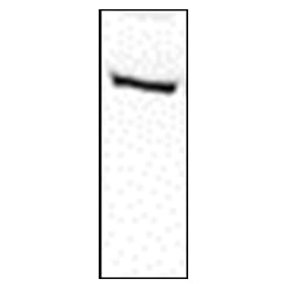
Western Blot: Src Antibody (5A18) [NBP1-19188]
Western Blot: Src Antibody (5A18) [NBP1-19188] - Western blot analysis of Src using 20 ug of whole cell lysate (Lane 2=HeLa, Lane 3=3T3, Lane 4=PC12) probed with with Src, mAb (5A18) at 1 ug/ml.![Immunohistochemistry: Src Antibody (5A18) [NBP1-19188]](https://resources.rndsystems.com/images/products/Src-Antibody-5A18-Immunohistochemistry-NBP1-19188-img0001.jpg "Immunohistochemistry: Src Antibody (5A18) [NBP1-19188]")
Immunohistochemistry: Src Antibody (5A18) [NBP1-19188]
Immunohistochemistry: Src Antibody (5A18) [NBP1-19188] - IHC Analysis: Human adrenal tissue stained with Src, mAb (5A18) at 20 ug/ml.Applications for Src Antibody (5A18) - BSA Free
Application
Recommended Usage
Immunohistochemistry
1:10-1:500
Immunohistochemistry-Paraffin
1:10-1:500
Western Blot
1 ug/ml
Application Notes
WB validation from a verified customer review.
See Simple Western Antibody Database for Simple Western validation: tested in A431 -/+ EGF; antibody dilution of 1:50; separated by size
See Simple Western Antibody Database for Simple Western validation: tested in A431 -/+ EGF; antibody dilution of 1:50; separated by size
Reviewed Applications
Read 2 reviews rated 5 using NBP1-19188 in the following applications:
Formulation, Preparation, and Storage
Purification
Protein G purified
Formulation
PBS (pH 7.2) and 50% Glycerol
Format
BSA Free
Preservative
0.09% Sodium Azide
Concentration
1.0 mg/ml
Shipping
The product is shipped with polar packs. Upon receipt, store it immediately at the temperature recommended below.
Stability & Storage
Store at -20C. Avoid freeze-thaw cycles.
Background: Src
Long Name
v-src Sarcoma [Schmidt-Ruppin A-2] Viral Oncogene Homolog
Alternate Names
ASV, c-Src, RSVgp4
Gene Symbol
SRC
UniProt
Additional Src Products
Product Documents for Src Antibody (5A18) - BSA Free
Certificate of Analysis
To download a Certificate of Analysis, please enter a lot or batch number in the search box below.
Product Specific Notices for Src Antibody (5A18) - BSA Free
This product is for research use only and is not approved for use in humans or in clinical diagnosis. Primary Antibodies are guaranteed for 1 year from date of receipt.
Citations for Src Antibody (5A18) - BSA Free
 Powered by Bioz
Customer Reviews for Src Antibody (5A18) - BSA Free (2)
5 out of 5
2 Customer Ratings
Have you used Src Antibody (5A18) - BSA Free?
Submit a review and receive an Amazon gift card!
$25/€18/£15/$25CAN/¥2500 Yen for a review with an image
$10/€7/£6/$10CAN/¥1110 Yen for a review without an image
Submit a review
Customer Images
Showing
1
-
2 的
2 reviews
Showing All
Filter By:
-
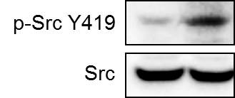
Application: Western BlotSample Tested: Breast cancer cellsSpecies: HumanVerified Customer | Posted 10/11/2018Human breast cancer cell line MDA-MB-231 was treated with carboplatin for 72 hours and the expression of p-Src at Y419 and total Src were detected by western blot.

-
Application: Western BlotSample Tested: Hela whole cell lysateSpecies: HumanVerified Customer | Posted 09/12/2012
There are no reviews that match your criteria.
Protocols
Find general support by application which include: protocols, troubleshooting, illustrated assays, videos and webinars.
- Antigen Retrieval Protocol (PIER)
- Antigen Retrieval for Frozen Sections Protocol
- Appropriate Fixation of IHC/ICC Samples
- Cellular Response to Hypoxia Protocols
- Chromogenic IHC Staining of Formalin-Fixed Paraffin-Embedded (FFPE) Tissue Protocol
- Chromogenic Immunohistochemistry Staining of Frozen Tissue
- ClariTSA™ Fluorophore Kits
- Detection & Visualization of Antibody Binding
- Fluorescent IHC Staining of Frozen Tissue Protocol
- Graphic Protocol for Heat-induced Epitope Retrieval
- Graphic Protocol for the Preparation and Fluorescent IHC Staining of Frozen Tissue Sections
- Graphic Protocol for the Preparation and Fluorescent IHC Staining of Paraffin-embedded Tissue Sections
- Graphic Protocol for the Preparation of Gelatin-coated Slides for Histological Tissue Sections
- IHC Sample Preparation (Frozen sections vs Paraffin)
- Immunofluorescent IHC Staining of Formalin-Fixed Paraffin-Embedded (FFPE) Tissue Protocol
- Immunohistochemistry (IHC) and Immunocytochemistry (ICC) Protocols
- Immunohistochemistry Frozen Troubleshooting
- Immunohistochemistry Paraffin Troubleshooting
- Preparing Samples for IHC/ICC Experiments
- Preventing Non-Specific Staining (Non-Specific Binding)
- Primary Antibody Selection & Optimization
- Protocol for Heat-Induced Epitope Retrieval (HIER)
- Protocol for Making a 4% Formaldehyde Solution in PBS
- Protocol for VisUCyte™ HRP Polymer Detection Reagent
- Protocol for the Preparation & Fixation of Cells on Coverslips
- Protocol for the Preparation and Chromogenic IHC Staining of Frozen Tissue Sections
- Protocol for the Preparation and Chromogenic IHC Staining of Frozen Tissue Sections - Graphic
- Protocol for the Preparation and Chromogenic IHC Staining of Paraffin-embedded Tissue Sections
- Protocol for the Preparation and Chromogenic IHC Staining of Paraffin-embedded Tissue Sections - Graphic
- Protocol for the Preparation and Fluorescent IHC Staining of Frozen Tissue Sections
- Protocol for the Preparation and Fluorescent IHC Staining of Paraffin-embedded Tissue Sections
- Protocol for the Preparation of Gelatin-coated Slides for Histological Tissue Sections
- R&D Systems Quality Control Western Blot Protocol
- TUNEL and Active Caspase-3 Detection by IHC/ICC Protocol
- The Importance of IHC/ICC Controls
- Troubleshooting Guide: Immunohistochemistry
- Troubleshooting Guide: Western Blot Figures
- Western Blot Conditions
- Western Blot Protocol
- Western Blot Protocol for Cell Lysates
- Western Blot Troubleshooting
- Western Blot Troubleshooting Guide
- View all Protocols, Troubleshooting, Illustrated assays and Webinars
Loading...
Associated Pathways


MAPK Signaling: Oxidative Stress Pathway
 MAPK Signaling Pathway: Mitogen Stimulation Pathway
 Pathogen or Damage-activated C-Type Lectin Receptor Signaling Pathways
 VEGF - VEGF R2 Signaling Pathways
 Wnt Signaling Pathways: beta-Catenin-dependent Wnt Signaling
