14-3-3 eta Antibody (3G12) - BSA Free
Novus Biologicals | Catalog # NBP1-92691
![Western Blot: 14-3-3 eta Antibody (3G12) [NBP1-92691]](https://resources.rndsystems.com/images/products/14-3-3-eta-Antibody-3G12-Western-Blot-NBP1-92691-img0006.jpg "Western Blot: 14-3-3 eta Antibody (3G12) [NBP1-92691]")
Key Product Details
Species Reactivity
Validated:
Human, Mouse, Rat, Bovine, Canine, Equine
Cited:
Human, Mouse
Applications
Validated:
Knockout Validated, Immunohistochemistry, Western Blot, Immunocytochemistry/ Immunofluorescence
Cited:
Knockout Validated, Western Blot, Immunocytochemistry/ Immunofluorescence
Label
Unconjugated
Antibody Source
Monoclonal Mouse IgG1 Clone # 3G12
Format
BSA Free
Loading...
Product Specifications
Immunogen
Full length recombinant human 14-3-3 eta expressed in and purified from E.coli [UniProt# Q04917].
Clonality
Monoclonal
Host
Mouse
Isotype
IgG1
Theoretical MW
28 kDa.
Disclaimer note: The observed molecular weight of the protein may vary from the listed predicted molecular weight due to post translational modifications, post translation cleavages, relative charges, and other experimental factors.
Disclaimer note: The observed molecular weight of the protein may vary from the listed predicted molecular weight due to post translational modifications, post translation cleavages, relative charges, and other experimental factors.
Scientific Data Images for 14-3-3 eta Antibody (3G12) - BSA Free
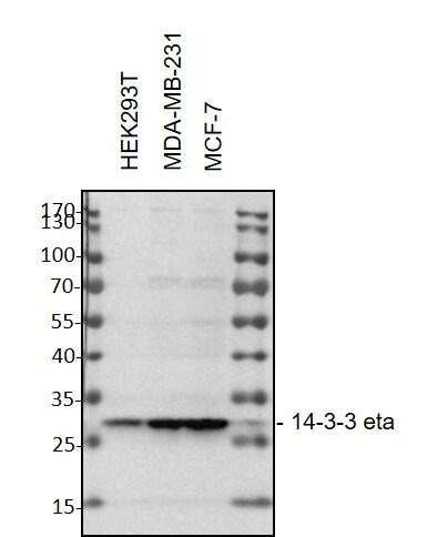
Western Blot: 14-3-3 eta Antibody (3G12) [NBP1-92691]
Western Blot: 14-3-3 eta Antibody (3G12) [NBP1-92691] - HEK293T, MDA-MB-231 and MCF-7 whole cell lysates, 30 ug/lane. 10% SDS-PAGE. Antibody at 1:1000, 4C overnight. WB image submitted by a verified customer review.![Immunocytochemistry/ Immunofluorescence: 14-3-3 eta Antibody (3G12) [NBP1-92691]](https://resources.rndsystems.com/images/products/14-3-3-eta-Antibody-3G12-Immunocytochemistry-Immunofluorescence-NBP1-92691-img0005.jpg "Immunocytochemistry/ Immunofluorescence: 14-3-3 eta Antibody (3G12) [NBP1-92691]")
Immunocytochemistry/ Immunofluorescence: 14-3-3 eta Antibody (3G12) [NBP1-92691]
Immunocytochemistry/Immunofluorescence: 14-3-3 eta Antibody (3G12) [NBP1-92691] - Analysis of HeLa cells stained with mouse mAb to 14.3.3, NBP1-92691, dilution 1:1,000 in red. Blue is DAPI staining of nuclear DNA. The NBP1-92691 antibody reveals the diffuse cytoplasmic distribution of 14.3.3 protein with higher concentration in the perinuclear region.![Western Blot: 14-3-3 eta Antibody (3G12) [NBP1-92691]](https://resources.rndsystems.com/images/products/14-3-3-eta-Antibody-3G12-Western-Blot-NBP1-92691-img0004.jpg "Western Blot: 14-3-3 eta Antibody (3G12) [NBP1-92691]")
Western Blot: 14-3-3 eta Antibody (3G12) [NBP1-92691]
Western Blot: 14-3-3 eta Antibody (3G12) [NBP1-92691] - analysis of neural tissue and cell lysates using Mouse mAb against 14-3-3n ( green). [1] protein standard, [2] Rat whole brain, [3] Mouse whole brain, [4] NIH/3T3, [5] Hek293, [6] HeLa, [7] SH-SY5Y, [8] C6 cells.Applications for 14-3-3 eta Antibody (3G12) - BSA Free
Application
Recommended Usage
Immunocytochemistry/ Immunofluorescence
1:1000
Immunohistochemistry
1:1000
Western Blot
1:5000
Application Notes
This 14-3-3 eta (3G12) antibody is useful for Immunocytochemistry/Immunofluorescence, immunohistochemistry, and Western blot, where a band can be seen at approximately 28 kDa. Knockout validation (PMID: 31640562).
Reviewed Applications
Read 1 review rated 5 using NBP1-92691 in the following applications:
Formulation, Preparation, and Storage
Purification
Immunogen affinity purified
Formulation
50% PBS, 50% glycerol
Format
BSA Free
Preservative
5mM Sodium Azide
Concentration
1 mg/ml
Shipping
The product is shipped with polar packs. Upon receipt, store it immediately at the temperature recommended below.
Stability & Storage
Store at 4C short term. Aliquot and store at -20C long term. Avoid freeze-thaw cycles.
Background: 14-3-3 eta
Alternate Names
1433 eta, Protein AS1, YWHA1, YWHAH
Entrez Gene IDs
7533 (Human)
Gene Symbol
YWHAH
UniProt
Additional 14-3-3 eta Products
Product Documents for 14-3-3 eta Antibody (3G12) - BSA Free
Certificate of Analysis
To download a Certificate of Analysis, please enter a lot or batch number in the search box below.
Product Specific Notices for 14-3-3 eta Antibody (3G12) - BSA Free
This product is for research use only and is not approved for use in humans or in clinical diagnosis. Primary Antibodies are guaranteed for 1 year from date of receipt.
Related Research Areas
Citations for 14-3-3 eta Antibody (3G12) - BSA Free
 Powered by Bioz
Customer Reviews for 14-3-3 eta Antibody (3G12) - BSA Free (1)
5 out of 5
1 Customer Rating
Have you used 14-3-3 eta Antibody (3G12) - BSA Free?
Submit a review and receive an Amazon gift card!
$25/€18/£15/$25CAN/¥2500 Yen for a review with an image
$10/€7/£6/$10CAN/¥1110 Yen for a review without an image
Submit a review
Customer Images
Showing
1
-
1 of
1 review
Showing All
Filter By:
-
Application: Western BlotSample Tested: 293T and MCF-7 cells-whole cell lysate and MDA-231 cell lysateSpecies: HumanVerified Customer | Posted 03/20/2021Western Blot: HEK293T, MDA-MB-231 and MCF-7 whole cell lysates were loaded with 30 ug/lane. 10% SDS-PAGE. Mouse Monoclonal 14-3-3 eta Antibody (3G12) Antibody (NBP1-92691) was used for primary antibody: 1:1000, 4℃, overnight.

There are no reviews that match your criteria.
Protocols
Find general support by application which include: protocols, troubleshooting, illustrated assays, videos and webinars.
- Antigen Retrieval Protocol (PIER)
- Antigen Retrieval for Frozen Sections Protocol
- Appropriate Fixation of IHC/ICC Samples
- Cellular Response to Hypoxia Protocols
- Chromogenic IHC Staining of Formalin-Fixed Paraffin-Embedded (FFPE) Tissue Protocol
- Chromogenic Immunohistochemistry Staining of Frozen Tissue
- ClariTSA™ Fluorophore Kits
- Detection & Visualization of Antibody Binding
- Fluorescent IHC Staining of Frozen Tissue Protocol
- Graphic Protocol for Heat-induced Epitope Retrieval
- Graphic Protocol for the Preparation and Fluorescent IHC Staining of Frozen Tissue Sections
- Graphic Protocol for the Preparation and Fluorescent IHC Staining of Paraffin-embedded Tissue Sections
- Graphic Protocol for the Preparation of Gelatin-coated Slides for Histological Tissue Sections
- ICC Cell Smear Protocol for Suspension Cells
- ICC Immunocytochemistry Protocol Videos
- ICC for Adherent Cells
- IHC Sample Preparation (Frozen sections vs Paraffin)
- Immunocytochemistry (ICC) Protocol
- Immunocytochemistry Troubleshooting
- Immunofluorescence of Organoids Embedded in Cultrex Basement Membrane Extract
- Immunofluorescent IHC Staining of Formalin-Fixed Paraffin-Embedded (FFPE) Tissue Protocol
- Immunohistochemistry (IHC) and Immunocytochemistry (ICC) Protocols
- Immunohistochemistry Frozen Troubleshooting
- Immunohistochemistry Paraffin Troubleshooting
- Preparing Samples for IHC/ICC Experiments
- Preventing Non-Specific Staining (Non-Specific Binding)
- Primary Antibody Selection & Optimization
- Protocol for Heat-Induced Epitope Retrieval (HIER)
- Protocol for Making a 4% Formaldehyde Solution in PBS
- Protocol for VisUCyte™ HRP Polymer Detection Reagent
- Protocol for the Fluorescent ICC Staining of Cell Smears - Graphic
- Protocol for the Fluorescent ICC Staining of Cultured Cells on Coverslips - Graphic
- Protocol for the Preparation & Fixation of Cells on Coverslips
- Protocol for the Preparation and Chromogenic IHC Staining of Frozen Tissue Sections
- Protocol for the Preparation and Chromogenic IHC Staining of Frozen Tissue Sections - Graphic
- Protocol for the Preparation and Chromogenic IHC Staining of Paraffin-embedded Tissue Sections
- Protocol for the Preparation and Chromogenic IHC Staining of Paraffin-embedded Tissue Sections - Graphic
- Protocol for the Preparation and Fluorescent ICC Staining of Cells on Coverslips
- Protocol for the Preparation and Fluorescent ICC Staining of Non-adherent Cells
- Protocol for the Preparation and Fluorescent ICC Staining of Stem Cells on Coverslips
- Protocol for the Preparation and Fluorescent IHC Staining of Frozen Tissue Sections
- Protocol for the Preparation and Fluorescent IHC Staining of Paraffin-embedded Tissue Sections
- Protocol for the Preparation of Gelatin-coated Slides for Histological Tissue Sections
- Protocol for the Preparation of a Cell Smear for Non-adherent Cell ICC - Graphic
- R&D Systems Quality Control Western Blot Protocol
- TUNEL and Active Caspase-3 Detection by IHC/ICC Protocol
- The Importance of IHC/ICC Controls
- Troubleshooting Guide: Immunohistochemistry
- Troubleshooting Guide: Western Blot Figures
- Western Blot Conditions
- Western Blot Protocol
- Western Blot Protocol for Cell Lysates
- Western Blot Troubleshooting
- Western Blot Troubleshooting Guide
- View all Protocols, Troubleshooting, Illustrated assays and Webinars
Loading...
Associated Pathways

