14-3-3 gamma [ac Val2] Antibody (KC21) - BSA Free
Novus Biologicals | Catalog # NB100-406
Key Product Details
Validated by
Biological Validation
Species Reactivity
Human
Applications
Western Blot, Immunocytochemistry/ Immunofluorescence, Simple Western
Label
Unconjugated
Antibody Source
Monoclonal Mouse IgG2A Clone # KC21
Format
BSA Free
Loading...
Product Specifications
Immunogen
N-terminal fragment of human 14-3-3 gamma where the N-terminal Val was acetylated [UniProt# P61981]
Modification
ac Val2
Localization
Cytoplasmic
Specificity
This antibody is specific for the naturally occurring human form of 14-3-3 gamma, where the N-terminal Met is removed, resulting in an acetylated N-terminal Val. Unprocessed (non-modified) 14-3-3 gamma is not recognized by this antibody.
Clonality
Monoclonal
Host
Mouse
Isotype
IgG2A
Theoretical MW
33 kDa.
Disclaimer note: The observed molecular weight of the protein may vary from the listed predicted molecular weight due to post translational modifications, post translation cleavages, relative charges, and other experimental factors.
Disclaimer note: The observed molecular weight of the protein may vary from the listed predicted molecular weight due to post translational modifications, post translation cleavages, relative charges, and other experimental factors.
Scientific Data Images for 14-3-3 gamma [ac Val2] Antibody (KC21) - BSA Free
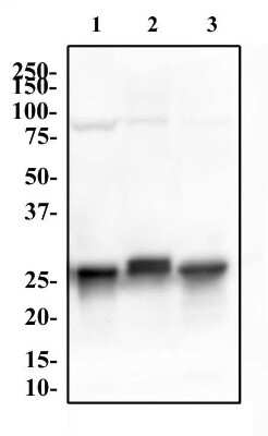
Western Blot: 14-3-3 gamma [ac Val2] Antibody (KC21) [NB100-406] - (1) HeLa, (2) PC12 and (3) NIH-3T3 whole cell lysates were separated by SDS-PAGE on a 12% gel and transferred to PVDF membrane. The membrane was probed with Anti-14-3-3 gamma [Ac Val2] antibody at 2 ug/ml for 1 hour, followed by incubation a 1:10,000 dilution of anti-rabbit HRP. The protein was detected at approximately 28 kDa after incubation with SuperSignal West Pico Chemiluminescent substrate.
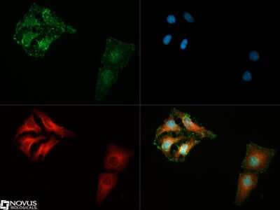
Immunocytochemistry/Immunofluorescence: 14-3-3 gamma [ac Val2] Antibody (KC21) [NB100-406] - 14-3-3 gamma [ac Val2] antibody was tested in SH-SY5Y cells with DyLight 488 (green). Nuclei and alpha-tubulin were counterstained with DAPI (blue) and DyLight 550 (red).
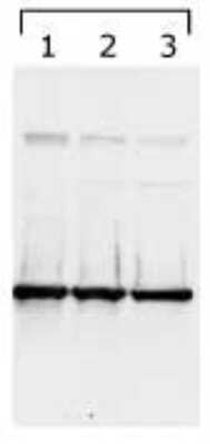

Simple Western: 14-3-3 gamma [ac Val2] Antibody (KC21) [NB100-406] - Lane view shows a specific band for 14-3-3 Gamma in 1.0 mg/ml of HeLa lysate. This experiment was performed under reducing conditions using the 12-230kDa separation system.
![14-3-3 gamma [ac Val2] Antibody (KC21) - BSA Free Immunocytochemistry/Immunofluorescence: 14-3-3 gamma [ac Val2] Antibody (KC21)](https://resources.rndsystems.com/images/products/antibody/nb100-406_mouse-monoclonal-14-3-3-gamma-ac-val2-antibody-kc21-231202683010.jpg "Immunocytochemistry/Immunofluorescence: 14-3-3 gamma [ac Val2] Antibody (KC21)")
Immunocytochemistry/Immunofluorescence: 14-3-3 gamma [ac Val2] Antibody (KC21)
14-3-3 gamma was detected in immersion fixed HeLa human cervix adenocarcinoma cell line using Mouse anti-14-3-3 gamma Protein G Purified Monoclonal Antibody (Catalog # NB100-406) at 1.0 µg/mL overnight at 4C. Cells were stained using DyLight 488-conjugated Anti-Mouse IgG (H+L) Cross-Absorbed Secondary Antibody (green), and counterstained with DAPI (blue). Cells were imaged using a 100X objective and digitally deconvolved.Applications for 14-3-3 gamma [ac Val2] Antibody (KC21) - BSA Free
Application
Recommended Usage
Immunocytochemistry/ Immunofluorescence
1:500
Simple Western
1:12.5
Western Blot
1:1000-1:3000
Formulation, Preparation, and Storage
Purification
Protein G purified
Formulation
PBS
Format
BSA Free
Preservative
0.02% Sodium Azide
Concentration
1.0 mg/ml
Shipping
The product is shipped with polar packs. Upon receipt, store it immediately at the temperature recommended below.
Stability & Storage
Store at 4C short term. Aliquot and store at -20C long term. Avoid freeze-thaw cycles.
Background: 14-3-3 gamma
Additional 14-3-3 gamma Products
Product Documents for 14-3-3 gamma [ac Val2] Antibody (KC21) - BSA Free
Certificate of Analysis
To download a Certificate of Analysis, please enter a lot or batch number in the search box below.
Product Specific Notices for 14-3-3 gamma [ac Val2] Antibody (KC21) - BSA Free
This product is for research use only and is not approved for use in humans or in clinical diagnosis. Primary Antibodies are guaranteed for 1 year from date of receipt.
Related Research Areas
Customer Reviews for 14-3-3 gamma [ac Val2] Antibody (KC21) - BSA Free
There are currently no reviews for this product. Be the first to review 14-3-3 gamma [ac Val2] Antibody (KC21) - BSA Free and earn rewards!
Have you used 14-3-3 gamma [ac Val2] Antibody (KC21) - BSA Free?
Submit a review and receive an Amazon gift card!
$25/€18/£15/$25CAN/¥2500 Yen for a review with an image
$10/€7/£6/$10CAN/¥1110 Yen for a review without an image
Submit a review
Protocols
View specific protocols for 14-3-3 gamma [ac Val2] Antibody (KC21) - BSA Free (NB100-406):
14-3-3 gamma [ac Val2] Antibody (KC21):
Culture cells to appropriate density in 35 mm culture dishes or 6-well plates.
1. Remove culture medium and add 10% formalin to the dish. Fix at room temperature for 30 minutes.
2. Remove the formalin and add ice cold methanol. Incubate for 5-10 minutes.
3. Remove methanol and add washing solution (i.e. PBS). Be sure to not let the specimen dry out. Wash three times for 10 minutes.
4. To block nonspecific antibody binding incubate in 10% normal goat serum from 1 hour to overnight at room temperature.
5. Add primary antibody at appropriate dilution and incubate at room temperature from 2 hours to overnight at room temperature.
6. Remove primary antibody and replace with washing solution. Wash three times for 10 minutes.
7. Add secondary antibody at appropriate dilution. Incubate for 1 hour at room temperature.
8. Remove antibody and replace with wash solution, then wash for 10 minutes. Add Hoechst 33258 to wash solution at 1:25,0000 and incubate for 10 minutes. Wash a third time for 10 minutes.
9. Cells can be viewed directly after washing. The plates can also be stored in PBS containing Azide covered in Parafilm (TM). Cells can also be cover-slipped using Fluoromount, with appropriate sealing.
*The above information is only intended as a guide. The researcher should determine what protocol best meets their needs. Please follow safe laboratory procedures.
14-3-3 gamma [ac Val2] Antibody (KC21):
1. Load protein on gel (ie: ~30 ug of HeLa whole cell control lysate) and run.
2. Transfer protein to nitrocellulose (Schleicher&Schuell, cat# BA83).
3. Block the membrane with 5% milk for 30 minutes at room temperature.
4. Incubate the membrane with anti-14-3-3 gamma [cat# NB 100-406] (1:1,000), overnight at 4C or 2 h at room temperature. Milk or PBS-T are good for this dilution.
5. Wash the membrane 3 times, 10 minutes per wash in PBS with 0.1% Tween-20 (PBS-T).
6. Incubate with secondary antibody for 30 minutes at room temperature. Use milk in PBS-T as a diluent.
7. Rinse the membrane 2 times with deionized water and place in an ECL working solution.
8. Expose to the film.
Find general support by application which include: protocols, troubleshooting, illustrated assays, videos and webinars.
- Appropriate Fixation of IHC/ICC Samples
- Cellular Response to Hypoxia Protocols
- ClariTSA™ Fluorophore Kits
- Detection & Visualization of Antibody Binding
- ICC Cell Smear Protocol for Suspension Cells
- ICC Immunocytochemistry Protocol Videos
- ICC for Adherent Cells
- Immunocytochemistry (ICC) Protocol
- Immunocytochemistry Troubleshooting
- Immunofluorescence of Organoids Embedded in Cultrex Basement Membrane Extract
- Immunohistochemistry (IHC) and Immunocytochemistry (ICC) Protocols
- Preparing Samples for IHC/ICC Experiments
- Preventing Non-Specific Staining (Non-Specific Binding)
- Primary Antibody Selection & Optimization
- Protocol for VisUCyte™ HRP Polymer Detection Reagent
- Protocol for the Fluorescent ICC Staining of Cell Smears - Graphic
- Protocol for the Fluorescent ICC Staining of Cultured Cells on Coverslips - Graphic
- Protocol for the Preparation and Fluorescent ICC Staining of Cells on Coverslips
- Protocol for the Preparation and Fluorescent ICC Staining of Non-adherent Cells
- Protocol for the Preparation and Fluorescent ICC Staining of Stem Cells on Coverslips
- Protocol for the Preparation of a Cell Smear for Non-adherent Cell ICC - Graphic
- R&D Systems Quality Control Western Blot Protocol
- TUNEL and Active Caspase-3 Detection by IHC/ICC Protocol
- The Importance of IHC/ICC Controls
- Troubleshooting Guide: Western Blot Figures
- Western Blot Conditions
- Western Blot Protocol
- Western Blot Protocol for Cell Lysates
- Western Blot Troubleshooting
- Western Blot Troubleshooting Guide
- View all Protocols, Troubleshooting, Illustrated assays and Webinars
Loading...
Associated Pathways

