58K Golgi Protein Antibody (58K-9) - BSA Free
Novus Biologicals | Catalog # NB600-412
![Western Blot: 58K Golgi Protein Antibody (58K-9) [NB600-412]](https://resources.rndsystems.com/images/products/58K-Golgi-Protein-Antibody-58K-9-Western-Blot-NB600-412-img0007.jpg "Western Blot: 58K Golgi Protein Antibody (58K-9) [NB600-412]")
Loading...
Key Product Details
Species Reactivity
Validated:
Human, Mouse, Rat, Bovine, Canine, Chicken
Cited:
Human, Mouse, Rat, Avian - Chicken
Predicted:
Chinese Hamster (92%). Backed by our 100% Guarantee.
Applications
Validated:
Immunohistochemistry, Immunohistochemistry-Paraffin, Immunohistochemistry-Frozen, Western Blot, Immunocytochemistry/ Immunofluorescence
Cited:
Immunohistochemistry-Frozen, Western Blot, Immunocytochemistry/ Immunofluorescence
Label
Unconjugated
Antibody Source
Monoclonal Mouse IgG1 Clone # 58K-9
Format
BSA Free
Loading...
Product Specifications
Immunogen
58K Golgi Protein purified from rat liver [UniProt# O88618]
Localization
Cytoplasmic and Golgi Apparatus
Marker
Golgi Apparatus Marker
Clonality
Monoclonal
Host
Mouse
Isotype
IgG1
Theoretical MW
58 kDa.
Disclaimer note: The observed molecular weight of the protein may vary from the listed predicted molecular weight due to post translational modifications, post translation cleavages, relative charges, and other experimental factors.
Disclaimer note: The observed molecular weight of the protein may vary from the listed predicted molecular weight due to post translational modifications, post translation cleavages, relative charges, and other experimental factors.
Scientific Data Images for 58K Golgi Protein Antibody (58K-9) - BSA Free
Western Blot: 58K Golgi Protein Antibody (58K-9) [NB600-412]
Western Blot: 58K Golgi Protein Antibody (58K-9) [NB600-412] - Analysis of 58K golgi protein expression in rat liver tissue using NB600-412.![Immunocytochemistry/ Immunofluorescence: 58K Golgi Protein Antibody (58K-9) [NB600-412]](https://resources.rndsystems.com/images/products/58K-Golgi-Protein-Antibody-58K-9-Immunocytochemistry-Immunofluorescence-NB600-412-img0010.jpg "Immunocytochemistry/ Immunofluorescence: 58K Golgi Protein Antibody (58K-9) [NB600-412]")
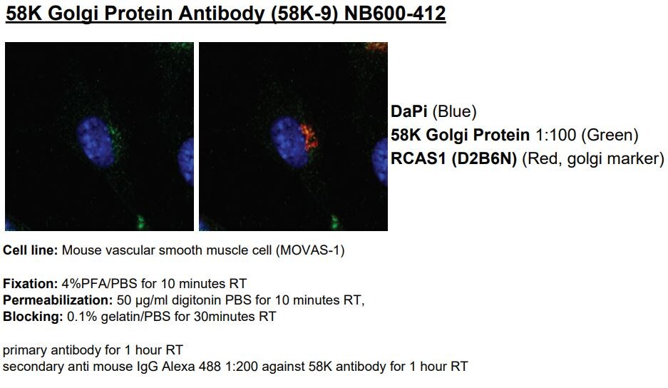
Immunocytochemistry/ Immunofluorescence: 58K Golgi Protein Antibody (58K-9) [NB600-412]
Immunocytochemistry/Immunofluorescence: 58K Golgi Protein Antibody (58K-9) [NB600-412] - HeLa cells, cultured on cover slips, were fixed with 10% formalin for 10 minutes and then permeabilized for 5 minutes using 1X TBS + 0.5% Triton-X100. The cells were then incubated with 1:200 dilution of anti-58K Golgi Protein antibody (clone 58K-9) for overnight at 4C and detected with an anti-mouse Dylight 488 (Green) secondary at a 1:500 dilution. Alpha tubulin (DM1A) [NB100-690] was used as a co-stain at a 1:1000 dilution and detected with an anti-mouse Dylight 550 (Red) at 1:500 dilution. Nuclei were counterstained with DAPI solution (Blue) [NBP2-31156]. Cells were imaged using a 40X objective. Antibody clone 58K-9 generated a specific signal in the Golgi apparatus of the cells.![Immunohistochemistry: 58K Golgi Protein Antibody (58K-9) [NB600-412]](https://resources.rndsystems.com/images/products/58K-Golgi-Protein-Antibody-58K-9-Immunohistochemistry-NB600-412-img0011.jpg "Immunohistochemistry: 58K Golgi Protein Antibody (58K-9) [NB600-412]")
Immunohistochemistry: 58K Golgi Protein Antibody (58K-9) [NB600-412]
58K-Golgi-Protein-Antibody-58K-9-Immunohistochemistry-NB600-412-img0011.jpg![Immunohistochemistry: 58K Golgi Protein Antibody (58K-9) [NB600-412]](https://resources.rndsystems.com/images/products/58K-Golgi-Protein-Antibody-58K-9-Immunohistochemistry-NB600-412-img0009.jpg "Immunohistochemistry: 58K Golgi Protein Antibody (58K-9) [NB600-412]")
Immunohistochemistry: 58K Golgi Protein Antibody (58K-9) [NB600-412]
Immunohistochemistry: 58K Golgi Protein Antibody (58K-9) [NB600-412] - Analysis of 58K Golgi Protein in mouse kidney using DAB with hematoxylin counterstain. [NB600-412] -")
Immunocytochemistry/Immunofluorescence: 58K Golgi Protein Antibody (58K-9) [NB600-412] -
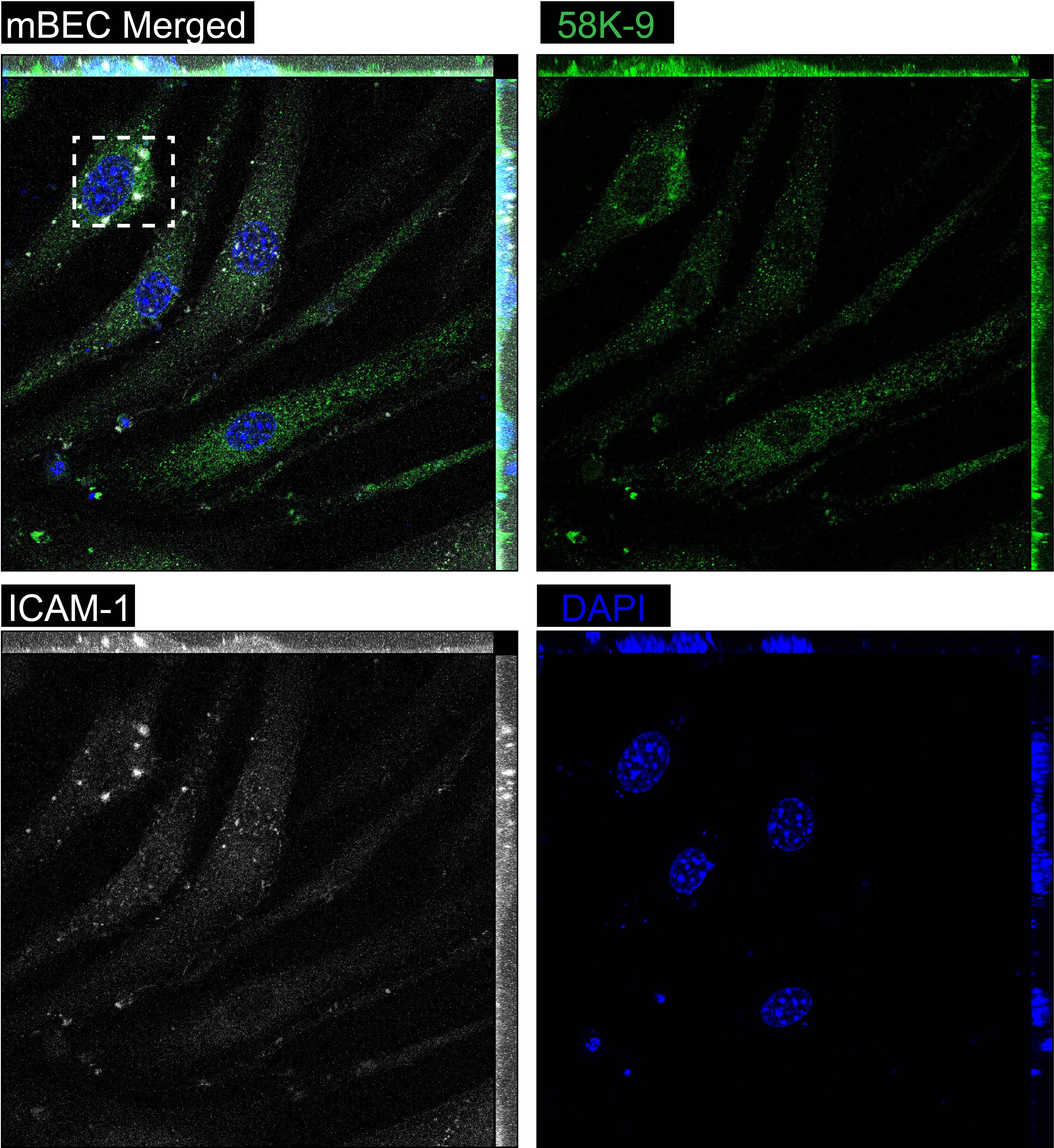
Immunocytochemistry/Immunofluorescence: 58K Golgi Protein Antibody (58K-9) [NB600-412] - Assessing Golgi morphology observed apoptosis in mBECs using Mouse 58K Golgi Protein at a dilution of 1:100 overnight at 4 °C. This image was taken on a Zeiss LSM 710 Confocal Microscope (META) with Zeiss Plan Apo 63x/1.40 oil. Image from verified customer review. [NB600-412] -")
Immunocytochemistry/ Immunofluorescence: 58K Golgi Protein Antibody (58K-9) [NB600-412] -
Immunocytochemistry/ Immunofluorescence: 58K Golgi Protein Antibody (58K-9) [NB600-412] - Co-localization of Neu5Gc staining with markers for endosomes & Golgi in BMD & DMD muscle.(A) BMD muscle co-stained for Neu5Gc (green) & clathrin (red), a marker of endosomes. Merged image on right shows overlap of Neu5Gc & clathrin expression in yellow. Arrow marks several examples of co-staining. (B) DMD muscle co-stained for Neu5Gc (green) with LAMP1, a lysosomal marker, 58K Golgi, a Golgi marker, or calnexin, & endoplasmic reticulum marker, all in red, & DAPI (blue). Arrow marks region of coincident staining (yellow) for Neu5Gc & 58K Golgi. Bar is 50 µm for all panels in A & B. Image collected & cropped by CiteAb from the following publication (https://pubmed.ncbi.nlm.nih.gov/24505439), licensed under a CC-BY license. Not internally tested by Novus Biologicals.Applications for 58K Golgi Protein Antibody (58K-9) - BSA Free
Application
Recommended Usage
Immunocytochemistry/ Immunofluorescence
1:50 - 1:250
Immunohistochemistry
1:100 - 1:200
Immunohistochemistry-Frozen
reported in scientific literature (PMID 24505439)
Immunohistochemistry-Paraffin
1:100 - 1:200
Western Blot
1:2000 - 1:5000
Application Notes
In Western blot a band is observed at ~58 kDa. Prior to immunostaining paraffin tissues, antigen retrieval with sodium citrate buffer (pH 6.0) is recommended. The observed molecular weight of the protein may vary from the listed predicted molecular weight due to post translational modifications, post translation cleavages, relative charges, and other experimental factors.
Reviewed Applications
Read 3 reviews rated 3.3 using NB600-412 in the following applications:
Formulation, Preparation, and Storage
Purification
Unpurified
Formulation
Ascites
Format
BSA Free
Preservative
0.1% Sodium Azide
Concentration
This product is unpurified. The exact concentration of antibody is not quantifiable.
Shipping
The product is shipped with polar packs. Upon receipt, store it immediately at the temperature recommended below.
Stability & Storage
Store at 4C short term. Aliquot and store at -20C long term. Avoid freeze-thaw cycles.
Background: 58K Golgi Protein
Alternate Names
formimidoyltransferase cyclodeaminase, formimidoyltransferase-cyclodeaminase, formiminotransferase cyclodeaminase, Formiminotransferase-cyclodeaminase, human formiminotransferase cyclodeaminase, EC 4.3.1.410formiminotransferase-cyclodeaminase, LCHC1
Gene Symbol
FTCD
UniProt
Additional 58K Golgi Protein Products
Product Documents for 58K Golgi Protein Antibody (58K-9) - BSA Free
Certificate of Analysis
To download a Certificate of Analysis, please enter a lot or batch number in the search box below.
Product Specific Notices for 58K Golgi Protein Antibody (58K-9) - BSA Free
This product is for research use only and is not approved for use in humans or in clinical diagnosis. Primary Antibodies are guaranteed for 1 year from date of receipt.
Citations for 58K Golgi Protein Antibody (58K-9) - BSA Free
 Powered by Bioz
Customer Reviews for 58K Golgi Protein Antibody (58K-9) - BSA Free (3)
3.3 out of 5
3 Customer Ratings
Have you used 58K Golgi Protein Antibody (58K-9) - BSA Free?
Submit a review and receive an Amazon gift card!
$25/€18/£15/$25CAN/¥2500 Yen for a review with an image
$10/€7/£6/$10CAN/¥1110 Yen for a review without an image
Submit a review
Customer Images
Showing
1
-
3 of
3 reviews
Showing All
Filter By:
-
Application: ImmunocytochemistrySample Tested: Mouse Brain Endothelial CellsSpecies: MouseVerified Customer | Posted 03/22/2023Primary mouse brain endothelial cells stained with 58K antibody (1:100). Staining is perinuclear but Golgi cannot be distinguished.
 Bio-Techne ResponseThank you for reviewing our product. We are sorry to hear that this product did not perform as expected. We have been in touch with the customer to resolve this issue according to our Product Guarantee and to the customer’s satisfaction.
 -
Application: ImmunocytochemistrySample Tested: Cultured Mouse brain endothelial cells, Cultured Brain Endothelial cells and Mouse Brain Endothelial cells (mBECs)Species: MouseVerified Customer | Posted 03/13/2023Assessing Golgi morphology observed apoptosis in mBECs using Mouse 58K Golgi Protein at a dilution of 1:100 overnight at 4 °C. This image was taken on a Zeiss LSM 710 Confocal Microscope (META) with Zeiss Plan Apo 63x/1.40 oil
-
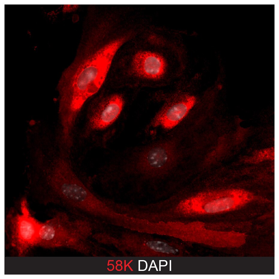
Application: ImmunocytochemistrySample Tested: mouse vascular smooth muscle cellSpecies: MouseVerified Customer | Posted 11/19/2018
There are no reviews that match your criteria.
Protocols
View specific protocols for 58K Golgi Protein Antibody (58K-9) - BSA Free (NB600-412):
Western Blot Protocol
1. Perform SDS-PAGE on samples to be analyzed, loading 30 ug of total protein per lane.
2. Transfer proteins to membrane according to the instructions provided by the manufacturer of the membrane and transfer apparatus.
3. Stain according to standard Ponceau S procedure (or similar product) to assess transfer success, and mark molecular weight standards where appropriate.
4. Rinse the blot.
5. Block the membrane using standard blocking buffer for at least 1 hour.
6. Wash the membrane in wash buffer three times for 10 minutes each.
7. Dilute primary antibody in blocking buffer and incubate 1 hour at room temperature.
8. Wash the membrane in wash buffer three times for 10 minutes each.
9. Apply the diluted HRP conjugated secondary antibody in blocking buffer (as per manufacturers instructions) and incubate 1 hour at room temperature.
10. Wash the blot in wash buffer three times for 10 minutes each (this step can be repeated as required to reduce background).
11. Apply the detection reagent of choice in accordance with the manufacturers instructions.
Note: Tween-20 can be added to the blocking or antibody dilution buffer at a final concentration of 0.05-0.2%.
Immunocytochemistry Protocol
Culture cells to appropriate density in 35 mm culture dishes or 6-well plates.
1. Remove culture medium and add 10% formalin to the dish. Fix at room temperature for 30 minutes.
2. Remove the formalin and add ice cold methanol. Incubate for 5-10 minutes.
3. Remove methanol and add washing solution (i.e. PBS). Be sure to not let the specimen dry out. Wash three times for 10 minutes.
4. To block nonspecific antibody binding incubate in 10% normal goat serum from 1 hour to overnight at room temperature.
5. Add primary antibody at appropriate dilution and incubate at room temperature from 2 hours to overnight at room temperature.
6. Remove primary antibody and replace with washing solution. Wash three times for 10 minutes.
7. Add secondary antibody at appropriate dilution. Incubate for 1 hour at room temperature.
8. Remove antibody and replace with wash solution, then wash for 10 minutes. Add Hoechst 33258 to wash solution at 1:25,0000 and incubate for 10 minutes. Wash a third time for 10 minutes.
9. Cells can be viewed directly after washing. The plates can also be stored in PBS containing Azide covered in Parafilm (TM). Cells can also be cover-slipped using Fluoromount, with appropriate sealing.
*The above information is only intended as a guide. The researcher should determine what protocol best meets their needs. Please follow safe laboratory procedures.
Find general support by application which include: protocols, troubleshooting, illustrated assays, videos and webinars.
- Antigen Retrieval Protocol (PIER)
- Antigen Retrieval for Frozen Sections Protocol
- Appropriate Fixation of IHC/ICC Samples
- Cellular Response to Hypoxia Protocols
- Chromogenic IHC Staining of Formalin-Fixed Paraffin-Embedded (FFPE) Tissue Protocol
- Chromogenic Immunohistochemistry Staining of Frozen Tissue
- ClariTSA™ Fluorophore Kits
- Detection & Visualization of Antibody Binding
- Fluorescent IHC Staining of Frozen Tissue Protocol
- Graphic Protocol for Heat-induced Epitope Retrieval
- Graphic Protocol for the Preparation and Fluorescent IHC Staining of Frozen Tissue Sections
- Graphic Protocol for the Preparation and Fluorescent IHC Staining of Paraffin-embedded Tissue Sections
- Graphic Protocol for the Preparation of Gelatin-coated Slides for Histological Tissue Sections
- ICC Cell Smear Protocol for Suspension Cells
- ICC Immunocytochemistry Protocol Videos
- ICC for Adherent Cells
- IHC Sample Preparation (Frozen sections vs Paraffin)
- Immunocytochemistry (ICC) Protocol
- Immunocytochemistry Troubleshooting
- Immunofluorescence of Organoids Embedded in Cultrex Basement Membrane Extract
- Immunofluorescent IHC Staining of Formalin-Fixed Paraffin-Embedded (FFPE) Tissue Protocol
- Immunohistochemistry (IHC) and Immunocytochemistry (ICC) Protocols
- Immunohistochemistry Frozen Troubleshooting
- Immunohistochemistry Paraffin Troubleshooting
- Preparing Samples for IHC/ICC Experiments
- Preventing Non-Specific Staining (Non-Specific Binding)
- Primary Antibody Selection & Optimization
- Protocol for Heat-Induced Epitope Retrieval (HIER)
- Protocol for Making a 4% Formaldehyde Solution in PBS
- Protocol for VisUCyte™ HRP Polymer Detection Reagent
- Protocol for the Fluorescent ICC Staining of Cell Smears - Graphic
- Protocol for the Fluorescent ICC Staining of Cultured Cells on Coverslips - Graphic
- Protocol for the Preparation & Fixation of Cells on Coverslips
- Protocol for the Preparation and Chromogenic IHC Staining of Frozen Tissue Sections
- Protocol for the Preparation and Chromogenic IHC Staining of Frozen Tissue Sections - Graphic
- Protocol for the Preparation and Chromogenic IHC Staining of Paraffin-embedded Tissue Sections
- Protocol for the Preparation and Chromogenic IHC Staining of Paraffin-embedded Tissue Sections - Graphic
- Protocol for the Preparation and Fluorescent ICC Staining of Cells on Coverslips
- Protocol for the Preparation and Fluorescent ICC Staining of Non-adherent Cells
- Protocol for the Preparation and Fluorescent ICC Staining of Stem Cells on Coverslips
- Protocol for the Preparation and Fluorescent IHC Staining of Frozen Tissue Sections
- Protocol for the Preparation and Fluorescent IHC Staining of Paraffin-embedded Tissue Sections
- Protocol for the Preparation of Gelatin-coated Slides for Histological Tissue Sections
- Protocol for the Preparation of a Cell Smear for Non-adherent Cell ICC - Graphic
- R&D Systems Quality Control Western Blot Protocol
- TUNEL and Active Caspase-3 Detection by IHC/ICC Protocol
- The Importance of IHC/ICC Controls
- Troubleshooting Guide: Immunohistochemistry
- Troubleshooting Guide: Western Blot Figures
- Western Blot Conditions
- Western Blot Protocol
- Western Blot Protocol for Cell Lysates
- Western Blot Troubleshooting
- Western Blot Troubleshooting Guide
- View all Protocols, Troubleshooting, Illustrated assays and Webinars
Loading...