ACE-2 Antibody (AC18F) - Azide and BSA Free
Novus Biologicals | Catalog # NBP2-80035
![Western Blot: ACE-2 Antibody (AC18F)Azide and BSA Free [NBP2-80035]](https://resources.rndsystems.com/images/products/ACE-2-Antibody-AC18F-Western-Blot-NBP2-80035-img0003.jpg "Western Blot: ACE-2 Antibody (AC18F)Azide and BSA Free [NBP2-80035]")
Key Product Details
Validated by
Biological Validation
Species Reactivity
Validated:
Human
Cited:
Human
Applications
Validated:
Western Blot, ELISA, Flow Cytometry, CyTOF-ready
Cited:
Western Blot, Flow Cytometry
Label
Unconjugated
Antibody Source
Monoclonal Mouse IgG1 kappa Clone # AC18F
Format
Azide and BSA Free
Loading...
Product Specifications
Immunogen
Recombinant human ACE-2.
Specificity
Recognizes human ACE-2. Does not detect recombinant human ACE-2 that has a tag at the C-terminus of the protein.
Clonality
Monoclonal
Host
Mouse
Isotype
IgG1 kappa
Scientific Data Images for ACE-2 Antibody (AC18F) - Azide and BSA Free
Western Blot: ACE-2 Antibody (AC18F)Azide and BSA Free [NBP2-80035]
Western Blot: ACE-2 Antibody (AC18F) [NBP2-80035] - Western blot analysis using anti-ACE2 (human), mAb (AC18F) at 1: 2,000 dilution. Lane 1. hACE2 (Fc protein). Lane 2. hACE2 (Ecto domain) (FLAG(R)-tagged). Lane 3. Other hGITR (Fc protein) (control).![Immunohistochemistry-Paraffin: ACE-2 Antibody (AC18F) - Azide and BSA Free [NBP2-80035]](https://resources.rndsystems.com/images/products/ACE-2-Antibody-AC18F-Immunohistochemistry-Paraffin-NBP2-80035-img0006.jpg "Immunohistochemistry-Paraffin: ACE-2 Antibody (AC18F) - Azide and BSA Free [NBP2-80035]")
Immunohistochemistry-Paraffin: ACE-2 Antibody (AC18F) - Azide and BSA Free [NBP2-80035]
Immunohistochemistry-Paraffin: ACE-2 Antibody (AC18F) [NBP2-80035] - ACE-2 staining in paraffin-embedded sections of hamster lung tissue using Mouse Anti-Human ACE-2 (AC18F) Monoclonal Antibody (Catalog # NBP2-80035) at 10 ug/ml overnight at 4 degrees C. Detection kit: VC001. Counterstain: Hematoxylin. Localization: respiratory bronchioles.![Flow Cytometry: ACE-2 Antibody (AC18F) - Azide and BSA Free [NBP2-80035]](https://resources.rndsystems.com/images/products/ACE-2-Antibody-AC18F-Flow-Cytometry-NBP2-80035-img0005.jpg "Flow Cytometry: ACE-2 Antibody (AC18F) - Azide and BSA Free [NBP2-80035]")
Flow Cytometry: ACE-2 Antibody (AC18F) - Azide and BSA Free [NBP2-80035]
Flow Cytometry: ACE-2 Antibody (AC18F) [NBP2-80035] - Detection of endogenous human ACE2 by anti-ACE2 (h), mAb (AC18F). HepG2 cell line treated with CellTase solution (Abeomics) before analysis and then stained with anti-ACE2 (h), mAb (AC18F) (red line) or an isotype control, mouse IgG1 (green line) at 1ug/10E6 cells each, revealed with a PE-conjugated goat anti-mouse IgG and then analyzed by flow cytometry.![Flow Cytometry: ACE-2 Antibody (AC18F) - Azide and BSA Free [NBP2-80035]](https://resources.rndsystems.com/images/products/ACE-2-Antibody-AC18F-Flow-Cytometry-NBP2-80035-img0001.jpg "Flow Cytometry: ACE-2 Antibody (AC18F) - Azide and BSA Free [NBP2-80035]")
Flow Cytometry: ACE-2 Antibody (AC18F) - Azide and BSA Free [NBP2-80035]
Flow Cytometry: ACE-2 Antibody (AC18F) [NBP2-80035] - HepG2 cells were stained significantly using anti-ACE2 (human), mAb (AC18F).Applications for ACE-2 Antibody (AC18F) - Azide and BSA Free
Application
Recommended Usage
ELISA
1:2000 - 1:10000
Flow Cytometry
1:1000
Western Blot
1:2000 - 1:10000
Application Notes
Western blot: Use ECL substrate. Suggested blocking and dilution buffer is PBST with 0.05% Tween 20 and 5% skim milk. Suggested incubation time is 1 hour at rooom temperature.
Reviewed Applications
Read 1 review rated 1 using NBP2-80035 in the following applications:
Flow Cytometry Panel Builder
Bio-Techne Knows Flow Cytometry
Save time and reduce costly mistakes by quickly finding compatible reagents using the Panel Builder Tool.
Advanced Features
- Spectra Viewer - Custom analysis of spectra from multiple fluorochromes
- Spillover Popups - Visualize the spectra of individual fluorochromes
- Antigen Density Selector - Match fluorochrome brightness with antigen density

Formulation, Preparation, and Storage
Purification
Protein G purified
Formulation
0.2 um-filtered solution in PBS, pH 7.4, 10% glycerol
Format
Azide and BSA Free
Preservative
No Preservative
Concentration
1 mg/ml
Shipping
The product is shipped with polar packs. Upon receipt, store it immediately at the temperature recommended below.
Stability & Storage
Store at 4C short term. Aliquot and store at -20C long term. Avoid freeze-thaw cycles.
Background: ACE-2
Genetic data from Drosophila, mice and rats show that the ACE2 protein is an essential regulator of heart function in vivo. ACE2 mRNA is found at high levels in testis, kidney and heart with moderate levels in colon, small intestine, and ovary. The ACE2 protein contributes to organ function through the renin-angiotensin system, playing a central role in vascular, renal, and myocardial physiology.
Virology research has demonstrated that the severe acute respiratory syndrome coronavirus (SARS-CoV) spike protein binds to its functional receptor, ACE2 (1). Vimentin is a type III intermediate filament protein expressed in mesenchymal cells that helps comprise the cytoskeleton in all animal cells. Studies have demonstrated that vimentin allows cell binding that allows the uptake of SARS-CoV spike protein into a host (2). This direct interaction of SARS-CoV with vimentin has been identified as an entry mechanism for the virus into a host, mediated by the SARS-CoV receptor ACE2 (1,2). It has been shown that ACE2 is the SARS-CoV-2 receptor required for cell entry and plays a physiological role in the replication of SARS-CoV in an infected host (1). Studies using human ACE2 antibody demonstrated a blockade of SARS-CoV-2 and ACE2 interaction, indicating an important physiological component of viral transmission and potential anti-viral therapeutic strategies (3).
References
1. Li, W., Moore, M. J., Vasilieva, N., Sui, J., Wong, S. K., Berne, M. A.,... Farzan, M. (2003). Angiotensin-converting enzyme 2 is a functional receptor for the SARS coronavirus. Nature, 426(6965), 450-454. doi:10.1038/nature02145
2. Yu YT, Chien SC, Chen IY, Lai CT, Tsay YG, Chang SC, Chang MF. (2016) Surface vimentin is critical for the cell entry of SARS-CoV. J Biomed Sci. doi: 10.1186/s12929-016-0234-7
3. Hoffmann, M., Kleine-Weber, H., Schroeder, S., Kruger, N., Herrler, T., Erichsen, S.,... Pohlmann, S. (2020). SARS-CoV-2 Cell Entry Depends on ACE2 and TMPRSS2 and Is Blocked by a Clinically Proven Protease Inhibitor. Cell. doi:10.1016/j.cell.2020.02.052
Long Name
Angiotensin I Converting Enzyme 2
Alternate Names
ACE2, ACEH
Gene Symbol
ACE2
Additional ACE-2 Products
Product Documents for ACE-2 Antibody (AC18F) - Azide and BSA Free
Certificate of Analysis
To download a Certificate of Analysis, please enter a lot or batch number in the search box below.
Product Specific Notices for ACE-2 Antibody (AC18F) - Azide and BSA Free
This product is for research use only and is not approved for use in humans or in clinical diagnosis. Primary Antibodies are guaranteed for 1 year from date of receipt.
Related Research Areas
Citations for ACE-2 Antibody (AC18F) - Azide and BSA Free
 Powered by Bioz
Customer Reviews for ACE-2 Antibody (AC18F) - Azide and BSA Free (1)
1 out of 5
1 Customer Rating
Have you used ACE-2 Antibody (AC18F) - Azide and BSA Free?
Submit a review and receive an Amazon gift card!
$25/€18/£15/$25CAN/¥2500 Yen for a review with an image
$10/€7/£6/$10CAN/¥1110 Yen for a review without an image
Submit a review
Customer Images
Showing
1
-
1 of
1 review
Showing All
Filter By:
-
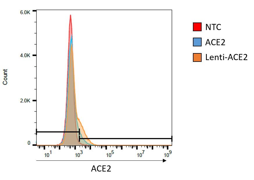
Application: Flow CytometrySample Tested: HEK293 cellsSpecies: HumanVerified Customer | Posted 07/01/2020Transfected HEK293 cells with 2 different types of ACE2 plasmids. Cells were harvested after 48hr & then stained (1:50) with alpha ACE2 Ab then stained with AlexaFlour647 secondary Ab.

There are no reviews that match your criteria.
Protocols
Find general support by application which include: protocols, troubleshooting, illustrated assays, videos and webinars.
- 7-Amino Actinomycin D (7-AAD) Cell Viability Flow Cytometry Protocol
- Cellular Response to Hypoxia Protocols
- ELISA Sample Preparation & Collection Guide
- ELISA Troubleshooting Guide
- Extracellular Membrane Flow Cytometry Protocol
- Flow Cytometry Protocol for Cell Surface Markers
- Flow Cytometry Protocol for Staining Membrane Associated Proteins
- Flow Cytometry Staining Protocols
- Flow Cytometry Troubleshooting Guide
- How to Run an R&D Systems DuoSet ELISA
- How to Run an R&D Systems Quantikine ELISA
- How to Run an R&D Systems Quantikine™ QuicKit™ ELISA
- Intracellular Flow Cytometry Protocol Using Alcohol (Methanol)
- Intracellular Flow Cytometry Protocol Using Detergents
- Intracellular Nuclear Staining Flow Cytometry Protocol Using Detergents
- Intracellular Staining Flow Cytometry Protocol Using Alcohol Permeabilization
- Intracellular Staining Flow Cytometry Protocol Using Detergents to Permeabilize Cells
- Propidium Iodide Cell Viability Flow Cytometry Protocol
- Protocol for Liperfluo
- Protocol for the Characterization of Human Th22 Cells
- Protocol for the Characterization of Human Th9 Cells
- Protocol: Annexin V and PI Staining by Flow Cytometry
- Protocol: Annexin V and PI Staining for Apoptosis by Flow Cytometry
- Quantikine HS ELISA Kit Assay Principle, Alkaline Phosphatase
- Quantikine HS ELISA Kit Principle, Streptavidin-HRP Polymer
- R&D Systems Quality Control Western Blot Protocol
- Sandwich ELISA (Colorimetric) – Biotin/Streptavidin Detection Protocol
- Sandwich ELISA (Colorimetric) – Direct Detection Protocol
- Troubleshooting Guide: ELISA
- Troubleshooting Guide: Fluorokine Flow Cytometry Kits
- Troubleshooting Guide: Western Blot Figures
- Western Blot Conditions
- Western Blot Protocol
- Western Blot Protocol for Cell Lysates
- Western Blot Troubleshooting
- Western Blot Troubleshooting Guide
- View all Protocols, Troubleshooting, Illustrated assays and Webinars
Loading...
Associated Pathways

