![Western Blot: ACSL3 Antibody [NBP1-89268]](https://resources.rndsystems.com/images/products/ACSL3-Antibody-Western-Blot-NBP1-89268-img0012.jpg "Western Blot: ACSL3 Antibody [NBP1-89268]")
Loading...
Key Product Details
Validated by
Orthogonal Validation
Species Reactivity
Validated:
Human
Predicted:
Mouse (91%), Rat (90%). Backed by our 100% Guarantee.
Applications
Immunohistochemistry, Immunohistochemistry-Paraffin, Western Blot
Label
Unconjugated
Antibody Source
Polyclonal Rabbit IgG
Format
BSA Free
Loading...
Product Specifications
Immunogen
This antibody was developed against Recombinant Protein corresponding to amino acids: MYNFQLVTLYATLGGPAIVHALNETEVTNIITSKELLQTKLKDIVSLVPRLRHIITVDGKPPTWSEFPKGIIVHTMAAVEALGAKASMENQPHSKPLPSDIAVIM
Clonality
Polyclonal
Host
Rabbit
Isotype
IgG
Scientific Data Images for ACSL3 Antibody - BSA Free
![Western Blot: ACSL3 Antibody [NBP1-89268]](https://resources.rndsystems.com/images/products/ACSL3-Antibody-Western-Blot-NBP1-89268-img0013.jpg "Western Blot: ACSL3 Antibody [NBP1-89268]")
Western Blot: ACSL3 Antibody [NBP1-89268]
Western Blot: ACSL3 Antibody [NBP1-89268] - Analysis in control (vector only transfected HEK293T lysate) and ACSL3 over-expression lysate (Co-expressed with a C-terminal myc-DDK tag (3.1 kDa) in mammalian HEK293T cells).![Immunohistochemistry-Paraffin: ACSL3 Antibody [NBP1-89268]](https://resources.rndsystems.com/images/products/ACSL3-Antibody-Immunohistochemistry-Paraffin-NBP1-89268-img0010.jpg "Immunohistochemistry-Paraffin: ACSL3 Antibody [NBP1-89268]")
Immunohistochemistry-Paraffin: ACSL3 Antibody [NBP1-89268]
Immunohistochemistry-Paraffin: ACSL3 Antibody [NBP1-89268] - Staining of human tonsil shows no positivity in lymphoid cells as expected.![Immunohistochemistry-Paraffin: ACSL3 Antibody [NBP1-89268]](https://resources.rndsystems.com/images/products/ACSL3-Antibody-Immunohistochemistry-Paraffin-NBP1-89268-img0007.jpg "Immunohistochemistry-Paraffin: ACSL3 Antibody [NBP1-89268]")
Immunohistochemistry-Paraffin: ACSL3 Antibody [NBP1-89268]
Immunohistochemistry-Paraffin: ACSL3 Antibody [NBP1-89268] - Staining of human placenta shows moderate cytoplasmic positivity in trophoblastic cells.![Immunohistochemistry-Paraffin: ACSL3 Antibody [NBP1-89268]](https://resources.rndsystems.com/images/products/ACSL3-Antibody-Immunohistochemistry-Paraffin-NBP1-89268-img0008.jpg "Immunohistochemistry-Paraffin: ACSL3 Antibody [NBP1-89268]")
Immunohistochemistry-Paraffin: ACSL3 Antibody [NBP1-89268]
Immunohistochemistry-Paraffin: ACSL3 Antibody [NBP1-89268] - Staining of human prostate shows moderate cytoplasmic positivity in glandular cells.![Immunohistochemistry-Paraffin: ACSL3 Antibody [NBP1-89268]](https://resources.rndsystems.com/images/products/ACSL3-Antibody-Immunohistochemistry-Paraffin-NBP1-89268-img0009.jpg "Immunohistochemistry-Paraffin: ACSL3 Antibody [NBP1-89268]")
Immunohistochemistry-Paraffin: ACSL3 Antibody [NBP1-89268]
Immunohistochemistry-Paraffin: ACSL3 Antibody [NBP1-89268] - Staining of human stomach shows moderate cytoplasmic positivity in glandular cells.Applications for ACSL3 Antibody - BSA Free
Application
Recommended Usage
Immunohistochemistry
1:20 - 1:50
Immunohistochemistry-Paraffin
1:20 - 1:50
Western Blot
0.04-0.4 ug/ml
Application Notes
For IHC-Paraffin, HIER pH 6 retrieval is recommended.
Reviewed Applications
Read 1 review rated 4 using NBP1-89268 in the following applications:
Formulation, Preparation, and Storage
Purification
Affinity purified
Formulation
PBS (pH 7.2) and 40% Glycerol
Format
BSA Free
Preservative
0.02% Sodium Azide
Concentration
Concentrations vary lot to lot. See vial label for concentration. If unlisted please contact technical services.
Shipping
The product is shipped with polar packs. Upon receipt, store it immediately at the temperature recommended below.
Stability & Storage
Store at 4C short term. Aliquot and store at -20C long term. Avoid freeze-thaw cycles.
Background: ACSL3
This isozyme is highly expressed in brain, and preferentially utilizes myristate, arachidonate, and eicosapentaenoate as substrates. The amino acid sequence of this isozyme is 92% identical to that of rat homolog. Two transcript variants encoding the same protein have been found for this gene.
Alternate Names
ACS3EC 6.2.1.3, acyl-CoA synthetase long-chain family member 3, FACL3lignoceroyl-CoA synthase, fatty-acid-Coenzyme A ligase, long-chain 3, LACS 3, LACS3, Long-chain acyl-CoA synthetase 3, long-chain-fatty-acid--CoA ligase 3, PRO2194
Gene Symbol
ACSL3
Additional ACSL3 Products
Product Documents for ACSL3 Antibody - BSA Free
Certificate of Analysis
To download a Certificate of Analysis, please enter a lot or batch number in the search box below.
Product Specific Notices for ACSL3 Antibody - BSA Free
This product is for research use only and is not approved for use in humans or in clinical diagnosis. Primary Antibodies are guaranteed for 1 year from date of receipt.
Customer Reviews for ACSL3 Antibody - BSA Free (1)
4 out of 5
1 Customer Rating
Have you used ACSL3 Antibody - BSA Free?
Submit a review and receive an Amazon gift card!
$25/€18/£15/$25CAN/¥2500 Yen for a review with an image
$10/€7/£6/$10CAN/¥1110 Yen for a review without an image
Submit a review
Customer Images
Showing
1
-
1 of
1 review
Showing All
Filter By:
-
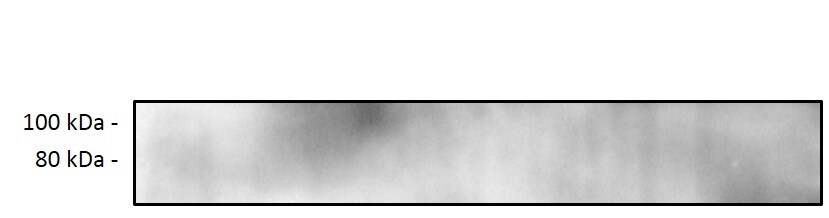
Application: Western BlotSample Tested: Adult intestineSpecies: MouseVerified Customer | Posted 11/01/2019Mouse small intestine was homogenised and protein content was quantified by a BCA assay. Twenty micrograms of protein were resolved on a 4-12% Bis-Tris gel and transferred to nitrocellulose membranes. Membranes were probed with primary antibody ACSL3 diluted 1:1000 in 5% BSA 2, before incubation with Anti-rabbit secondary horseradish peroxidase-conjugated antibody 1:5000. Blots were visualised with Immobilon Western Chemiluminescence HRP Substrate and imaged with Syngene chemiluminescence imaging system.

There are no reviews that match your criteria.
Protocols
Find general support by application which include: protocols, troubleshooting, illustrated assays, videos and webinars.
- Antigen Retrieval Protocol (PIER)
- Antigen Retrieval for Frozen Sections Protocol
- Appropriate Fixation of IHC/ICC Samples
- Cellular Response to Hypoxia Protocols
- Chromogenic IHC Staining of Formalin-Fixed Paraffin-Embedded (FFPE) Tissue Protocol
- Chromogenic Immunohistochemistry Staining of Frozen Tissue
- ClariTSA™ Fluorophore Kits
- Detection & Visualization of Antibody Binding
- Fluorescent IHC Staining of Frozen Tissue Protocol
- Graphic Protocol for Heat-induced Epitope Retrieval
- Graphic Protocol for the Preparation and Fluorescent IHC Staining of Frozen Tissue Sections
- Graphic Protocol for the Preparation and Fluorescent IHC Staining of Paraffin-embedded Tissue Sections
- Graphic Protocol for the Preparation of Gelatin-coated Slides for Histological Tissue Sections
- IHC Sample Preparation (Frozen sections vs Paraffin)
- Immunofluorescent IHC Staining of Formalin-Fixed Paraffin-Embedded (FFPE) Tissue Protocol
- Immunohistochemistry (IHC) and Immunocytochemistry (ICC) Protocols
- Immunohistochemistry Frozen Troubleshooting
- Immunohistochemistry Paraffin Troubleshooting
- Preparing Samples for IHC/ICC Experiments
- Preventing Non-Specific Staining (Non-Specific Binding)
- Primary Antibody Selection & Optimization
- Protocol for Heat-Induced Epitope Retrieval (HIER)
- Protocol for Making a 4% Formaldehyde Solution in PBS
- Protocol for VisUCyte™ HRP Polymer Detection Reagent
- Protocol for the Preparation & Fixation of Cells on Coverslips
- Protocol for the Preparation and Chromogenic IHC Staining of Frozen Tissue Sections
- Protocol for the Preparation and Chromogenic IHC Staining of Frozen Tissue Sections - Graphic
- Protocol for the Preparation and Chromogenic IHC Staining of Paraffin-embedded Tissue Sections
- Protocol for the Preparation and Chromogenic IHC Staining of Paraffin-embedded Tissue Sections - Graphic
- Protocol for the Preparation and Fluorescent IHC Staining of Frozen Tissue Sections
- Protocol for the Preparation and Fluorescent IHC Staining of Paraffin-embedded Tissue Sections
- Protocol for the Preparation of Gelatin-coated Slides for Histological Tissue Sections
- R&D Systems Quality Control Western Blot Protocol
- TUNEL and Active Caspase-3 Detection by IHC/ICC Protocol
- The Importance of IHC/ICC Controls
- Troubleshooting Guide: Immunohistochemistry
- Troubleshooting Guide: Western Blot Figures
- Western Blot Conditions
- Western Blot Protocol
- Western Blot Protocol for Cell Lysates
- Western Blot Troubleshooting
- Western Blot Troubleshooting Guide
- View all Protocols, Troubleshooting, Illustrated assays and Webinars
Loading...