ACSL5 Antibody (5H8) - Azide and BSA Free
Novus Biologicals | Catalog # H00051703-M01
![Western Blot: ACSL5 Antibody (5H8) [H00051703-M01]](https://resources.rndsystems.com/images/products/ACSL5-Antibody-5H8-Western-Blot-H00051703-M01-img0012.jpg "Western Blot: ACSL5 Antibody (5H8) [H00051703-M01]")
Loading...
Key Product Details
Species Reactivity
Human
Applications
Immunohistochemistry, Immunohistochemistry-Paraffin, Western Blot, ELISA, Sandwich ELISA, Immunoprecipitation
Label
Unconjugated
Antibody Source
Monoclonal Mouse IgG1 kappa Clone # 5H8
Format
Azide and BSA Free
Loading...
Product Specifications
Immunogen
ACSL5 (NP_057318, 91 a.a. ~ 186 a.a) partial recombinant protein with GST tag. MW of the GST tag alone is 26 KDa. PQPVLPLLDLNNQSVGIEGGARKGVSQKNNDLTSCCFSDAKTMYEVFQRGLAVSDNGPCLGYRKPNQPYRWLSYKQVSDRAEYLGSCLLHKGYKSS
Reactivity Notes
Human. Other species not tested.
Specificity
ACSL5 - acyl-CoA synthetase long-chain family member 5
Clonality
Monoclonal
Host
Mouse
Isotype
IgG1 kappa
Scientific Data Images for ACSL5 Antibody (5H8) - Azide and BSA Free
Western Blot: ACSL5 Antibody (5H8) [H00051703-M01]
Western Blot: ACSL5 Antibody (5H8) [H00051703-M01] - ACSL5 monoclonal antibody (M01), clone 5H8 Analysis of ACSL5 expression in HepG2.![Immunohistochemistry-Paraffin: ACSL5 Antibody (5H8) [H00051703-M01]](https://resources.rndsystems.com/images/products/ACSL5-Antibody-5H8-Immunohistochemistry-Paraffin-H00051703-M01-img0009.jpg "Immunohistochemistry-Paraffin: ACSL5 Antibody (5H8) [H00051703-M01]")
Immunohistochemistry-Paraffin: ACSL5 Antibody (5H8) [H00051703-M01]
Immunohistochemistry-Paraffin: ACSL5 Antibody (5H8) [H00051703-M01] - Analysis of monoclonal antibody to ACSL5 on formalin-fixed paraffin-embedded human colon. Antibody concentration 3 ug/ml.![Western Blot: ACSL5 Antibody (5H8) [H00051703-M01]](https://resources.rndsystems.com/images/products/ACSL5-Antibody-5H8-Western-Blot-H00051703-M01-img0013.jpg "Western Blot: ACSL5 Antibody (5H8) [H00051703-M01]")
Western Blot: ACSL5 Antibody (5H8) [H00051703-M01]
Western Blot: ACSL5 Antibody (5H8) [H00051703-M01] - Analysis of ACSL5 expression in transfected 293T cell line by ACSL5 monoclonal antibody (M01), clone 5H8.Lane 1: ACSL5 transfected lysate(82.3 KDa).Lane 2: Non-transfected lysate.![Immunoprecipitation: ACSL5 Antibody (5H8) [H00051703-M01]](https://resources.rndsystems.com/images/products/ACSL5-Antibody-5H8-Immunoprecipitation-H00051703-M01-img0010.jpg "Immunoprecipitation: ACSL5 Antibody (5H8) [H00051703-M01]")
Immunoprecipitation: ACSL5 Antibody (5H8) [H00051703-M01]
Immunoprecipitation: ACSL5 Antibody (5H8) [H00051703-M01] - Analysis of ACSL5 transfected lysate using anti-ACSL5 monoclonal antibody and Protein A Magnetic Bead, and immunoblotted with ACSL5 MaxPab rabbit polyclonal antibody.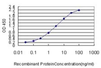
Sandwich ELISA: ACSL5 Antibody (5H8) [H00051703-M01] - Detection limit for recombinant GST tagged ACSL5 is approximately 0.1ng/ml as a capture antibody.
 [H00051703-M01] -")
Western Blot: ACSL5 Antibody (5H8) [H00051703-M01] -
h00051703-m01_mouse-monoclonal-acsl5-antibody-5h8-25520231539511.jpg [H00051703-M01] -")
Western Blot: ACSL5 Antibody (5H8) [H00051703-M01] -
Western Blot: ACSL5 Antibody (5H8) [H00051703-M01] - Effects of IGF1R localization over cell metabolism. (A) U87Empty, U87WT, & U87Mut cells were cultured 24 h in serum-free medium or with 50-nM IGF1 stimulation (IGF1). To test the specificity of the response, pre-incubation (1 h) with 0.5 μM OSI906 was also performed (IGF1+ OSI). (A, B) GLUT1 mRNA expression was calculated by rqPCR by the relative quantitation method. (A) Values are presented as fold change compared to control conditions (serum-free). (B) IGF1 stimuli comparison between cell lines. The results are presented as fold change due to IGF1 stimulation over basal conditions (serum-free). Values are expressed as mean ± SD of three independent experiments performed in triplicates (*p < 0.05, **p < 0.005, ***p < 0.001, ****p < 0.0001, ANOVA, Tukey’s post-test). (C) LDH enzyme activity was measured & normalized to DNA content (mg). The results are expressed as mean ± SD of three independent experiments (**p < 0.005, ANOVA, Tukey’s post-test). (D) Representative western blotting (n = 3) for U87Empty, U87WT, & U87Mut cell protein extracts. The membranes were blotted with anti-pPDC (line 1), anti-FAS (line 3), anti-ACSL5 (line 5), & anti-pS6K (line 7). Each blot was stripped & reprobed with anti-beta actin (lines 2, 4, & 6) or antitotal-S6K (line 8). The relative quantification of the bands is shown under each line. Image collected & cropped by CiteAb from the following publication (https://pubmed.ncbi.nlm.nih.gov/35574033), licensed under a CC-BY license. Not internally tested by Novus Biologicals.Applications for ACSL5 Antibody (5H8) - Azide and BSA Free
Application
Recommended Usage
Western Blot
1:500
Application Notes
Antibody reactive against cell lysate and recombinant protein for Western Blot. Has also been used for immunohistochemistry (paraffin) and ELISA.
Formulation, Preparation, and Storage
Purification
IgG purified
Formulation
In 1x PBS, pH 7.4
Format
Azide and BSA Free
Preservative
No Preservative
Concentration
Concentrations vary lot to lot. See vial label for concentration. If unlisted please contact technical services.
Shipping
The product is shipped with polar packs. Upon receipt, store it immediately at the temperature recommended below.
Stability & Storage
Aliquot and store at -20C or -80C. Avoid freeze-thaw cycles.
Background: ACSL5
Alternate Names
ACS2, ACS5FACL5 for fatty acid coenzyme A ligase 5, acyl-CoA synthetase long-chain family member 5, EC 6.2.1, EC 6.2.1.3, FACL5fatty acid coenzyme A ligase 5, fatty-acid-Coenzyme A ligase, long-chain 5, LACS 5, Long-chain acyl-CoA synthetase 5, long-chain fatty acid coenzyme A ligase 5, long-chain-fatty-acid--CoA ligase 5
Entrez Gene IDs
51703 (Human)
Gene Symbol
ACSL5
OMIM
605677 (Human)
UniProt
Additional ACSL5 Products
Product Documents for ACSL5 Antibody (5H8) - Azide and BSA Free
Certificate of Analysis
To download a Certificate of Analysis, please enter a lot or batch number in the search box below.
Product Specific Notices for ACSL5 Antibody (5H8) - Azide and BSA Free
This product is produced by and distributed for Abnova, a company based in Taiwan.
This product is for research use only and is not approved for use in humans or in clinical diagnosis. Primary Antibodies are guaranteed for 1 year from date of receipt.
Citations for ACSL5 Antibody (5H8) - Azide and BSA Free
 Powered by Bioz
Customer Reviews for ACSL5 Antibody (5H8) - Azide and BSA Free
There are currently no reviews for this product. Be the first to review ACSL5 Antibody (5H8) - Azide and BSA Free and earn rewards!
Have you used ACSL5 Antibody (5H8) - Azide and BSA Free?
Submit a review and receive an Amazon gift card!
$25/€18/£15/$25CAN/¥2500 Yen for a review with an image
$10/€7/£6/$10CAN/¥1110 Yen for a review without an image
Submit a review
Protocols
Find general support by application which include: protocols, troubleshooting, illustrated assays, videos and webinars.
- Antigen Retrieval Protocol (PIER)
- Antigen Retrieval for Frozen Sections Protocol
- Appropriate Fixation of IHC/ICC Samples
- Cellular Response to Hypoxia Protocols
- Chromogenic IHC Staining of Formalin-Fixed Paraffin-Embedded (FFPE) Tissue Protocol
- Chromogenic Immunohistochemistry Staining of Frozen Tissue
- ClariTSA™ Fluorophore Kits
- Detection & Visualization of Antibody Binding
- ELISA Sample Preparation & Collection Guide
- ELISA Troubleshooting Guide
- Fluorescent IHC Staining of Frozen Tissue Protocol
- Graphic Protocol for Heat-induced Epitope Retrieval
- Graphic Protocol for the Preparation and Fluorescent IHC Staining of Frozen Tissue Sections
- Graphic Protocol for the Preparation and Fluorescent IHC Staining of Paraffin-embedded Tissue Sections
- Graphic Protocol for the Preparation of Gelatin-coated Slides for Histological Tissue Sections
- How to Run an R&D Systems DuoSet ELISA
- How to Run an R&D Systems Quantikine ELISA
- How to Run an R&D Systems Quantikine™ QuicKit™ ELISA
- IHC Sample Preparation (Frozen sections vs Paraffin)
- Immunofluorescent IHC Staining of Formalin-Fixed Paraffin-Embedded (FFPE) Tissue Protocol
- Immunohistochemistry (IHC) and Immunocytochemistry (ICC) Protocols
- Immunohistochemistry Frozen Troubleshooting
- Immunohistochemistry Paraffin Troubleshooting
- Immunoprecipitation Protocol
- Preparing Samples for IHC/ICC Experiments
- Preventing Non-Specific Staining (Non-Specific Binding)
- Primary Antibody Selection & Optimization
- Protocol for Heat-Induced Epitope Retrieval (HIER)
- Protocol for Making a 4% Formaldehyde Solution in PBS
- Protocol for VisUCyte™ HRP Polymer Detection Reagent
- Protocol for the Preparation & Fixation of Cells on Coverslips
- Protocol for the Preparation and Chromogenic IHC Staining of Frozen Tissue Sections
- Protocol for the Preparation and Chromogenic IHC Staining of Frozen Tissue Sections - Graphic
- Protocol for the Preparation and Chromogenic IHC Staining of Paraffin-embedded Tissue Sections
- Protocol for the Preparation and Chromogenic IHC Staining of Paraffin-embedded Tissue Sections - Graphic
- Protocol for the Preparation and Fluorescent IHC Staining of Frozen Tissue Sections
- Protocol for the Preparation and Fluorescent IHC Staining of Paraffin-embedded Tissue Sections
- Protocol for the Preparation of Gelatin-coated Slides for Histological Tissue Sections
- Quantikine HS ELISA Kit Assay Principle, Alkaline Phosphatase
- Quantikine HS ELISA Kit Principle, Streptavidin-HRP Polymer
- R&D Systems Quality Control Western Blot Protocol
- Sandwich ELISA (Colorimetric) – Biotin/Streptavidin Detection Protocol
- Sandwich ELISA (Colorimetric) – Direct Detection Protocol
- TUNEL and Active Caspase-3 Detection by IHC/ICC Protocol
- The Importance of IHC/ICC Controls
- Troubleshooting Guide: ELISA
- Troubleshooting Guide: Immunohistochemistry
- Troubleshooting Guide: Western Blot Figures
- Western Blot Conditions
- Western Blot Protocol
- Western Blot Protocol for Cell Lysates
- Western Blot Troubleshooting
- Western Blot Troubleshooting Guide
- View all Protocols, Troubleshooting, Illustrated assays and Webinars
Loading...