![Western Blot: Adenosine A2aR Antibody [NB300-597]](https://resources.rndsystems.com/images/products/Adenosine-A2a-R-Antibody-Western-Blot-NB300-597-img0011.jpg "Western Blot: Adenosine A2aR Antibody [NB300-597]")
Loading...
Key Product Details
Species Reactivity
Validated:
Human, Mouse, Rat, Canine
Cited:
Human
Applications
Validated:
Immunohistochemistry, Immunohistochemistry-Paraffin, Western Blot, Flow Cytometry, Immunocytochemistry/ Immunofluorescence, Simple Western, Immunoprecipitation
Cited:
Western Blot, Flow Cytometry, IF/IHC
Label
Unconjugated
Antibody Source
Polyclonal Rabbit IgG
Loading...
Product Specifications
Immunogen
Synthetic peptide corresponding to residues E(373) S H G D M G L P D V E L L S H E L K(391) of canine A2aAR.
Localization
Cell Membrane
Specificity
Detects adenosine receptor A2a. This does not detect other AR subtypes.
Clonality
Polyclonal
Host
Rabbit
Isotype
IgG
Theoretical MW
45 kDa.
Disclaimer note: The observed molecular weight of the protein may vary from the listed predicted molecular weight due to post translational modifications, post translation cleavages, relative charges, and other experimental factors.
Disclaimer note: The observed molecular weight of the protein may vary from the listed predicted molecular weight due to post translational modifications, post translation cleavages, relative charges, and other experimental factors.
Scientific Data Images for Adenosine A2aR Antibody
Western Blot: Adenosine A2aR Antibody [NB300-597]
Western Blot: Adenosine A2aR Antibody [NB300-597] - Analysis of 25 ug of human placenta (lane 1), HepG2 (lane 2), Hela (lane 3) and mouse liver (lane 4) cell lysates.![Simple Western: Adenosine A2aR Antibody [NB300-597]](https://resources.rndsystems.com/images/products/Adenosine-A2a-R-Antibody-Simple-Western-NB300-597-img0006.jpg "Simple Western: Adenosine A2aR Antibody [NB300-597]")
Simple Western: Adenosine A2aR Antibody [NB300-597]
Simple Western: Adenosine A2aR Antibody [NB300-597] - Simple Western lane view shows a specific band for Adenosine A2a R in 0.5 mg/ml of HeLa lysate. This experiment was performed under reducing conditions using the 12-230 kDa separation system.![Immunohistochemistry-Paraffin: Adenosine A2aR Antibody [NB300-597]](https://resources.rndsystems.com/images/products/Adenosine-A2a-R-Antibody-Immunohistochemistry-Paraffin-NB300-597-img0010.jpg "Immunohistochemistry-Paraffin: Adenosine A2aR Antibody [NB300-597]")
Immunohistochemistry-Paraffin: Adenosine A2aR Antibody [NB300-597]
Immunohistochemistry-Paraffin: Adenosine A2aR Antibody [NB300-597] - Analysis showing positive staining in the cytoplasm and membrane of Human placenta tissue (right) compared with a negative control in the absence of primary antibody (left).![Immunocytochemistry/ Immunofluorescence: Adenosine A2aR Antibody [NB300-597]](https://resources.rndsystems.com/images/products/Adenosine-A2a-R-Antibody-Immunocytochemistry-Immunofluorescence-NB300-597-img0007.jpg "Immunocytochemistry/ Immunofluorescence: Adenosine A2aR Antibody [NB300-597]")
Immunocytochemistry/ Immunofluorescence: Adenosine A2aR Antibody [NB300-597]
Immunocytochemistry/Immunofluorescence: Adenosine A2aR Antibody [NB300-597] - Analysis of Adenosine Receptor A2a (green) showing staining in the cytoplasm of U251 cells (right) compared to a negative control without primary antibody (left). F-actin (red) was stained with a flourescent red phalloidin and nuclei (blue) were stained with Hoechst or DAPI![Immunocytochemistry/ Immunofluorescence: Adenosine A2aR Antibody [NB300-597]](https://resources.rndsystems.com/images/products/Adenosine-A2a-R-Antibody-Immunocytochemistry-NB300-597-img0012.jpg "Immunocytochemistry/ Immunofluorescence: Adenosine A2aR Antibody [NB300-597]")
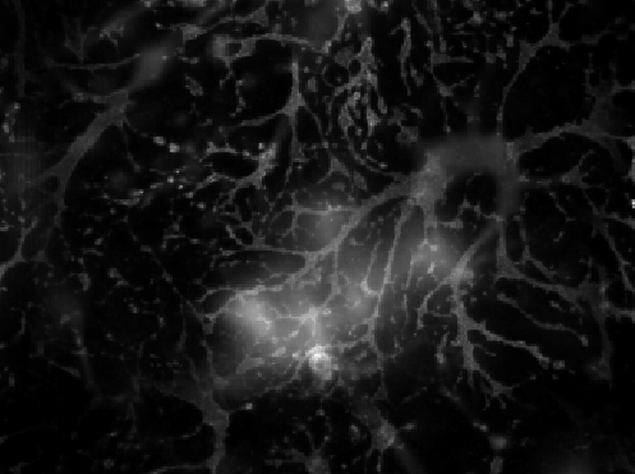
Immunocytochemistry/ Immunofluorescence: Adenosine A2aR Antibody [NB300-597]
Immunocytochemistry/Immunofluorescence: Adenosine A2aR Antibody [NB300-597] - Mixed cortical rat neurons culture (21 DIV) stained for A2A labelled with Alexa647. The staining visualised the astrocytes in culture. This image was submitted via customer Review.![Immunohistochemistry-Paraffin: Adenosine A2aR Antibody [NB300-597]](https://resources.rndsystems.com/images/products/Adenosine-A2a-R-Antibody-Immunohistochemistry-Paraffin-NB300-597-img0008.jpg "Immunohistochemistry-Paraffin: Adenosine A2aR Antibody [NB300-597]")
Immunohistochemistry-Paraffin: Adenosine A2aR Antibody [NB300-597]
Immunohistochemistry-Paraffin: Adenosine A2aR Antibody [NB300-597] - Staining in the cytoplasm and membrane of Mouse testis tissue (right) compared with a negative control in the absence of primary antibody (left).![Immunohistochemistry-Paraffin: Adenosine A2aR Antibody [NB300-597]](https://resources.rndsystems.com/images/products/Adenosine-A2a-R-Antibody-Immunohistochemistry-Paraffin-NB300-597-img0009.jpg "Immunohistochemistry-Paraffin: Adenosine A2aR Antibody [NB300-597]")
Immunohistochemistry-Paraffin: Adenosine A2aR Antibody [NB300-597]
Immunohistochemistry-Paraffin: Adenosine A2aR Antibody [NB300-597] - Analysis showing positive staining in the cytoplasm and membrane of Human testis tissue (right) compared with a negative control in the absence of primary antibody (left).Applications for Adenosine A2aR Antibody
Application
Recommended Usage
Immunocytochemistry/ Immunofluorescence
1:10 - 1:100
Immunohistochemistry
1:10-1:500
Immunohistochemistry-Paraffin
1:20-1:200
Immunoprecipitation
1:10-1:500
Simple Western
1:20
Western Blot
1:1000
Application Notes
Use in Flow reported in scientific literature (PMID:34536555). WB: Detects an approx. 45 kDa protein from canine striatum representing A2aAR.
See Simple Western Antibody Database for Simple Western validation: tested in HeLa lysate (0.5 mg/ml); antibody dilution of 1:20; separated by size; detects a band at 51 kDa
See Simple Western Antibody Database for Simple Western validation: tested in HeLa lysate (0.5 mg/ml); antibody dilution of 1:20; separated by size; detects a band at 51 kDa
Reviewed Applications
Read 1 review rated 5 using NB300-597 in the following applications:
Flow Cytometry Panel Builder
Bio-Techne Knows Flow Cytometry
Save time and reduce costly mistakes by quickly finding compatible reagents using the Panel Builder Tool.
Advanced Features
- Spectra Viewer - Custom analysis of spectra from multiple fluorochromes
- Spillover Popups - Visualize the spectra of individual fluorochromes
- Antigen Density Selector - Match fluorochrome brightness with antigen density

Formulation, Preparation, and Storage
Purification
Immunogen affinity purified
Formulation
PBS with 1 mg/ml BSA
Preservative
0.05% Sodium Azide
Concentration
0.69 mg/ml
Shipping
The product is shipped with polar packs. Upon receipt, store it immediately at the temperature recommended below.
Stability & Storage
Store at -20C. Avoid freeze-thaw cycles.
Background: Adenosine A2aR
Long Name
Adenosine A2A Receptor
Alternate Names
A2aR, Adenosine A2a R, ADORA2A, RDC8
Gene Symbol
ADORA2A
Additional Adenosine A2aR Products
Product Documents for Adenosine A2aR Antibody
Certificate of Analysis
To download a Certificate of Analysis, please enter a lot or batch number in the search box below.
Product Specific Notices for Adenosine A2aR Antibody
This product is for research use only and is not approved for use in humans or in clinical diagnosis. Primary Antibodies are guaranteed for 1 year from date of receipt.
Related Research Areas
Citations for Adenosine A2aR Antibody
 Powered by Bioz
Customer Reviews for Adenosine A2aR Antibody (1)
5 out of 5
1 Customer Rating
Have you used Adenosine A2aR Antibody?
Submit a review and receive an Amazon gift card!
$25/€18/£15/$25CAN/¥2500 Yen for a review with an image
$10/€7/£6/$10CAN/¥1110 Yen for a review without an image
Submit a review
Customer Images
Showing
1
-
1 of
1 review
Showing All
Filter By:
-
Application: ImmunocytochemistrySample Tested: Primary rat cortical neuronsSpecies: RatVerified Customer | Posted 06/27/2017Mixed cortical rat culture (21 DIV) stained for A2A labelled with Alexa647. The staining visualised the astrocytes in culture.Fixation Solution and Conditions: 4% paraformaldehyde in PBS, 15 minutes RT Blocking Solution & Duration: 3% BSA in PBS with 0.2% saponin, 30 minutes RT Primary Antibody Diluent and Dilutions Tested: 1:120 in 1% BSA in PBS with 0.2% saponin, over night 4°C Secondary Antibody Manufacturer, Host Species, Dilution, & Diluent: Thermo donkey anti-rabbit Alexa647, 1:500 in PBS with 0.2% saponin, 1 hour, RT

There are no reviews that match your criteria.
Protocols
Find general support by application which include: protocols, troubleshooting, illustrated assays, videos and webinars.
- 7-Amino Actinomycin D (7-AAD) Cell Viability Flow Cytometry Protocol
- Antigen Retrieval Protocol (PIER)
- Antigen Retrieval for Frozen Sections Protocol
- Appropriate Fixation of IHC/ICC Samples
- Cellular Response to Hypoxia Protocols
- Chromogenic IHC Staining of Formalin-Fixed Paraffin-Embedded (FFPE) Tissue Protocol
- Chromogenic Immunohistochemistry Staining of Frozen Tissue
- ClariTSA™ Fluorophore Kits
- Detection & Visualization of Antibody Binding
- Extracellular Membrane Flow Cytometry Protocol
- Flow Cytometry Protocol for Cell Surface Markers
- Flow Cytometry Protocol for Staining Membrane Associated Proteins
- Flow Cytometry Staining Protocols
- Flow Cytometry Troubleshooting Guide
- Fluorescent IHC Staining of Frozen Tissue Protocol
- Graphic Protocol for Heat-induced Epitope Retrieval
- Graphic Protocol for the Preparation and Fluorescent IHC Staining of Frozen Tissue Sections
- Graphic Protocol for the Preparation and Fluorescent IHC Staining of Paraffin-embedded Tissue Sections
- Graphic Protocol for the Preparation of Gelatin-coated Slides for Histological Tissue Sections
- ICC Cell Smear Protocol for Suspension Cells
- ICC Immunocytochemistry Protocol Videos
- ICC for Adherent Cells
- IHC Sample Preparation (Frozen sections vs Paraffin)
- Immunocytochemistry (ICC) Protocol
- Immunocytochemistry Troubleshooting
- Immunofluorescence of Organoids Embedded in Cultrex Basement Membrane Extract
- Immunofluorescent IHC Staining of Formalin-Fixed Paraffin-Embedded (FFPE) Tissue Protocol
- Immunohistochemistry (IHC) and Immunocytochemistry (ICC) Protocols
- Immunohistochemistry Frozen Troubleshooting
- Immunohistochemistry Paraffin Troubleshooting
- Immunoprecipitation Protocol
- Intracellular Flow Cytometry Protocol Using Alcohol (Methanol)
- Intracellular Flow Cytometry Protocol Using Detergents
- Intracellular Nuclear Staining Flow Cytometry Protocol Using Detergents
- Intracellular Staining Flow Cytometry Protocol Using Alcohol Permeabilization
- Intracellular Staining Flow Cytometry Protocol Using Detergents to Permeabilize Cells
- Preparing Samples for IHC/ICC Experiments
- Preventing Non-Specific Staining (Non-Specific Binding)
- Primary Antibody Selection & Optimization
- Propidium Iodide Cell Viability Flow Cytometry Protocol
- Protocol for Heat-Induced Epitope Retrieval (HIER)
- Protocol for Liperfluo
- Protocol for Making a 4% Formaldehyde Solution in PBS
- Protocol for VisUCyte™ HRP Polymer Detection Reagent
- Protocol for the Characterization of Human Th22 Cells
- Protocol for the Characterization of Human Th9 Cells
- Protocol for the Fluorescent ICC Staining of Cell Smears - Graphic
- Protocol for the Fluorescent ICC Staining of Cultured Cells on Coverslips - Graphic
- Protocol for the Preparation & Fixation of Cells on Coverslips
- Protocol for the Preparation and Chromogenic IHC Staining of Frozen Tissue Sections
- Protocol for the Preparation and Chromogenic IHC Staining of Frozen Tissue Sections - Graphic
- Protocol for the Preparation and Chromogenic IHC Staining of Paraffin-embedded Tissue Sections
- Protocol for the Preparation and Chromogenic IHC Staining of Paraffin-embedded Tissue Sections - Graphic
- Protocol for the Preparation and Fluorescent ICC Staining of Cells on Coverslips
- Protocol for the Preparation and Fluorescent ICC Staining of Non-adherent Cells
- Protocol for the Preparation and Fluorescent ICC Staining of Stem Cells on Coverslips
- Protocol for the Preparation and Fluorescent IHC Staining of Frozen Tissue Sections
- Protocol for the Preparation and Fluorescent IHC Staining of Paraffin-embedded Tissue Sections
- Protocol for the Preparation of Gelatin-coated Slides for Histological Tissue Sections
- Protocol for the Preparation of a Cell Smear for Non-adherent Cell ICC - Graphic
- Protocol: Annexin V and PI Staining by Flow Cytometry
- Protocol: Annexin V and PI Staining for Apoptosis by Flow Cytometry
- R&D Systems Quality Control Western Blot Protocol
- TUNEL and Active Caspase-3 Detection by IHC/ICC Protocol
- The Importance of IHC/ICC Controls
- Troubleshooting Guide: Fluorokine Flow Cytometry Kits
- Troubleshooting Guide: Immunohistochemistry
- Troubleshooting Guide: Western Blot Figures
- Western Blot Conditions
- Western Blot Protocol
- Western Blot Protocol for Cell Lysates
- Western Blot Troubleshooting
- Western Blot Troubleshooting Guide
- View all Protocols, Troubleshooting, Illustrated assays and Webinars
Loading...