![Western Blot: AGO1/EIF2C1 Antibody [NB100-2817]](https://resources.rndsystems.com/images/products/AGO1-EIF2C1-Antibody-Western-Blot-NB100-2817-img0002.jpg "Western Blot: AGO1/EIF2C1 Antibody [NB100-2817]")
Loading...
Key Product Details
Species Reactivity
Validated:
Human
Cited:
Human
Predicted:
Canine (100%), Mouse (100%), Rat (100%). Backed by our 100% Guarantee.
Applications
Validated:
Immunohistochemistry, Immunohistochemistry-Paraffin, Western Blot, Peptide ELISA, Immunocytochemistry/ Immunofluorescence
Cited:
Western Blot, Immunocytochemistry/ Immunofluorescence
Label
Unconjugated
Antibody Source
Polyclonal Goat IgG
Loading...
Product Specifications
Immunogen
Peptide with sequence C-KNASYNLDPYIQEF corresponding to internal region according to NP_036331.1.
Specificity
This product is not expected to cross-react with EIF2C2, EIF2C3 and EIF2C4.
Clonality
Polyclonal
Host
Goat
Isotype
IgG
Scientific Data Images for AGO1/EIF2C1 Antibody
Western Blot: AGO1/EIF2C1 Antibody [NB100-2817]
Western Blot: AGO1/EIF2C1 Antibody [NB100-2817] - HEK293 overexpressing AGO1/EIF2C1 and probed with NB100-2817 (non-transfected HEK293 in lane B).![Immunohistochemistry-Paraffin: AGO1/EIF2C1 Antibody [NB100-2817]](https://resources.rndsystems.com/images/products/AGO1-EIF2C1-Antibody-Immunohistochemistry-Paraffin-NB100-2817-img0004.jpg "Immunohistochemistry-Paraffin: AGO1/EIF2C1 Antibody [NB100-2817]")
Immunohistochemistry-Paraffin: AGO1/EIF2C1 Antibody [NB100-2817]
Immunohistochemistry-Paraffin: AGO1/EIF2C1 Antibody [NB100-2817] - Staining of paraffin embedded Human Placenta. Antibody at 2.5 ug/mL. Steamed antigen retrieval with citrate buffer pH 6, AP-staining.![Immunohistochemistry-Paraffin: AGO1/EIF2C1 Antibody [NB100-2817]](https://resources.rndsystems.com/images/products/AGO1-EIF2C1-Antibody-Immunohistochemistry-Paraffin-NB100-2817-img0003.jpg "Immunohistochemistry-Paraffin: AGO1/EIF2C1 Antibody [NB100-2817]")
Immunohistochemistry-Paraffin: AGO1/EIF2C1 Antibody [NB100-2817]
Immunohistochemistry-Paraffin: AGO1/EIF2C1 Antibody [NB100-2817] - Staining of paraffin embedded Human Small Intestine. Antibody at 2.5 ug/mL. Steamed antigen retrieval with citrate buffer pH 6, AP-staining.
Western Blot: AGO1/EIF2C1 Antibody [NB100-2817] -
AGO1x localizes to the nuclear regionRepresentative immunofluorescence images showing the subcellular distribution of AGO1x (red) relative to nuclear and cytoplasmic markers. DAPI was used to mark the nucleus (blue). The co‐stained subcellular marker is indicated in each panel in green. SC 35, Lsm4, alpha ‐tubulin, ERP72, p54 (NRB), and nucleolin serve as markers for nuclear speckles, cytosol and nucleus, cytosol, endoplasmic reticulum, paraspeckle, and nucleolus, respectively.Mean (+/- SD) pixel intensities of AGO1x staining in nucleolus and nucleoplasm, computed from z‐stack images of MDA‐MB-231 cells (n = 20). The P‐value was determined using a paired two‐tailed t‐test.Representative AGO1x and AGO1 blot from MDA-MB‐231 cell fractions. GAPDH and hnRNP C1/C2 served as markers of purity of the individual fractions.Source data are available online for this figure. Image collected and cropped by CiteAb from the following open publication (https://pubmed.ncbi.nlm.nih.gov/32812257), licensed under a CC-BY license. Not internally tested by Novus Biologicals.
Western Blot: AGO1/EIF2C1 Antibody [NB100-2817] -
AGO1x antibody targets specifically its cognate protein and not the canonical AGO1AWestern blot analysis of multiple cell lines demonstrates that in addition to the canonical AGO1 protein band, a characteristic second band of higher MW is revealed by the AGO1 antibody.BHigher MW band observed in (A) corresponds to AGO1x protein. Representative Western blot shows that the intensity of the higher MW band is sensitive to ectopic overexpression (using the pIRES‐Neo vector) of FLAG‐tagged AGO1x but not of FLAG‐tagged AGO1. Expression of the corresponding isoform is confirmed with a blot for FLAG expression. The overexpression constructs are indicated with labels above the blots. Protein ladders show that the higher MW band corresponds to approximately 100 kDa.C, DWestern blot with AGO1x antibody demonstrates its specificity for the AGO1x isoform stably expressed from pCDH‐FLAG-tagged plasmids. Middle and lower panels depict AGO1x levels in individual samples, at low and high exposure, respectively, of the same blot. The higher exposure was used to better assess the expression level in untransfected (control) samples.E, FRepresentative images of MDA‐MB-231 stained with AGO1 (green) and AGO1x (red) antibodies under conditions of endogenous expression or knockdown with an siRNA pool targeting the transcript that encodes both isoforms (E). An AGO1x overexpression system, where FLAG‐AGO1x was stably integrated into MDA‐MB-231 cells, was also tested (F). DAPI was used to mark the nucleus (blue).GWestern blot analysis of AGO1x protein level in cells treated with either siAGO1 or siControl confirms the imaging results from panel E.Source data are available online for this figure. Image collected and cropped by CiteAb from the following open publication (https://pubmed.ncbi.nlm.nih.gov/32812257), licensed under a CC-BY license. Not internally tested by Novus Biologicals.
Western Blot: AGO1/EIF2C1 Antibody [NB100-2817] -
Evidence of AGO1 transcript translational readthrough and of AGO1x expressionATop: schema of analyzed TR regions (purple), located downstream of the annotated open reading frame (gray), between the annotated stop codon (red triangle) and the next in‐frame stop codon (orange triangle); Bottom: histogram of average PhastCons conservation scores (x‐axis) of putative TR regions of all RefSeq‐annotated transcripts. The scores of the four human Argonaute protein family members are highlighted.BMultiple sequence alignment of the AGO1 putative TR region across vertebrates.CMultiple sequence alignment of the corresponding predicted amino acid sequence. The unique peptide targeted by the polyclonal antibody is indicated by the red line. The green and blue lines indicate peptide sequences obtained after tryptic digestion, in which cleavage is exclusively at arginine (R) and lysine (K) (further described below in panels E and F). Red asterisks indicate stop codons.DWestern blot showing AGO1x expression in three cell lines. For comparison, a parallel blot was probed with an antibody directed against canonical AGO1. Tubulin served as loading control.E, FAnnotated MS/MS spectrum of peptides specific for the endogenous AGO1x, “QNAVTSLDR", depicted in green (E) and “LSKPQELCHPNPEEAR", depicted in blue (F). The Mascot ion score (text color corresponds to peptides marked in Fig 1C for reference) as well as the annotated fragments (blue = y‐ions; red = b‐ions) together with the corresponding amino acids is indicated. Image collected and cropped by CiteAb from the following open publication (https://pubmed.ncbi.nlm.nih.gov/32812257), licensed under a CC-BY license. Not internally tested by Novus Biologicals.
Immunocytochemistry/ Immunofluorescence: AGO1/EIF2C1 Antibody [NB100-2817] -
AGO1x antibody targets specifically its cognate protein and not the canonical AGO1AWestern blot analysis of multiple cell lines demonstrates that in addition to the canonical AGO1 protein band, a characteristic second band of higher MW is revealed by the AGO1 antibody.BHigher MW band observed in (A) corresponds to AGO1x protein. Representative Western blot shows that the intensity of the higher MW band is sensitive to ectopic overexpression (using the pIRES‐Neo vector) of FLAG‐tagged AGO1x but not of FLAG‐tagged AGO1. Expression of the corresponding isoform is confirmed with a blot for FLAG expression. The overexpression constructs are indicated with labels above the blots. Protein ladders show that the higher MW band corresponds to approximately 100 kDa.C, DWestern blot with AGO1x antibody demonstrates its specificity for the AGO1x isoform stably expressed from pCDH‐FLAG-tagged plasmids. Middle and lower panels depict AGO1x levels in individual samples, at low and high exposure, respectively, of the same blot. The higher exposure was used to better assess the expression level in untransfected (control) samples.E, FRepresentative images of MDA‐MB-231 stained with AGO1 (green) and AGO1x (red) antibodies under conditions of endogenous expression or knockdown with an siRNA pool targeting the transcript that encodes both isoforms (E). An AGO1x overexpression system, where FLAG‐AGO1x was stably integrated into MDA‐MB-231 cells, was also tested (F). DAPI was used to mark the nucleus (blue).GWestern blot analysis of AGO1x protein level in cells treated with either siAGO1 or siControl confirms the imaging results from panel E.Source data are available online for this figure. Image collected and cropped by CiteAb from the following open publication (https://pubmed.ncbi.nlm.nih.gov/32812257), licensed under a CC-BY license. Not internally tested by Novus Biologicals.Applications for AGO1/EIF2C1 Antibody
Application
Recommended Usage
Immunohistochemistry-Paraffin
2.5 ug/mL
Peptide ELISA
Detection limit 1:16000
Western Blot
0.3 - 1 ug/mL
Application Notes
Use in Immunocytochemistry/Immunofluorescence reported in scientific literature (PMID:32812257)WB: In transfected HEK293 transiently expressing human EIF2C1bands of approx. 110 Knockdown Validateda band and 35 Knockdown Validateda band are observed. The 110 Knockdown Validateda band is not observed in the non-transfected HEK293. The calculated molecular size is 97.2 Knockdown Validateda band according to NP_036331.1
Reviewed Applications
Read 1 review rated 4 using NB100-2817 in the following applications:
Formulation, Preparation, and Storage
Purification
Immunogen affinity purified
Formulation
Tris saline (20 mM Tris pH 7.3, 150 mM NaCl), 0.5% BSA
Preservative
0.02% Sodium Azide
Concentration
0.5 mg/ml
Shipping
The product is shipped with polar packs. Upon receipt, store it immediately at the temperature recommended below.
Stability & Storage
Store at -20C. Avoid freeze-thaw cycles.
Background: AGO1/EIF2C1
Alternate Names
AGO1Q99, argonaute 1, Argonaute1, DKFZp686M13167, EIF2C, eIF2C 1, eIF-2C 1, Eukaryotic translation initiation factor 2C 1, eukaryotic translation initiation factor 2C, 1, GERP95, Golgi Endoplasmic Reticulum protein 95 kDa, hAgo1, protein argonaute-1, Putative RNA-binding protein Q99
Entrez Gene IDs
26523 (Human)
Gene Symbol
AGO1
UniProt
Additional AGO1/EIF2C1 Products
Product Documents for AGO1/EIF2C1 Antibody
Certificate of Analysis
To download a Certificate of Analysis, please enter a lot or batch number in the search box below.
Product Specific Notices for AGO1/EIF2C1 Antibody
This product is for research use only and is not approved for use in humans or in clinical diagnosis. Primary Antibodies are guaranteed for 1 year from date of receipt.
Citations for AGO1/EIF2C1 Antibody
 Powered by Bioz
Customer Reviews for AGO1/EIF2C1 Antibody (1)
4 out of 5
1 Customer Rating
Have you used AGO1/EIF2C1 Antibody?
Submit a review and receive an Amazon gift card!
$25/€18/£15/$25CAN/¥2500 Yen for a review with an image
$10/€7/£6/$10CAN/¥1110 Yen for a review without an image
Submit a review
Customer Images
Showing
1
-
1 of
1 review
Showing All
Filter By:
-
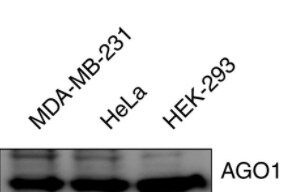
Application: Western BlotSample Tested: 3 human cancer cell linesSpecies: HumanVerified Customer | Posted 05/07/2021Western Blot of AGO1 in three cell lines.

There are no reviews that match your criteria.
Protocols
Find general support by application which include: protocols, troubleshooting, illustrated assays, videos and webinars.
- Antigen Retrieval Protocol (PIER)
- Antigen Retrieval for Frozen Sections Protocol
- Appropriate Fixation of IHC/ICC Samples
- Cellular Response to Hypoxia Protocols
- Chromogenic IHC Staining of Formalin-Fixed Paraffin-Embedded (FFPE) Tissue Protocol
- Chromogenic Immunohistochemistry Staining of Frozen Tissue
- ClariTSA™ Fluorophore Kits
- Detection & Visualization of Antibody Binding
- ELISA Sample Preparation & Collection Guide
- ELISA Troubleshooting Guide
- Fluorescent IHC Staining of Frozen Tissue Protocol
- Graphic Protocol for Heat-induced Epitope Retrieval
- Graphic Protocol for the Preparation and Fluorescent IHC Staining of Frozen Tissue Sections
- Graphic Protocol for the Preparation and Fluorescent IHC Staining of Paraffin-embedded Tissue Sections
- Graphic Protocol for the Preparation of Gelatin-coated Slides for Histological Tissue Sections
- How to Run an R&D Systems DuoSet ELISA
- How to Run an R&D Systems Quantikine ELISA
- How to Run an R&D Systems Quantikine™ QuicKit™ ELISA
- ICC Cell Smear Protocol for Suspension Cells
- ICC Immunocytochemistry Protocol Videos
- ICC for Adherent Cells
- IHC Sample Preparation (Frozen sections vs Paraffin)
- Immunocytochemistry (ICC) Protocol
- Immunocytochemistry Troubleshooting
- Immunofluorescence of Organoids Embedded in Cultrex Basement Membrane Extract
- Immunofluorescent IHC Staining of Formalin-Fixed Paraffin-Embedded (FFPE) Tissue Protocol
- Immunohistochemistry (IHC) and Immunocytochemistry (ICC) Protocols
- Immunohistochemistry Frozen Troubleshooting
- Immunohistochemistry Paraffin Troubleshooting
- Preparing Samples for IHC/ICC Experiments
- Preventing Non-Specific Staining (Non-Specific Binding)
- Primary Antibody Selection & Optimization
- Protocol for Heat-Induced Epitope Retrieval (HIER)
- Protocol for Making a 4% Formaldehyde Solution in PBS
- Protocol for VisUCyte™ HRP Polymer Detection Reagent
- Protocol for the Fluorescent ICC Staining of Cell Smears - Graphic
- Protocol for the Fluorescent ICC Staining of Cultured Cells on Coverslips - Graphic
- Protocol for the Preparation & Fixation of Cells on Coverslips
- Protocol for the Preparation and Chromogenic IHC Staining of Frozen Tissue Sections
- Protocol for the Preparation and Chromogenic IHC Staining of Frozen Tissue Sections - Graphic
- Protocol for the Preparation and Chromogenic IHC Staining of Paraffin-embedded Tissue Sections
- Protocol for the Preparation and Chromogenic IHC Staining of Paraffin-embedded Tissue Sections - Graphic
- Protocol for the Preparation and Fluorescent ICC Staining of Cells on Coverslips
- Protocol for the Preparation and Fluorescent ICC Staining of Non-adherent Cells
- Protocol for the Preparation and Fluorescent ICC Staining of Stem Cells on Coverslips
- Protocol for the Preparation and Fluorescent IHC Staining of Frozen Tissue Sections
- Protocol for the Preparation and Fluorescent IHC Staining of Paraffin-embedded Tissue Sections
- Protocol for the Preparation of Gelatin-coated Slides for Histological Tissue Sections
- Protocol for the Preparation of a Cell Smear for Non-adherent Cell ICC - Graphic
- Quantikine HS ELISA Kit Assay Principle, Alkaline Phosphatase
- Quantikine HS ELISA Kit Principle, Streptavidin-HRP Polymer
- R&D Systems Quality Control Western Blot Protocol
- Sandwich ELISA (Colorimetric) – Biotin/Streptavidin Detection Protocol
- Sandwich ELISA (Colorimetric) – Direct Detection Protocol
- TUNEL and Active Caspase-3 Detection by IHC/ICC Protocol
- The Importance of IHC/ICC Controls
- Troubleshooting Guide: ELISA
- Troubleshooting Guide: Immunohistochemistry
- Troubleshooting Guide: Western Blot Figures
- Western Blot Conditions
- Western Blot Protocol
- Western Blot Protocol for Cell Lysates
- Western Blot Troubleshooting
- Western Blot Troubleshooting Guide
- View all Protocols, Troubleshooting, Illustrated assays and Webinars
Loading...