AIF-1/Iba1 Antibody - Azide and BSA Free
Novus Biologicals | Catalog # NBP2-19019
![Immunohistochemistry-Paraffin: AIF-1/Iba1 Antibody [NBP2-19019]](https://resources.rndsystems.com/images/products/AIF-1-Iba1-Antibody-Immunohistochemistry-Paraffin-NBP2-19019-img0020.jpg "Immunohistochemistry-Paraffin: AIF-1/Iba1 Antibody [NBP2-19019]")
Loading...
Key Product Details
Species Reactivity
Validated:
Human, Mouse, Rat
Cited:
Human, Mouse, Rat, Primate - Macaca mulatta (Rhesus Macaque)
Predicted:
Porcine (100%), Rhesus Macaque (100%). Backed by our 100% Guarantee.
Applications
Validated:
Immunohistochemistry, Immunohistochemistry-Paraffin, Immunohistochemistry-Frozen, Immunohistochemistry Free-Floating, Western Blot, Flow Cytometry, Immunocytochemistry/ Immunofluorescence, Simple Western
Cited:
Immunohistochemistry, Immunohistochemistry-Paraffin, Immunohistochemistry-Frozen, Western Blot, Immunocytochemistry/ Immunofluorescence, IF/IHC
Label
Unconjugated
Antibody Source
Polyclonal Rabbit IgG
Format
Azide and BSA Free
Loading...
Product Specifications
Immunogen
Carrier-protein conjugated synthetic peptide encompassing a sequence within the C-terminus region of human AIF-1/Iba1. The exact sequence is proprietary.
Marker
pan-Microglia Marker
Clonality
Polyclonal
Host
Rabbit
Isotype
IgG
Theoretical MW
17 kDa.
Disclaimer note: The observed molecular weight of the protein may vary from the listed predicted molecular weight due to post translational modifications, post translation cleavages, relative charges, and other experimental factors.
Disclaimer note: The observed molecular weight of the protein may vary from the listed predicted molecular weight due to post translational modifications, post translation cleavages, relative charges, and other experimental factors.
Scientific Data Images for AIF-1/Iba1 Antibody - Azide and BSA Free
Immunohistochemistry-Paraffin: AIF-1/Iba1 Antibody [NBP2-19019]
Immunohistochemistry-Paraffin: AIF-1/Iba1 Antibody [NBP2-19019] - Mouse fore brain. Iba1 antibody dilution: 1:500. Antigen Retrieval: Trilogy™ (EDTA based, pH 8.0) buffer, 15min![Immunocytochemistry/ Immunofluorescence: AIF-1/Iba1 Antibody [NBP2-19019]](https://resources.rndsystems.com/images/products/AIF-1-Iba1-Antibody-Immunocytochemistry-Immunofluorescence-NBP2-19019-img0002.jpg "Immunocytochemistry/ Immunofluorescence: AIF-1/Iba1 Antibody [NBP2-19019]")
Immunocytochemistry/ Immunofluorescence: AIF-1/Iba1 Antibody [NBP2-19019]
Immunocytochemistry/Immunofluorescence: AIF-1/Iba1 Antibody [NBP2-19019] - AIF-1/Iba1 Antibody (1 1:1000, Red) expression in dorsal root ganglia. Green fluorescent Nissl was used as counter-staining. Image submitted by a verified customer review.![Immunohistochemistry-Paraffin: AIF-1/Iba1 Antibody [NBP2-19019]](https://resources.rndsystems.com/images/products/AIF-1-Iba1-Antibody-Immunohistochemistry-Paraffin-NBP2-19019-img0007.jpg "Immunohistochemistry-Paraffin: AIF-1/Iba1 Antibody [NBP2-19019]")
Immunohistochemistry-Paraffin: AIF-1/Iba1 Antibody [NBP2-19019]
Immunohistochemistry-Paraffin: AIF-1/Iba1 Antibody [NBP2-19019] - Mouse cerebellum stained by AIF-1/Iba1 Antibody diluted at 1:500. Antigen Retrieval: Citrate buffer, pH 6.0, 15 min.![Immunohistochemistry-Paraffin: AIF-1/Iba1 Antibody [NBP2-19019]](https://resources.rndsystems.com/images/products/AIF-1-Iba1-Antibody-Immunohistochemistry-Paraffin-NBP2-19019-img0010.jpg "Immunohistochemistry-Paraffin: AIF-1/Iba1 Antibody [NBP2-19019]")
Immunohistochemistry-Paraffin: AIF-1/Iba1 Antibody [NBP2-19019]
Immunohistochemistry-Paraffin: AIF-1/Iba1 Antibody [NBP2-19019] - Rat brain stained by AIF-1/Iba1 Antibody diluted at 1:500. Antigen Retrieval: Citrate buffer, pH 6.0, 15 min.![Immunohistochemistry-Paraffin: AIF-1/Iba1 Antibody [NBP2-19019]](https://resources.rndsystems.com/images/products/AIF-1-Iba1-Antibody-Immunohistochemistry-Paraffin-NBP2-19019-img0011.jpg "Immunohistochemistry-Paraffin: AIF-1/Iba1 Antibody [NBP2-19019]")
Immunohistochemistry-Paraffin: AIF-1/Iba1 Antibody [NBP2-19019]
Immunohistochemistry-Paraffin: AIF-1/Iba1 Antibody [NBP2-19019] - Rat cerebellum stained by AIF-1/Iba1 Antibody diluted at 1:500. Antigen Retrieval: Citrate buffer, pH 6.0, 15 min.![Immunohistochemistry-Paraffin: AIF-1/Iba1 Antibody [NBP2-19019]](https://resources.rndsystems.com/images/products/AIF-1-Iba1-Antibody-Immunohistochemistry-Paraffin-NBP2-19019-img0016.jpg "Immunohistochemistry-Paraffin: AIF-1/Iba1 Antibody [NBP2-19019]")
Immunohistochemistry-Paraffin: AIF-1/Iba1 Antibody [NBP2-19019]
Immunohistochemistry-Paraffin: AIF-1/Iba1 Antibody [NBP2-19019] - Rat hind brain. Iba1 antibody dilution: 1:500. Antigen Retrieval: Trilogy™ (EDTA based, pH 8.0) buffer, 15min![Immunohistochemistry-Paraffin: AIF-1/Iba1 Antibody [NBP2-19019]](https://resources.rndsystems.com/images/products/AIF-1-Iba1-Antibody-Immunohistochemistry-Paraffin-NBP2-19019-img0018.jpg "Immunohistochemistry-Paraffin: AIF-1/Iba1 Antibody [NBP2-19019]")
Immunohistochemistry-Paraffin: AIF-1/Iba1 Antibody [NBP2-19019]
Immunohistochemistry-Paraffin: AIF-1/Iba1 Antibody [NBP2-19019] - Analysis of paraffin-embedded human hepatoma, using at 1:500 dilution.Iba1 antibody detects AIF-1/Iba1 in the cytosol of Kupffer cell in human hepatoma by immunohistochemical analysis.
Immunohistochemistry: AIF-1/Iba1 Antibody [NBP2-19019] -
nbp2-19019_rabbit-polyclonal-aif-1-iba1-antibody-2552023153956.jpg
Immunocytochemistry/ Immunofluorescence: AIF-1/Iba1 Antibody [NBP2-19019] -
Immunocytochemistry/ Immunofluorescence: AIF-1/Iba1 Antibody [NBP2-19019] - LCN2 mainly expressed on neurons. a Representative immunofluorescence images of LCN2 (green), GFAP (red), NeuN (red) & IbaI (red) staining in sections of the hippocampus after surgery. Long scale bar, 600 µm; short scale bar, 25 µm. b Percentage quantification of LCN2 positive cells (n = 3 mice/group). c Cartoon depicting MACS sorting procedures for isolating CD11b+ & CD11b − cells. d Relative expression of cx3cr1 in CD11b + & CD11b − cells after sorting from all samples. d Relative expression level of Lcn2 mRNA in CD11b + & CD11b − cells from hippocampus 6 h after surgery. e Relative expression level of IL-6 mRNA in CD11b + cells. Data are represented as mean ± SEM. **** P < 0.0001 Image collected & cropped by CiteAb from the following publication (https://pubmed.ncbi.nlm.nih.gov/35413913), licensed under a CC-BY license. Not internally tested by Novus Biologicals.
Immunocytochemistry/ Immunofluorescence: AIF-1/Iba1 Antibody [NBP2-19019] -
Immunocytochemistry/ Immunofluorescence: AIF-1/Iba1 Antibody [NBP2-19019] - Knockdown of LCN2 prevents microglial activation & neuroinflammation. a Representative immunofluorescence images of hippocampal sections with IbaI+ cells (White). Scale bars, 150 µm. b Quantification of the microglia density in (A) (n = 4 mice/group, 2–3 slides/mice). c Quantification of the microglia Ramification index at 24 h after surgery (n = 72 cells from 4 mice/group). d Number of process intersections with shells at distances (in 2 µm in increments) from the soma by Sholl analysis at 24 h after surgery (n = 72 cells from 4 mice/group). e Tissue ELISA of IL-6 at 24 h after surgery (n = 5 mice/group). f Relative expression level of tnf-alpha, IL-6 and/or IL-1 beta 6 h after application of rLCN2 (500 ng/mL) on BV-2 (n = 4–6/group) & primary microglia cells (n = 3/group). Vehicle: PBS buffer. Data represent mean ± SEM; n = 4/group. * P < 0.05; *** P < 0.001 Image collected & cropped by CiteAb from the following publication (https://pubmed.ncbi.nlm.nih.gov/35413913), licensed under a CC-BY license. Not internally tested by Novus Biologicals.
Immunohistochemistry: AIF-1/Iba1 Antibody [NBP2-19019] -
Immunohistochemistry: AIF-1/Iba1 Antibody [NBP2-19019] - AIF-1/Iba1 antibody detects AIF-1/Iba1 protein at cell membrane and cytoplasm by immunohistochemical analysis.Sample: Frozen-sectioned mouse brain.
Green: AIF-1/Iba1 stained by AIF-1/Iba1 antibody (NBP2-19019) diluted at 1:500.
Blue: Fluoroshield with DAPI.
Antigen Retrieval: ice-cold MeOH for 5 min

Western Blot: AIF-1/Iba1 Antibody [NBP2-19019] -
Western Blot: AIF-1/Iba1 Antibody [NBP2-19019] - Rat tissue extract (50 ug) was separated by 15% SDS-PAGE, and the membrane was blotted with AIF-1/Iba1 antibody (NBP2-19019) diluted at 1:1000.
Immunohistochemistry-Paraffin: AIF-1/Iba1 Antibody [NBP2-19019] -
Immunohistochemistry-Paraffin: AIF-1/Iba1 Antibody [NBP2-19019] - AIF-1/Iba1 antibody detects AIF-1/Iba1 protein by immunohistochemical analysis.Sample: Paraffin-embedded rat tissues.
AIF-1/Iba1 stained by AIF-1/Iba1 antibody (NBP2-19019) diluted at 1:100.
Antigen Retrieval: Citrate buffer, pH 6.0, 15 min

Immunocytochemistry/ Immunofluorescence: AIF-1/Iba1 Antibody [NBP2-19019] -
Immunocytochemistry/ Immunofluorescence: AIF-1/Iba1 Antibody [NBP2-19019] - AIF-1/Iba1 antibody detects AIF-1/Iba1 protein at cell membrane by immunofluorescent analysis.Sample: THP-1 cells were fixed in 4% paraformaldehyde at RT for 15 min.
Green: AIF-1/Iba1 stained by AIF-1/Iba1 antibody (NBP2-19019) diluted at 1:500.
Blue: Fluoroshield with DAPI.

Immunohistochemistry-Paraffin: AIF-1/Iba1 Antibody [NBP2-19019] -
Immunohistochemistry-Paraffin: AIF-1/Iba1 Antibody [NBP2-19019] - AIF-1/Iba1 antibody detects AIF-1/Iba1 protein at cell membrane and cytoplasm by immunohistochemical analysis.Sample: Paraffin-embedded mouse brain.
AIF-1/Iba1 stained by AIF-1/Iba1 antibody (NBP2-19019) diluted at 1:500.
Antigen Retrieval: Citrate buffer, pH 6.0, 15 min

Flow Cytometry: AIF-1/Iba1 Antibody [NBP2-19019] -
Iba1 antibody (NBP2-19019) detects AIF1 protein by flow cytometry analysis.Sample: THP-1 cell.
Black: Unlabelled sample was used as a control.
Red: Iba1 antibody (NBP2-19019) dilution: 1:50.
Acquisition of 20,000 events were collected for FACS analysis.

Immunohistochemistry-Frozen: AIF-1/Iba1 Antibody [NBP2-19019] -
Iba1 antibody detects Iba1 protein expression at microglias by immunohistochemical analysis.Sample: Frozen sectioned E13.5 Rat brain.
Green: Iba1 protein stained by Iba1 antibody (NBP2-19019) diluted at 1:250.
Red: beta Tubulin 3/ TUJ1, a mature neuron marker, stained by beta Tubulin 3/ TUJ1 antibody [GT11710] diluted at 1:500.
Blue: Fluoroshield with DAPI.

Western Blot: AIF-1/Iba1 Antibody [NBP2-19019] -
Mouse tissue extract (50 ug) was separated by 15% SDS-PAGE, and the membrane was blotted with Iba1 antibody (NBP2-19019) diluted at 1:1000.
Western Blot: AIF-1/Iba1 Antibody [NBP2-19019] -
Various whole cell extracts (30 ug) were separated by 15% SDS-PAGE, and the membrane was blotted with Iba1 antibody (NBP2-19019) diluted at 1:1000. The HRP-conjugated anti-rabbit IgG antibody was used to detect the primary antibody. Corresponding RNA expression data for the same cell lines are based on Human Protein Atlas program.
Western Blot: AIF-1/Iba1 Antibody [NBP2-19019] -
Various tissue extracts (50 ug) were separated by 15% SDS-PAGE, and the membrane was blotted with Iba1 antibody (NBP2-19019) diluted at 1:1000. The HRP-conjugated anti-rabbit IgG antibody was used to detect the primary antibody.Applications for AIF-1/Iba1 Antibody - Azide and BSA Free
Application
Recommended Usage
Flow Cytometry
1:50-1:200
Immunocytochemistry/ Immunofluorescence
1:100-1:1000
Immunohistochemistry
1:100-1000
Immunohistochemistry Free-Floating
Assay dependent
Immunohistochemistry-Frozen
1:100-1:1000
Immunohistochemistry-Paraffin
1:100-1:1000
Simple Western
1:50
Western Blot
1:500-1:10000
Application Notes
Simple Western Separated by Size, antibody dilution of 1:50, detects a band at 25 kDa.
Reviewed Applications
Read 4 reviews rated 3.8 using NBP2-19019 in the following applications:
Flow Cytometry Panel Builder
Bio-Techne Knows Flow Cytometry
Save time and reduce costly mistakes by quickly finding compatible reagents using the Panel Builder Tool.
Advanced Features
- Spectra Viewer - Custom analysis of spectra from multiple fluorochromes
- Spillover Popups - Visualize the spectra of individual fluorochromes
- Antigen Density Selector - Match fluorochrome brightness with antigen density

Formulation, Preparation, and Storage
Purification
Antigen Affinity-purified
Formulation
PBS
Format
Azide and BSA Free
Preservative
No Preservative
Concentration
Concentrations vary lot to lot. See vial label for concentration. If unlisted please contact technical services.
Shipping
The product is shipped with polar packs. Upon receipt, store it immediately at the temperature recommended below.
Stability & Storage
Aliquot and store at -20C or -80C. Avoid freeze-thaw cycles.
Background: AIF-1/Iba1
Several cellular functions have been associated with AIF-1/Iba1 expression including cell growth, cell migration, actin bundling, membrane ruffling, and phagocytic activity (2, 4). Iba1 induces Rac signaling through a PLC-gamma dependent pathway (1). Rac, a member of the Rho family of small GTPases, localizes with Iba1 and F-actin in membrane ruffles and phagocytic cups and plays a role in microglia activation. AIF-1/Iba1 induction in macrophages and microglia occur in association with immunological inflammatory processes in various disease states including endometriosis, cerebral infarction and rheumatoid arthritis (5). Immunodetection of Iba1 through flow cytometry, immunohistochemical or immunocytochemical applications is commonly used for identification and analysis of microglia.
References
1. Imai, Y., & Kohsaka, S. (2002). Intracellular signaling in M-CSF-induced microglia activation: Role of Iba1. GLIA. https://doi.org/10.1002/glia.10149
2. Deininger, M. H., Meyermann, R., & Schluesener, H. J. (2002). The allograft inflammatory factor-1 family of proteins. FEBS Letters. https://doi.org/10.1016/S0014-5793(02)02430-4
3. Utans, U., Quist, W. C., Mcmanus, B. M., Wilson, J. E., Arceci, R. J., Wallace, A. F., & Russell, M. E. (1996). Allograft inflammatory factor-1: A cytokine-responsive macrophage molecule expressed in transplanted human hearts. Transplantation. https://doi.org/10.1097/00007890-199605150-00018
4. Franco-Bocanegra, McAuley, Nicoll, & Boche. (2019). Molecular Mechanisms of Microglial Motility: Changes in Ageing and Alzheimer's Disease. Cells. https://doi.org/10.3390/cells8060639
5. Kimura, M., Kawahito, Y., Obayashi, H., Ohta, M., Hara, H., Adachi, T.,... Yoshikawa, T. (2007). A Critical Role for Allograft Inflammatory Factor-1 in the Pathogenesis of Rheumatoid Arthritis. The Journal of Immunology. https://doi.org/10.4049/jimmunol.178.5.3316
Long Name
Allograft Inflammatory Factor 1
Alternate Names
AIF1, IBA1, IRT1
Entrez Gene IDs
199 (Human)
Gene Symbol
AIF1
Additional AIF-1/Iba1 Products
Product Documents for AIF-1/Iba1 Antibody - Azide and BSA Free
Certificate of Analysis
To download a Certificate of Analysis, please enter a lot or batch number in the search box below.
Product Specific Notices for AIF-1/Iba1 Antibody - Azide and BSA Free
This product is for research use only and is not approved for use in humans or in clinical diagnosis. Primary Antibodies are guaranteed for 1 year from date of receipt.
Related Research Areas
Citations for AIF-1/Iba1 Antibody - Azide and BSA Free
 Powered by Bioz
Customer Reviews for AIF-1/Iba1 Antibody - Azide and BSA Free (4)
3.8 out of 5
4 Customer Ratings
Have you used AIF-1/Iba1 Antibody - Azide and BSA Free?
Submit a review and receive an Amazon gift card!
$25/€18/£15/$25CAN/¥2500 Yen for a review with an image
$10/€7/£6/$10CAN/¥1110 Yen for a review without an image
Submit a review
Customer Images
Showing
1
-
4 of
4 reviews
Showing All
Filter By:
-
Application: Immunohistochemistry-FrozenSample Tested: Brain (hypothalamus) tissueSpecies: SheepVerified Customer | Posted 03/13/2020Photomicrograph (40x magnification) of sheep hypothalamus stained using IBA-1 (NBP2-19019) at dilution of 1:100. No positive staining was detected.To test, we used free-floating sheep hypothalamic brain sections from two animals. We did dilutions of 1:100 to 1:1000 and did not see any staining in our tissue. We also employed an antigen retrieval step in one of our runs, and it did not yield any different results.
 Bio-Techne ResponseThis review was submitted through the legacy Novus Innovators Program, reflecting a new species or application tested on a primary antibody.
 -
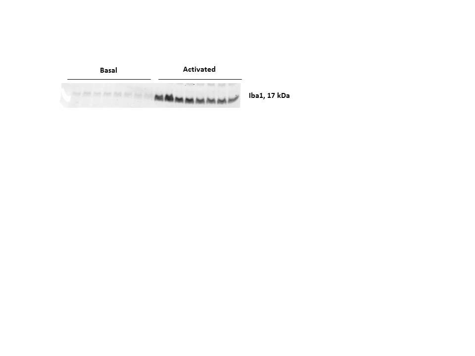
Application: Western BlotSample Tested: Mouse forebrain lysateSpecies: MouseVerified Customer | Posted 12/18/2017Iba1 expression in mouse forebrain lysate under basal and activated conditions
-
Application: ImmunocytochemistrySample Tested: C57BL/6 mouse retinaSpecies: MouseVerified Customer | Posted 04/01/2017
-
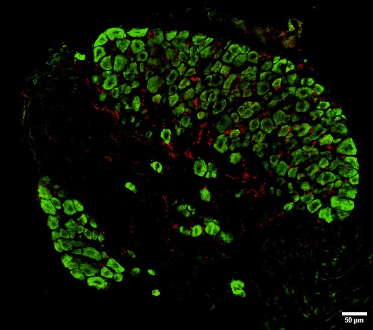
Application: ImmunocytochemistrySample Tested: Dorsal root gangliaSpecies: MouseVerified Customer | Posted 03/10/2017AIF-1/Iba1 (Red, NBP2-19019) expression in dorsal root ganglia. Green fluorescent Nissl was used as counter-staining.Blocking and antibody solution: BSA1%/Triton 02% in PBS Dilution AIF1: 1/1000 Secondary antibody: Alexa 555 Counter-staining: Nissl
There are no reviews that match your criteria.
Protocols
Find general support by application which include: protocols, troubleshooting, illustrated assays, videos and webinars.
- 7-Amino Actinomycin D (7-AAD) Cell Viability Flow Cytometry Protocol
- Antigen Retrieval Protocol (PIER)
- Antigen Retrieval for Frozen Sections Protocol
- Appropriate Fixation of IHC/ICC Samples
- Cellular Response to Hypoxia Protocols
- Chromogenic IHC Staining of Formalin-Fixed Paraffin-Embedded (FFPE) Tissue Protocol
- Chromogenic Immunohistochemistry Staining of Frozen Tissue
- ClariTSA™ Fluorophore Kits
- Detection & Visualization of Antibody Binding
- Extracellular Membrane Flow Cytometry Protocol
- Flow Cytometry Protocol for Cell Surface Markers
- Flow Cytometry Protocol for Staining Membrane Associated Proteins
- Flow Cytometry Staining Protocols
- Flow Cytometry Troubleshooting Guide
- Fluorescent IHC Staining of Frozen Tissue Protocol
- Graphic Protocol for Heat-induced Epitope Retrieval
- Graphic Protocol for the Preparation and Fluorescent IHC Staining of Frozen Tissue Sections
- Graphic Protocol for the Preparation and Fluorescent IHC Staining of Paraffin-embedded Tissue Sections
- Graphic Protocol for the Preparation of Gelatin-coated Slides for Histological Tissue Sections
- ICC Cell Smear Protocol for Suspension Cells
- ICC Immunocytochemistry Protocol Videos
- ICC for Adherent Cells
- IHC Sample Preparation (Frozen sections vs Paraffin)
- Immunocytochemistry (ICC) Protocol
- Immunocytochemistry Troubleshooting
- Immunofluorescence of Organoids Embedded in Cultrex Basement Membrane Extract
- Immunofluorescent IHC Staining of Formalin-Fixed Paraffin-Embedded (FFPE) Tissue Protocol
- Immunohistochemistry (IHC) and Immunocytochemistry (ICC) Protocols
- Immunohistochemistry Frozen Troubleshooting
- Immunohistochemistry Paraffin Troubleshooting
- Intracellular Flow Cytometry Protocol Using Alcohol (Methanol)
- Intracellular Flow Cytometry Protocol Using Detergents
- Intracellular Nuclear Staining Flow Cytometry Protocol Using Detergents
- Intracellular Staining Flow Cytometry Protocol Using Alcohol Permeabilization
- Intracellular Staining Flow Cytometry Protocol Using Detergents to Permeabilize Cells
- Preparing Samples for IHC/ICC Experiments
- Preventing Non-Specific Staining (Non-Specific Binding)
- Primary Antibody Selection & Optimization
- Propidium Iodide Cell Viability Flow Cytometry Protocol
- Protocol for Heat-Induced Epitope Retrieval (HIER)
- Protocol for Liperfluo
- Protocol for Making a 4% Formaldehyde Solution in PBS
- Protocol for VisUCyte™ HRP Polymer Detection Reagent
- Protocol for the Characterization of Human Th22 Cells
- Protocol for the Characterization of Human Th9 Cells
- Protocol for the Fluorescent ICC Staining of Cell Smears - Graphic
- Protocol for the Fluorescent ICC Staining of Cultured Cells on Coverslips - Graphic
- Protocol for the Preparation & Fixation of Cells on Coverslips
- Protocol for the Preparation and Chromogenic IHC Staining of Frozen Tissue Sections
- Protocol for the Preparation and Chromogenic IHC Staining of Frozen Tissue Sections - Graphic
- Protocol for the Preparation and Chromogenic IHC Staining of Paraffin-embedded Tissue Sections
- Protocol for the Preparation and Chromogenic IHC Staining of Paraffin-embedded Tissue Sections - Graphic
- Protocol for the Preparation and Fluorescent ICC Staining of Cells on Coverslips
- Protocol for the Preparation and Fluorescent ICC Staining of Non-adherent Cells
- Protocol for the Preparation and Fluorescent ICC Staining of Stem Cells on Coverslips
- Protocol for the Preparation and Fluorescent IHC Staining of Frozen Tissue Sections
- Protocol for the Preparation and Fluorescent IHC Staining of Paraffin-embedded Tissue Sections
- Protocol for the Preparation of Gelatin-coated Slides for Histological Tissue Sections
- Protocol for the Preparation of a Cell Smear for Non-adherent Cell ICC - Graphic
- Protocol: Annexin V and PI Staining by Flow Cytometry
- Protocol: Annexin V and PI Staining for Apoptosis by Flow Cytometry
- R&D Systems Quality Control Western Blot Protocol
- TUNEL and Active Caspase-3 Detection by IHC/ICC Protocol
- The Importance of IHC/ICC Controls
- Troubleshooting Guide: Fluorokine Flow Cytometry Kits
- Troubleshooting Guide: Immunohistochemistry
- Troubleshooting Guide: Western Blot Figures
- Western Blot Conditions
- Western Blot Protocol
- Western Blot Protocol for Cell Lysates
- Western Blot Troubleshooting
- Western Blot Troubleshooting Guide
- View all Protocols, Troubleshooting, Illustrated assays and Webinars
FAQs for AIF-1/Iba1 Antibody - Azide and BSA Free
Showing
1
-
1 of
1 FAQ
Showing All
-
Q: For prepared antibody NBP2-19019, what is the corresponding amino acid sequence?
A:
The AIF-1/Iba1 antibody (NBP2-19019) was generated using a carrier-protein conjugated synthetic peptide encompassing a sequence within the C-terminus region of human Iba1. The exact sequence is proprietary, but we can share that it is within aa 97-147 (Isoform 1) - https://www.uniprot.org/uniprot/P55008). The full-length human form for this one is 147aa total.
Using the exact sequence, there is 100% identity to - https://www.uniprot.org/uniprot/Q5TM25
Loading...