ALDH1A2 Antibody - BSA Free
Novus Biologicals | Catalog # NBP1-87158
![Immunohistochemistry-Paraffin: ALDH1A2 Antibody [NBP1-87158]](https://resources.rndsystems.com/images/products/ALDH1A2-Antibody-Immunohistochemistry-Paraffin-NBP1-87158-img0012.jpg "Immunohistochemistry-Paraffin: ALDH1A2 Antibody [NBP1-87158]")
Key Product Details
Validated by
Species Reactivity
Validated:
Cited:
Predicted:
Applications
Validated:
Cited:
Label
Antibody Source
Format
Product Specifications
Immunogen
Marker
Clonality
Host
Isotype
Theoretical MW
Disclaimer note: The observed molecular weight of the protein may vary from the listed predicted molecular weight due to post translational modifications, post translation cleavages, relative charges, and other experimental factors.
Scientific Data Images for ALDH1A2 Antibody - BSA Free
![Western Blot: ALDH1A2 Antibody [NBP1-87158]](https://resources.rndsystems.com/images/products/ALDH1A2-Antibody-Western-Blot-NBP1-87158-img0013.jpg "Western Blot: ALDH1A2 Antibody [NBP1-87158]")
Western Blot: ALDH1A2 Antibody [NBP1-87158]
Western Blot: ALDH1A2 Antibody [NBP1-87158] - Analysis in human cell line K562.![Immunohistochemistry-Paraffin: ALDH1A2 Antibody [NBP1-87158]](https://resources.rndsystems.com/images/products/ALDH1A2-Antibody-Immunohistochemistry-Paraffin-NBP1-87158-img0008.jpg "Immunohistochemistry-Paraffin: ALDH1A2 Antibody [NBP1-87158]")
Immunohistochemistry-Paraffin: ALDH1A2 Antibody [NBP1-87158]
Immunohistochemistry-Paraffin: ALDH1A2 Antibody [NBP1-87158] - Staining of human endometrium shows moderate to strong cytoplasmic positivity in cells in endometrial stroma.![Immunohistochemistry-Paraffin: ALDH1A2 Antibody [NBP1-87158]](https://resources.rndsystems.com/images/products/ALDH1A2-Antibody-Immunohistochemistry-Paraffin-NBP1-87158-img0009.jpg "Immunohistochemistry-Paraffin: ALDH1A2 Antibody [NBP1-87158]")
Immunohistochemistry-Paraffin: ALDH1A2 Antibody [NBP1-87158]
Immunohistochemistry-Paraffin: ALDH1A2 Antibody [NBP1-87158] - Staining of human fallopian tube shows moderate to strong cytoplasmic positivity in glandular cells.![Immunohistochemistry-Paraffin: ALDH1A2 Antibody [NBP1-87158]](https://resources.rndsystems.com/images/products/ALDH1A2-Antibody-Immunohistochemistry-Paraffin-NBP1-87158-img0010.jpg "Immunohistochemistry-Paraffin: ALDH1A2 Antibody [NBP1-87158]")
Immunohistochemistry-Paraffin: ALDH1A2 Antibody [NBP1-87158]
Immunohistochemistry-Paraffin: ALDH1A2 Antibody [NBP1-87158] - Staining of human skin shows weak cytoplasmic positivity in a subset of keratinocytes.![Immunohistochemistry-Paraffin: ALDH1A2 Antibody [NBP1-87158]](https://resources.rndsystems.com/images/products/ALDH1A2-Antibody-Immunohistochemistry-Paraffin-NBP1-87158-img0011.jpg "Immunohistochemistry-Paraffin: ALDH1A2 Antibody [NBP1-87158]")
Immunohistochemistry-Paraffin: ALDH1A2 Antibody [NBP1-87158]
Immunohistochemistry-Paraffin: ALDH1A2 Antibody [NBP1-87158] - Staining of human testis shows moderate to strong cytoplasmic positivity in cells in seminiferous ducts.![Simple Western: ALDH1A2 Antibody [NBP1-87158]](https://resources.rndsystems.com/images/products/ALDH1A2-Antibody-Simple-Western-NBP1-87158-img0006.jpg "Simple Western: ALDH1A2 Antibody [NBP1-87158]")
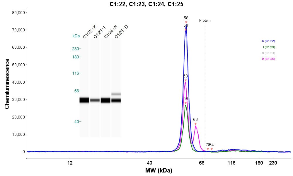
Simple Western: ALDH1A2 Antibody [NBP1-87158]
Simple Western: ALDH1A2 Antibody [NBP1-87158] - Detection of ALDH1A2 in K562 cells using ALDH1A2 antibody at a dilution of 1:50. K (lane 1) = Wild Type K562, I (lane 2) = Imatinib Resistant K562, N (lane 3) = Nilotinib Resistant K562 and D (lane 4) = Dasatinib Resistant K562. Image submitted by a verified customer review.
Immunohistochemistry: ALDH1A2 Antibody [NBP1-87158] -
Immunohistochemistry of candidate biomarkers in prostate cancer. Representative immunohistochemical staining of ACPP, ADAM9, ALDH1A2, CASR, CCND1, CCPG1, CD34, CD44, CD44v6, CHGA, CHMP1A, EI24, ENO2, GADD45B, HA, HAS2, HES6, HMMR, HOXC6, HYAL1, IGF1, IQCK, MAP4K4, MKI67, PAGE4, PLIN2, PTEN, SIAH2, SMAD4, SOX9, SPP1, SYP, & TP53 from prostate cancer tissue microarrays. Scale bar represents 50 μm. Image collected & cropped by CiteAb from the following publication (https://bmccancer.biomedcentral.com/articles/10.1186/1471-2407-14-244), licensed under a CC-BY license. Not internally tested by Novus Biologicals.Applications for ALDH1A2 Antibody - BSA Free
Immunohistochemistry
Immunohistochemistry-Paraffin
Simple Western
Western Blot
See Simple Western Antibody Database for Simple Western validation: Tested in Wild Type K562 cells, Imatinib Resistant K562 cells, Nilotinib Resistant K562 cells and Dasatinib Resistant K56 cells, separated by Size, antibody dilution of 1:50
Reviewed Applications
Read 1 review rated 5 using NBP1-87158 in the following applications:
Formulation, Preparation, and Storage
Purification
Formulation
Format
Preservative
Concentration
Shipping
Stability & Storage
Background: ALDH1A2
Long Name
Alternate Names
Entrez Gene IDs
Gene Symbol
UniProt
Additional ALDH1A2 Products
Product Documents for ALDH1A2 Antibody - BSA Free
Certificate of Analysis
To download a Certificate of Analysis, please enter a lot or batch number in the search box below.
Product Specific Notices for ALDH1A2 Antibody - BSA Free
This product is for research use only and is not approved for use in humans or in clinical diagnosis. Primary Antibodies are guaranteed for 1 year from date of receipt.
Related Research Areas
Citations for ALDH1A2 Antibody - BSA Free
 Powered by Bioz
Customer Reviews for ALDH1A2 Antibody - BSA Free (1)
Have you used ALDH1A2 Antibody - BSA Free?
Submit a review and receive an Amazon gift card!
$25/€18/£15/$25CAN/¥2500 Yen for a review with an image
$10/€7/£6/$10CAN/¥1110 Yen for a review without an image
Submit a review
Customer Images
-
Application: Simple WesternSample Tested: k562 cellsSpecies: HumanVerified Customer | Posted 08/18/2016Simple Western analysis of untreated or treated K562 cells using ALDH1A2 antibody

There are no reviews that match your criteria.
Protocols
Find general support by application which include: protocols, troubleshooting, illustrated assays, videos and webinars.
- Antigen Retrieval Protocol (PIER)
- Antigen Retrieval for Frozen Sections Protocol
- Appropriate Fixation of IHC/ICC Samples
- Cellular Response to Hypoxia Protocols
- Chromogenic IHC Staining of Formalin-Fixed Paraffin-Embedded (FFPE) Tissue Protocol
- Chromogenic Immunohistochemistry Staining of Frozen Tissue
- ClariTSA™ Fluorophore Kits
- Detection & Visualization of Antibody Binding
- Fluorescent IHC Staining of Frozen Tissue Protocol
- Graphic Protocol for Heat-induced Epitope Retrieval
- Graphic Protocol for the Preparation and Fluorescent IHC Staining of Frozen Tissue Sections
- Graphic Protocol for the Preparation and Fluorescent IHC Staining of Paraffin-embedded Tissue Sections
- Graphic Protocol for the Preparation of Gelatin-coated Slides for Histological Tissue Sections
- IHC Sample Preparation (Frozen sections vs Paraffin)
- Immunofluorescent IHC Staining of Formalin-Fixed Paraffin-Embedded (FFPE) Tissue Protocol
- Immunohistochemistry (IHC) and Immunocytochemistry (ICC) Protocols
- Immunohistochemistry Frozen Troubleshooting
- Immunohistochemistry Paraffin Troubleshooting
- Preparing Samples for IHC/ICC Experiments
- Preventing Non-Specific Staining (Non-Specific Binding)
- Primary Antibody Selection & Optimization
- Protocol for Heat-Induced Epitope Retrieval (HIER)
- Protocol for Making a 4% Formaldehyde Solution in PBS
- Protocol for VisUCyte™ HRP Polymer Detection Reagent
- Protocol for the Preparation & Fixation of Cells on Coverslips
- Protocol for the Preparation and Chromogenic IHC Staining of Frozen Tissue Sections
- Protocol for the Preparation and Chromogenic IHC Staining of Frozen Tissue Sections - Graphic
- Protocol for the Preparation and Chromogenic IHC Staining of Paraffin-embedded Tissue Sections
- Protocol for the Preparation and Chromogenic IHC Staining of Paraffin-embedded Tissue Sections - Graphic
- Protocol for the Preparation and Fluorescent IHC Staining of Frozen Tissue Sections
- Protocol for the Preparation and Fluorescent IHC Staining of Paraffin-embedded Tissue Sections
- Protocol for the Preparation of Gelatin-coated Slides for Histological Tissue Sections
- R&D Systems Quality Control Western Blot Protocol
- TUNEL and Active Caspase-3 Detection by IHC/ICC Protocol
- The Importance of IHC/ICC Controls
- Troubleshooting Guide: Immunohistochemistry
- Troubleshooting Guide: Western Blot Figures
- Western Blot Conditions
- Western Blot Protocol
- Western Blot Protocol for Cell Lysates
- Western Blot Troubleshooting
- Western Blot Troubleshooting Guide
- View all Protocols, Troubleshooting, Illustrated assays and Webinars
FAQs for ALDH1A2 Antibody - BSA Free
-
Q: I am looking for an anti-human ALDH1A2 antibody to use for flow cytometry and I’m wondering if anyone has used NBP1-87158 for this purpose?
A: Unfortunately we have not yet investigated the use of product NBP1-87158 in flow cytometry yet. This antibody is validated for use in IHC and ICC, so it is very likely that it would work in flow as well. Since we are not able to cover this application with our guarantee, I can offer you our Innovator's Reward Program.