![Immunohistochemistry: alpha-Synuclein Antibody [NBP2-25146]](https://resources.rndsystems.com/images/products/alpha-Synuclein-Antibody-Immunofluorescence-NBP2-25146-img0010.jpg "Immunohistochemistry: alpha-Synuclein Antibody [NBP2-25146]")
Loading...
Key Product Details
Species Reactivity
Human, Mouse, Rat, Porcine, Bovine, Equine
Applications
Immunohistochemistry, Immunohistochemistry Free-Floating, Western Blot, Immunocytochemistry/ Immunofluorescence
Label
Unconjugated
Antibody Source
Polyclonal Chicken IgY
Format
BSA Free
Loading...
Product Specifications
Immunogen
This alpha-Synuclein Antibody was developed against Full length recombinant human alpha-Synuclein with the epitope from amino acids 61-95.
Localization
Cytoplasm. Membrane. Nucleus. Cell junction > synapse. Note: Membrane-bound in dopaminergic neurons.
Clonality
Polyclonal
Host
Chicken
Isotype
IgY
Theoretical MW
15 kDa.
Disclaimer note: The observed molecular weight of the protein may vary from the listed predicted molecular weight due to post translational modifications, post translation cleavages, relative charges, and other experimental factors.
Disclaimer note: The observed molecular weight of the protein may vary from the listed predicted molecular weight due to post translational modifications, post translation cleavages, relative charges, and other experimental factors.
Scientific Data Images for alpha-Synuclein Antibody - BSA Free
Immunohistochemistry: alpha-Synuclein Antibody [NBP2-25146]
Immunohistochemistry: alpha-Synuclein Antibody [NBP2-25146] - Immunofluorescent analysis of a section of rat cerebellum stained with chicken pAb to alpha-synuclein, NBP2-25146, dilution 1:3,000 in red, and costained with rabbit pAb to GFAP, dilution 1:5,000 in green. The blue is DAPI staining of nuclear DNA. Following transcardial perfusion of rat with 4% paraformaldehyde, brain was post fixed for 24 hours, cut to 45uM, and free-floating sections were stained with above antibodies. The alpha-synuclein protein is concentrated in presynaptic regions in the granular and molecular layers, while GFAP antibody stains the network of Bergmann and astroglial cells.![Western Blot: alpha-Synuclein Antibody [NBP2-25146]](https://resources.rndsystems.com/images/products/alpha-Synuclein-Antibody-Western-Blot-NBP2-25146-img0009.jpg "Western Blot: alpha-Synuclein Antibody [NBP2-25146]")
Western Blot: alpha-Synuclein Antibody [NBP2-25146]
Western Blot: alpha-Synuclein Antibody [NBP2-25146] - Western blot analysis of different tissue lysates using chicken pAb to alpha-synuclein, NBP2-25146, dilution 1:2,000 in green: [1] protein standard (red), [2] rat brain, [3] rat spinal cord, [4] mouse brain, [5] mouse spinal cord. The strong band at about 15kDa corresponds to the alpha-synuclein protein in brain extracts, which are rich in synapses, while a weaker band is seen in spinal cord extracts where synapses are a more minor component.![Immunocytochemistry/ Immunofluorescence: alpha-Synuclein Antibody [NBP2-25146]](https://resources.rndsystems.com/images/products/Synuclein-alpha-Antibody-Immunocytochemistry-Immunofluorescence-NBP2-25146-img0008.jpg "Immunocytochemistry/ Immunofluorescence: alpha-Synuclein Antibody [NBP2-25146]")
Immunocytochemistry/ Immunofluorescence: alpha-Synuclein Antibody [NBP2-25146]
Immunocytochemistry/Immunofluorescence: Synuclein-alpha Antibody [NBP2-25146] - Mixed rat neuron-glial cultures stained with NBP2-25146, polyclonal antibody to alpha-Synuclein (red) and monoclonal antibody to MAP2 (NBP2-25156, green). The alpha-Synuclein antibody stains vesicular structures, the perikarya and processes of the neurons in this image. Note that some of the neuronal perikarya contain much more alpha-Synuclein than others. The blue channel shows the localization of DNA.![Immunocytochemistry/ Immunofluorescence: alpha-Synuclein Antibody [NBP2-25146]](https://resources.rndsystems.com/images/products/Synuclein-alpha-Antibody-Immunocytochemistry-Immunofluorescence-NBP2-25146-img0006.jpg "Immunocytochemistry/ Immunofluorescence: alpha-Synuclein Antibody [NBP2-25146]")
Immunocytochemistry/ Immunofluorescence: alpha-Synuclein Antibody [NBP2-25146]
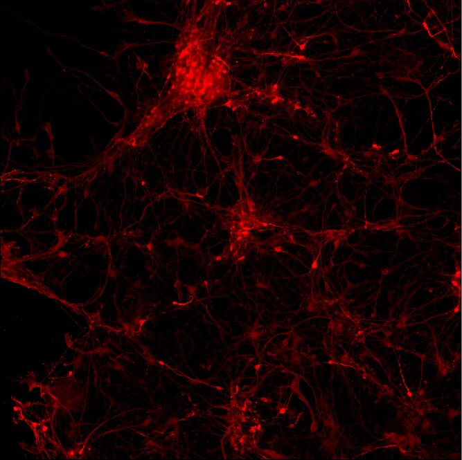
Immunocytochemistry/Immunofluorescence: alpha-Synuclein Antibody [NBP2-25146] - Human iPS derived neurons, fixed in 4% formaldehyde solution. NBP2-25146 diluted 1:1500, followed by staining with Alexa anti chicken-594 (1:1000) secondary antibody. All antibodies were diluted in PBS/BSA 3% (w/v) ) Triton X-100 0,3 % (v/v). Images taken by an Arrayscan (Cellomics). Image from verified customer review.Applications for alpha-Synuclein Antibody - BSA Free
Application
Recommended Usage
Immunocytochemistry/ Immunofluorescence
1:1000
Immunohistochemistry
1:1000
Immunohistochemistry Free-Floating
1:1000
Western Blot
1:2000
Application Notes
This alpha-Synuclein antibody is useful for Immunocytochemistry/Immunofluorescence and Western Blot, where a band can be seen at ~15 kDa.
Reviewed Applications
Read 1 review rated 4 using NBP2-25146 in the following applications:
Formulation, Preparation, and Storage
Purification
IgY purified
Formulation
Supplied as a concentrated total IgY preparation from egg yolk, dialyzed against PBS with added preservative.
Format
BSA Free
Preservative
0.035% Sodium Azide
Concentration
1 mg/ml
Shipping
The product is shipped with polar packs. Upon receipt, store it immediately at the temperature recommended below.
Stability & Storage
Store at 4C short term. Aliquot and store at -20C long term. Avoid freeze-thaw cycles.
Background: alpha-Synuclein
A number of studies have revealed that alpha-synuclein aggregation is a hallmark feature in a number of neurodegenerative diseases, referred to as synucleinopathies (2-4). Alpha-synuclein protein aggregates are a large component of Lewy bodies that are present in Parkinson's disease (PD), Lewy body dementia (LBD), and multiple system atrophy (1-6). Research has shown phosphorylation of alpha-synuclein at Ser129 moves the protein from the nucleus to the cytoplasm and promotes fibril formation associated with synucleinopathies (1,2,5). Recent studies also suggest that alpha-synuclein accumulation can prevent mitochondrial import machinery causing mitochondrial dysfunction that is often observed in neurodegeneration (5). It is thought that preventing alpha-synuclein aggregation may prevent PD, thus alpha-synuclein is a target for many potential therapeutic interventions aimed at decreasing aggregate formation or increasing clearance (1,2,4-6).
References
1. Villar-Pique, A., Lopes da Fonseca, T., & Outeiro, T. F. (2016). Structure, function and toxicity of alpha-synuclein: the Bermuda triangle in synucleinopathies. Journal of neurochemistry. https://doi.org/10.1111/jnc.13249
2. Emamzadeh F. N. (2016). Alpha-synuclein structure, functions, and interactions. Journal of research in medical sciences : the official journal of Isfahan University of Medical Sciences. https://doi.org/10.4103/1735-1995.181989
3. Burre J. (2015). The Synaptic Function of alpha-Synuclein. Journal of Parkinson's disease. https://doi.org/10.3233/JPD-150642
4. Lashuel, H. A., Overk, C. R., Oueslati, A., & Masliah, E. (2013). The many faces of alpha-synuclein: from structure and toxicity to therapeutic target. Nature reviews. Neuroscience. https://doi.org/10.1038/nrn3406
5. Rocha, E. M., De Miranda, B., & Sanders, L. H. (2018). Alpha-synuclein: Pathology, mitochondrial dysfunction and neuroinflammation in Parkinson's disease. Neurobiology of disease. https://doi.org/10.1016/j.nbd.2017.04.004
6. O'Leary, E. I., & Lee, J. C. (2019). Interplay between alpha-synuclein amyloid formation and membrane structure. Biochimica et biophysica acta. Proteins and proteomics. https://doi.org/10.1016/j.bbapap.2018.09.012
Alternate Names
NACP, PARK1, PARK4, SNCA, Synuclein-alpha
Gene Symbol
SNCA
OMIM
163890 (Human)
UniProt
Additional alpha-Synuclein Products
Product Documents for alpha-Synuclein Antibody - BSA Free
Certificate of Analysis
To download a Certificate of Analysis, please enter a lot or batch number in the search box below.
Product Specific Notices for alpha-Synuclein Antibody - BSA Free
Chicken products cannot be exported to Canada.
This product is for research use only and is not approved for use in humans or in clinical diagnosis. Primary Antibodies are guaranteed for 1 year from date of receipt.
Related Research Areas
Customer Reviews for alpha-Synuclein Antibody - BSA Free (1)
4 out of 5
1 Customer Rating
Have you used alpha-Synuclein Antibody - BSA Free?
Submit a review and receive an Amazon gift card!
$25/€18/£15/$25CAN/¥2500 Yen for a review with an image
$10/€7/£6/$10CAN/¥1110 Yen for a review without an image
Submit a review
Customer Images
Showing
1
-
1 of
1 review
Showing All
Filter By:
-
Application: ImmunocytochemistrySample Tested:Species: HumanVerified Customer | Posted 12/23/2014human iPS derived neurons, fixed in 4% formaldehyde solution

There are no reviews that match your criteria.
Protocols
Find general support by application which include: protocols, troubleshooting, illustrated assays, videos and webinars.
- Antigen Retrieval Protocol (PIER)
- Antigen Retrieval for Frozen Sections Protocol
- Appropriate Fixation of IHC/ICC Samples
- Cellular Response to Hypoxia Protocols
- Chromogenic IHC Staining of Formalin-Fixed Paraffin-Embedded (FFPE) Tissue Protocol
- Chromogenic Immunohistochemistry Staining of Frozen Tissue
- ClariTSA™ Fluorophore Kits
- Detection & Visualization of Antibody Binding
- Fluorescent IHC Staining of Frozen Tissue Protocol
- Graphic Protocol for Heat-induced Epitope Retrieval
- Graphic Protocol for the Preparation and Fluorescent IHC Staining of Frozen Tissue Sections
- Graphic Protocol for the Preparation and Fluorescent IHC Staining of Paraffin-embedded Tissue Sections
- Graphic Protocol for the Preparation of Gelatin-coated Slides for Histological Tissue Sections
- ICC Cell Smear Protocol for Suspension Cells
- ICC Immunocytochemistry Protocol Videos
- ICC for Adherent Cells
- IHC Sample Preparation (Frozen sections vs Paraffin)
- Immunocytochemistry (ICC) Protocol
- Immunocytochemistry Troubleshooting
- Immunofluorescence of Organoids Embedded in Cultrex Basement Membrane Extract
- Immunofluorescent IHC Staining of Formalin-Fixed Paraffin-Embedded (FFPE) Tissue Protocol
- Immunohistochemistry (IHC) and Immunocytochemistry (ICC) Protocols
- Immunohistochemistry Frozen Troubleshooting
- Immunohistochemistry Paraffin Troubleshooting
- Preparing Samples for IHC/ICC Experiments
- Preventing Non-Specific Staining (Non-Specific Binding)
- Primary Antibody Selection & Optimization
- Protocol for Heat-Induced Epitope Retrieval (HIER)
- Protocol for Making a 4% Formaldehyde Solution in PBS
- Protocol for VisUCyte™ HRP Polymer Detection Reagent
- Protocol for the Fluorescent ICC Staining of Cell Smears - Graphic
- Protocol for the Fluorescent ICC Staining of Cultured Cells on Coverslips - Graphic
- Protocol for the Preparation & Fixation of Cells on Coverslips
- Protocol for the Preparation and Chromogenic IHC Staining of Frozen Tissue Sections
- Protocol for the Preparation and Chromogenic IHC Staining of Frozen Tissue Sections - Graphic
- Protocol for the Preparation and Chromogenic IHC Staining of Paraffin-embedded Tissue Sections
- Protocol for the Preparation and Chromogenic IHC Staining of Paraffin-embedded Tissue Sections - Graphic
- Protocol for the Preparation and Fluorescent ICC Staining of Cells on Coverslips
- Protocol for the Preparation and Fluorescent ICC Staining of Non-adherent Cells
- Protocol for the Preparation and Fluorescent ICC Staining of Stem Cells on Coverslips
- Protocol for the Preparation and Fluorescent IHC Staining of Frozen Tissue Sections
- Protocol for the Preparation and Fluorescent IHC Staining of Paraffin-embedded Tissue Sections
- Protocol for the Preparation of Gelatin-coated Slides for Histological Tissue Sections
- Protocol for the Preparation of a Cell Smear for Non-adherent Cell ICC - Graphic
- R&D Systems Quality Control Western Blot Protocol
- TUNEL and Active Caspase-3 Detection by IHC/ICC Protocol
- The Importance of IHC/ICC Controls
- Troubleshooting Guide: Immunohistochemistry
- Troubleshooting Guide: Western Blot Figures
- Western Blot Conditions
- Western Blot Protocol
- Western Blot Protocol for Cell Lysates
- Western Blot Troubleshooting
- Western Blot Troubleshooting Guide
- View all Protocols, Troubleshooting, Illustrated assays and Webinars
FAQs for alpha-Synuclein Antibody - BSA Free
Showing
1
-
1 of
1 FAQ
Showing All
-
Q: I'm looking for an alpha-Synuclein antibody with an epitope located in the first half (N-terminus) of the protein - preferably a monoclonal antibody. Can you help me with that?
A:
Please take a look at NB110-57475. It has been validated for human, rat and mouse and the applications ICC and WB and the epitope it detects is in the N terminal.
Loading...