alpha Tubulin Antibody (TU-01) - BSA Free
Novus Biologicals | Catalog # NB500-333
![Western Blot: alpha Tubulin Antibody (TU-01)BSA Free [NB500-333]](https://resources.rndsystems.com/images/products/Tubulin-Antibody-TU-01-Western-Blot-NB500-333-img0007.jpg "Western Blot: alpha Tubulin Antibody (TU-01)BSA Free [NB500-333]")
Key Product Details
Species Reactivity
Validated:
Human, Mouse, Porcine, A. thaliana, Eukaryote, Invertebrate, Plant, Turkey, Yeast
Cited:
Plant
Applications
Validated:
Immunohistochemistry, Immunohistochemistry-Paraffin, Western Blot, Flow (Intracellular), Immunocytochemistry/ Immunofluorescence, Immunoprecipitation
Cited:
Western Blot
Label
Unconjugated
Antibody Source
Monoclonal Mouse IgG1 Clone # TU-01
Format
BSA Free
Loading...
Product Specifications
Immunogen
This alpha Tubulin Antibody (TU-01) was developed against a fraction of tubulin purified from pig brain by two cycles of polymerization-depolymerization.
Reactivity Notes
Plant reactivity reported in scientific literature (PMID: 26594217).
Localization
Major constituent of microtubules.
Marker
Microtubule Marker
Specificity
This alpha Tubulin Antibody (TU-01) recognizes the defined epitope (aa 65-97) on N-terminal structureal domain of alpha tubulin.
Clonality
Monoclonal
Host
Mouse
Isotype
IgG1
Theoretical MW
50 kDa.
Disclaimer note: The observed molecular weight of the protein may vary from the listed predicted molecular weight due to post translational modifications, post translation cleavages, relative charges, and other experimental factors.
Disclaimer note: The observed molecular weight of the protein may vary from the listed predicted molecular weight due to post translational modifications, post translation cleavages, relative charges, and other experimental factors.
Scientific Data Images for alpha Tubulin Antibody (TU-01) - BSA Free
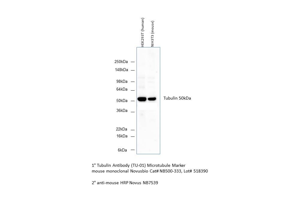
Western Blot: alpha Tubulin Antibody (TU-01)BSA Free [NB500-333]
Western Blot: alpha Tubulin Antibody (TU-01) [NB500-333] - Analysis of Tubulin in HEK293T and NIH/3T3 cells using Tubulin antibody; alpha tubulin molecular weight: 50 kDa. Image from verified customer review.![Immunocytochemistry/ Immunofluorescence: alpha Tubulin Antibody (TU-01) - BSA Free [NB500-333]](https://resources.rndsystems.com/images/products/Tubulin-Antibody-TU-01-Immunocytochemistry-Immunofluorescence-NB500-333-img0001.jpg "Immunocytochemistry/ Immunofluorescence: alpha Tubulin Antibody (TU-01) - BSA Free [NB500-333]")
Immunocytochemistry/ Immunofluorescence: alpha Tubulin Antibody (TU-01) - BSA Free [NB500-333]
Immunocytochemistry/Immunofluorescence: alpha Tubulin Antibody (TU-01) [NB500-333] - Staining of 3T3 mouse embryonal fibroblast cell line using anti-alpha-tubulin (TU-01; green) and anti-Vimentin (VI-01; red). Nucleus is stained with DAPI (blue).![Immunoprecipitation: alpha Tubulin Antibody (TU-01) - BSA Free [NB500-333]](https://resources.rndsystems.com/images/products/Tubulin-Antibody-TU-01-Immunoprecipitation-NB500-333-img0006.jpg "Immunoprecipitation: alpha Tubulin Antibody (TU-01) - BSA Free [NB500-333]")
Immunoprecipitation: alpha Tubulin Antibody (TU-01) - BSA Free [NB500-333]
Immunoprecipitation: alpha Tubulin Antibody (TU-01) [NB500-333] - alpha Tubulin from HeLa and RAJI cell lysate by antibody TU-16 and its detection by antibody TU-01. IgM heavy chain (76-92 kDa) and IgM light chain (25-30 kDa) indicated. Molecular weight of alpha tubulin is around 50 kDa. L = lysate IPr = immunoprecipitate (reducing conditions) IPn = immunoprecipitate (non-reducing conditions)![Western Blot: alpha Tubulin Antibody (TU-01)BSA Free [NB500-333]](https://resources.rndsystems.com/images/products/Tubulin-Antibody-TU-01-Western-Blot-NB500-333-img0008.jpg "Western Blot: alpha Tubulin Antibody (TU-01)BSA Free [NB500-333]")
Western Blot: alpha Tubulin Antibody (TU-01)BSA Free [NB500-333]
Western Blot: alpha Tubulin Antibody (TU-01) [NB500-333] - Analysis of alpha-tubulin (molecular weight of 50 kDa) in porcine brain lysate by antibody TU-01 biotin.![Immunocytochemistry/ Immunofluorescence: alpha Tubulin Antibody (TU-01) - BSA Free [NB500-333]](https://resources.rndsystems.com/images/products/Tubulin-Antibody-TU-01-Immunocytochemistry-Immunofluorescence-NB500-333-img0005.jpg "Immunocytochemistry/ Immunofluorescence: alpha Tubulin Antibody (TU-01) - BSA Free [NB500-333]")
Immunocytochemistry/ Immunofluorescence: alpha Tubulin Antibody (TU-01) - BSA Free [NB500-333]
Immunocytochemistry/Immunofluorescence: alpha Tubulin Antibody (TU-01) [NB500-333] - Tubulin filament in HeLa. Image from verified customer review.![Western Blot: alpha Tubulin Antibody (TU-01)BSA Free [NB500-333]](https://resources.rndsystems.com/images/products/Tubulin-Antibody-TU-01-Western-Blot-NB500-333-img0004.jpg "Western Blot: alpha Tubulin Antibody (TU-01)BSA Free [NB500-333]")
Western Blot: alpha Tubulin Antibody (TU-01)BSA Free [NB500-333]
Western Blot: alpha Tubulin Antibody (TU-01) [NB500-333] - Use of anti-alpha-tubulin antibody TU-01 as a loading control (A) in an Western blotting experiment revealing the staining pattern of lysates of various cell lines by a newly developed monoclonal antibody (B). Alpha tubulin molecular weight: 50 kDa.![Immunocytochemistry/ Immunofluorescence: alpha Tubulin Antibody (TU-01) - BSA Free [NB500-333]](https://resources.rndsystems.com/images/products/Tubulin-Antibody-TU-01-Immunocytochemistry-Immunofluorescence-NB500-333-img0002.jpg "Immunocytochemistry/ Immunofluorescence: alpha Tubulin Antibody (TU-01) - BSA Free [NB500-333]")
Immunocytochemistry/ Immunofluorescence: alpha Tubulin Antibody (TU-01) - BSA Free [NB500-333]
Immunocytochemistry/Immunofluorescence: alpha Tubulin Antibody (TU-01) [NB500-333] - Staining of HeLa human cervix carcinoma cell line using anti-alpha-tubulin (TU-01; red). Nucleus is stained with DAPI (blue). - BSA Free [NB500-333] -")
Western Blot: alpha Tubulin Antibody (TU-01) - BSA Free [NB500-333] -
Exo70 is redistributed into PSD in the hippocampus of mTBI mice. a Schematic representation of subcellular fractionation proteins. b Example of subcellular fractionation. Hippocampus from two-month-old male mice was fractionated and microsome, PSD, and nonPSD fractions were obtained. 20 ug of protein samples were resolved in a 10% SDS-PAGE and transferred to PVDF membranes. Membranes were incubated with the respective antibodies shown in the figure. Membranes were stripped and tested again with the indicated antibodies. c Proteins distribution were analyzed with densitometric analysis by comparing signal intensity from each fraction with homogenized. Mean values +/- SEM are shown. d Cortex and Hippocampus (f) from Sham and mTBI mice were fractionated and analyzed by western blot using Exo70, PDI, Actin, and Tubulin antibodies. PDI/Actin/Tubulin was used as loading controls. 30 ug of protein samples were used. e, g The graph shows the Exo70 densitometric analysis normalized with loading controls. Values represent means +/- SEM, n = 3 mice per experimental group. Statistical differences were determined by an unpaired t-test comparing Sham and mTBI. *p < 0.05, **p < 0.01 Image collected and cropped by CiteAb from the following open publication (https://pubmed.ncbi.nlm.nih.gov/33593425), licensed under a CC-BY license. Not internally tested by Novus Biologicals.Applications for alpha Tubulin Antibody (TU-01) - BSA Free
Application
Recommended Usage
Flow (Intracellular)
1-4 ug/ml
Immunocytochemistry/ Immunofluorescence
1:10-1:500
Immunohistochemistry
1:10-1:500
Immunohistochemistry-Paraffin
5 ug/ml
Immunoprecipitation
1:10-1:500
Western Blot
1-2 ug/ml
Application Notes
Western blotting: Recommended dilution in reducing conditions.
Reviewed Applications
Read 1 review rated 5 using NB500-333 in the following applications:
Flow Cytometry Panel Builder
Bio-Techne Knows Flow Cytometry
Save time and reduce costly mistakes by quickly finding compatible reagents using the Panel Builder Tool.
Advanced Features
- Spectra Viewer - Custom analysis of spectra from multiple fluorochromes
- Spillover Popups - Visualize the spectra of individual fluorochromes
- Antigen Density Selector - Match fluorochrome brightness with antigen density

Formulation, Preparation, and Storage
Purification
Protein A purified
Formulation
Phosphate buffered saline (PBS), pH 7.4
Format
BSA Free
Preservative
15mM Sodium Azide
Concentration
1.0 mg/ml
Shipping
The product is shipped with polar packs. Upon receipt, store it immediately at the temperature recommended below.
Stability & Storage
Store at 4C. Do not freeze.
Background: alpha Tubulin
Tyrosine ligase adds a C-terminal tyrosine to monomeric alpha tubulin. Assembled microtubules can again be detyrosinated by a cytoskeleton associated carboxypeptidase. Detyrosinated alpha tubulin is referred to as Glu-tubulin. Another post-translational modification of detyrosinated alpha tubulin is C-terminal polyglutamylation which is characteristic for microtubules in neuronal cells and the mitotic spindle.
Like GAPDH and beta-actin, alpha/beta tubulin is often used as a loading control in immunoblot applications (1). Alpha/beta tubulin is also good for counterstaining microtubules in immunofluorescence (2).
References
1. Hannen, R., Selmansberger, M., Hauswald, M., Pagenstecher, A., Nist, A., Stiewe, T.,... Bartsch, J. W. (2019). Comparative Transcriptomic Analysis of Temozolomide Resistant Primary GBM Stem-Like Cells and Recurrent GBM Identifies Up-Regulation of the Carbonic Anhydrase CA2 Gene as Resistance Factor. Cancers (Basel), 11(7). doi:10.3390/cancers11070921
2. Nel, M., Joubert, A. M., Dohle, W., Potter, B. V., & Theron, A. E. (2018). Modes of cell death induced by tetrahydroisoquinoline-based analogs in MDA-MB-231 breast and A549 lung cancer cell lines. Drug Des Devel Ther, 12, 1881-1904. doi:10.2147/dddt.S152718
Long Name
Tubulin Alpha 1a
Alternate Names
Alpha-Tubulin 3, B-ALPHA-1, LIS3, TUBA1A, TUBA3, Tubulin B-Alpha-1
Entrez Gene IDs
20766 (Mouse)
Gene Symbol
TUBA1A
UniProt
Additional alpha Tubulin Products
Product Documents for alpha Tubulin Antibody (TU-01) - BSA Free
Certificate of Analysis
To download a Certificate of Analysis, please enter a lot or batch number in the search box below.
Product Specific Notices for alpha Tubulin Antibody (TU-01) - BSA Free
This product is for research use only and is not approved for use in humans or in clinical diagnosis. Primary Antibodies are guaranteed for 1 year from date of receipt.
Related Research Areas
Citations for alpha Tubulin Antibody (TU-01) - BSA Free
 Powered by Bioz
Customer Reviews for alpha Tubulin Antibody (TU-01) - BSA Free (1)
5 out of 5
1 Customer Rating
Have you used alpha Tubulin Antibody (TU-01) - BSA Free?
Submit a review and receive an Amazon gift card!
$25/€18/£15/$25CAN/¥2500 Yen for a review with an image
$10/€7/£6/$10CAN/¥1110 Yen for a review without an image
Submit a review
Customer Images
Showing
1
-
1 of
1 review
Showing All
Filter By:
-
Application: Western BlotSample Tested: HEK293T and NIH3T3 whole cell lysateSpecies: HumanVerified Customer | Posted 03/15/2016Tubulin Antibody (NB500-333)

There are no reviews that match your criteria.
Protocols
Find general support by application which include: protocols, troubleshooting, illustrated assays, videos and webinars.
- 7-Amino Actinomycin D (7-AAD) Cell Viability Flow Cytometry Protocol
- Antigen Retrieval Protocol (PIER)
- Antigen Retrieval for Frozen Sections Protocol
- Appropriate Fixation of IHC/ICC Samples
- Cellular Response to Hypoxia Protocols
- Chromogenic IHC Staining of Formalin-Fixed Paraffin-Embedded (FFPE) Tissue Protocol
- Chromogenic Immunohistochemistry Staining of Frozen Tissue
- ClariTSA™ Fluorophore Kits
- Detection & Visualization of Antibody Binding
- Extracellular Membrane Flow Cytometry Protocol
- Flow Cytometry Protocol for Cell Surface Markers
- Flow Cytometry Protocol for Staining Membrane Associated Proteins
- Flow Cytometry Staining Protocols
- Flow Cytometry Troubleshooting Guide
- Fluorescent IHC Staining of Frozen Tissue Protocol
- Graphic Protocol for Heat-induced Epitope Retrieval
- Graphic Protocol for the Preparation and Fluorescent IHC Staining of Frozen Tissue Sections
- Graphic Protocol for the Preparation and Fluorescent IHC Staining of Paraffin-embedded Tissue Sections
- Graphic Protocol for the Preparation of Gelatin-coated Slides for Histological Tissue Sections
- ICC Cell Smear Protocol for Suspension Cells
- ICC Immunocytochemistry Protocol Videos
- ICC for Adherent Cells
- IHC Sample Preparation (Frozen sections vs Paraffin)
- Immunocytochemistry (ICC) Protocol
- Immunocytochemistry Troubleshooting
- Immunofluorescence of Organoids Embedded in Cultrex Basement Membrane Extract
- Immunofluorescent IHC Staining of Formalin-Fixed Paraffin-Embedded (FFPE) Tissue Protocol
- Immunohistochemistry (IHC) and Immunocytochemistry (ICC) Protocols
- Immunohistochemistry Frozen Troubleshooting
- Immunohistochemistry Paraffin Troubleshooting
- Immunoprecipitation Protocol
- Intracellular Flow Cytometry Protocol Using Alcohol (Methanol)
- Intracellular Flow Cytometry Protocol Using Detergents
- Intracellular Nuclear Staining Flow Cytometry Protocol Using Detergents
- Intracellular Staining Flow Cytometry Protocol Using Alcohol Permeabilization
- Intracellular Staining Flow Cytometry Protocol Using Detergents to Permeabilize Cells
- Preparing Samples for IHC/ICC Experiments
- Preventing Non-Specific Staining (Non-Specific Binding)
- Primary Antibody Selection & Optimization
- Propidium Iodide Cell Viability Flow Cytometry Protocol
- Protocol for Heat-Induced Epitope Retrieval (HIER)
- Protocol for Liperfluo
- Protocol for Making a 4% Formaldehyde Solution in PBS
- Protocol for VisUCyte™ HRP Polymer Detection Reagent
- Protocol for the Characterization of Human Th22 Cells
- Protocol for the Characterization of Human Th9 Cells
- Protocol for the Fluorescent ICC Staining of Cell Smears - Graphic
- Protocol for the Fluorescent ICC Staining of Cultured Cells on Coverslips - Graphic
- Protocol for the Preparation & Fixation of Cells on Coverslips
- Protocol for the Preparation and Chromogenic IHC Staining of Frozen Tissue Sections
- Protocol for the Preparation and Chromogenic IHC Staining of Frozen Tissue Sections - Graphic
- Protocol for the Preparation and Chromogenic IHC Staining of Paraffin-embedded Tissue Sections
- Protocol for the Preparation and Chromogenic IHC Staining of Paraffin-embedded Tissue Sections - Graphic
- Protocol for the Preparation and Fluorescent ICC Staining of Cells on Coverslips
- Protocol for the Preparation and Fluorescent ICC Staining of Non-adherent Cells
- Protocol for the Preparation and Fluorescent ICC Staining of Stem Cells on Coverslips
- Protocol for the Preparation and Fluorescent IHC Staining of Frozen Tissue Sections
- Protocol for the Preparation and Fluorescent IHC Staining of Paraffin-embedded Tissue Sections
- Protocol for the Preparation of Gelatin-coated Slides for Histological Tissue Sections
- Protocol for the Preparation of a Cell Smear for Non-adherent Cell ICC - Graphic
- Protocol: Annexin V and PI Staining by Flow Cytometry
- Protocol: Annexin V and PI Staining for Apoptosis by Flow Cytometry
- R&D Systems Quality Control Western Blot Protocol
- TUNEL and Active Caspase-3 Detection by IHC/ICC Protocol
- The Importance of IHC/ICC Controls
- Troubleshooting Guide: Fluorokine Flow Cytometry Kits
- Troubleshooting Guide: Immunohistochemistry
- Troubleshooting Guide: Western Blot Figures
- Western Blot Conditions
- Western Blot Protocol
- Western Blot Protocol for Cell Lysates
- Western Blot Troubleshooting
- Western Blot Troubleshooting Guide
- View all Protocols, Troubleshooting, Illustrated assays and Webinars
FAQs for alpha Tubulin Antibody (TU-01) - BSA Free
Showing
1
-
2 of
2 FAQs
Showing All
-
Q: We would like to stain cilia with an acetylated alpha tubulin antibody in our cells, but I am unsure if this antibody will be able to conclusively differentiate cilia from other structures such as spindle pole bodies. Does anyone know what acetylated alpha tubulin antibodies might bind to apart from cilia?
A: Acetylated alpha tubulin is found in relatively stable microtubules. It is best practice to use this marker together with a centrosome/centriole marker, which will stain the basal bodies at the base of the cilium. After that, it is relatively straightforward to identify the acetylated alpha tubulin signal that corresponds to the cilium.
-
Q: Will this alpha tubulin antibody recognize both isoforms of alpha tubulin?
A: The epitope for this alpha tubulin antibody lies on the C-terminus of the protein and the difference between the two major isoforms is within the first 35 aa of the N-terminus so this alpha tubulin antibody will recognize both isoforms.
-
Q: We would like to stain cilia with an acetylated alpha tubulin antibody in our cells, but I am unsure if this antibody will be able to conclusively differentiate cilia from other structures such as spindle pole bodies. Does anyone know what acetylated alpha tubulin antibodies might bind to apart from cilia?
A: Acetylated alpha tubulin is found in relatively stable microtubules. It is best practice to use this marker together with a centrosome/centriole marker, which will stain the basal bodies at the base of the cilium. After that, it is relatively straightforward to identify the acetylated alpha tubulin signal that corresponds to the cilium.
-
Q: Will this alpha tubulin antibody recognize both isoforms of alpha tubulin?
A: The epitope for this alpha tubulin antibody lies on the C-terminus of the protein and the difference between the two major isoforms is within the first 35 aa of the N-terminus so this alpha tubulin antibody will recognize both isoforms.
Loading...