AlphaB Crystallin/CRYAB Antibody (OTI6A9)
Novus Biologicals | Catalog # NBP1-47708
Key Product Details
Species Reactivity
Human, Mouse, Rat
Applications
Validated:
Immunohistochemistry, Immunohistochemistry-Paraffin, Western Blot, Flow Cytometry, Immunocytochemistry/ Immunofluorescence, Immunoprecipitation
Cited:
Immunocytochemistry/ Immunofluorescence
Label
Unconjugated
Antibody Source
Monoclonal Mouse IgG1 Clone # OTI6A9
Loading...
Product Specifications
Immunogen
Full length human recombinant protein of human CRYAB (NP_001876) produced in HEK293T cell.
Reactivity Notes
Please note that this antibody is reactive to Mouse and derived from the same host, Mouse. Mouse-On-Mouse blocking reagent may be needed for IHC and ICC experiments to reduce high background signal. You can find these reagents under catalog numbers PK-2200-NB and MP-2400-NB. Please contact Technical Support if you have any questions.
Specificity
This antibody is specific for Homo sapiens crystallin, alpha B (CRYAB).
Clonality
Monoclonal
Host
Mouse
Isotype
IgG1
Theoretical MW
20 kDa.
Disclaimer note: The observed molecular weight of the protein may vary from the listed predicted molecular weight due to post translational modifications, post translation cleavages, relative charges, and other experimental factors.
Disclaimer note: The observed molecular weight of the protein may vary from the listed predicted molecular weight due to post translational modifications, post translation cleavages, relative charges, and other experimental factors.
Scientific Data Images for AlphaB Crystallin/CRYAB Antibody (OTI6A9)
![Western Blot: AlphaB Crystallin/CRYAB Antibody (OTI6A9) [NBP1-47708]](https://resources.rndsystems.com/images/products/AlphaB-Crystallin-CRYAB-Antibody-OTI6A9-Western-Blot-NBP1-47708-img0008.jpg "Western Blot: AlphaB Crystallin/CRYAB Antibody (OTI6A9) [NBP1-47708]")
Western Blot: AlphaB Crystallin/CRYAB Antibody (OTI6A9) [NBP1-47708]
Western Blot: AlphaB Crystallin/CRYAB Antibody (OTI6A9) [NBP1-47708] - Analysis of extracts (35ug) from 9 different cell lines by usin g anti-Crystallin AB monoclonal antibody (HepG2: human; HeLa: human; SVT2: mouse; A549: human; COS7: monkey; Jurkat: human; MDCK: canine; PC12: rat; MCF7: human).![Immunocytochemistry/ Immunofluorescence: AlphaB Crystallin/CRYAB Antibody (OTI6A9) [NBP1-47708]](https://resources.rndsystems.com/images/products/AlphaB-Crystallin-CRYAB-Antibody-OTI6A9-Immunocytochemistry-Immunofluorescence-NBP1-47708-img0011.jpg "Immunocytochemistry/ Immunofluorescence: AlphaB Crystallin/CRYAB Antibody (OTI6A9) [NBP1-47708]")
Immunocytochemistry/ Immunofluorescence: AlphaB Crystallin/CRYAB Antibody (OTI6A9) [NBP1-47708]
Immunocytochemistry/Immunofluorescence: AlphaB Crystallin/CRYAB Antibody (OTI6A9) [NBP1-47708] - Staining of COS7 cells transiently transfected by pCMV6-ENTRY Crystallin AB.![Immunohistochemistry-Paraffin: AlphaB Crystallin/CRYAB Antibody (OTI6A9) [NBP1-47708]](https://resources.rndsystems.com/images/products/AlphaB-Crystallin-CRYAB-Antibody-OTI6A9-Immunohistochemistry-Paraffin-NBP1-47708-img0013.jpg "Immunohistochemistry-Paraffin: AlphaB Crystallin/CRYAB Antibody (OTI6A9) [NBP1-47708]")
Immunohistochemistry-Paraffin: AlphaB Crystallin/CRYAB Antibody (OTI6A9) [NBP1-47708]
Immunohistochemistry-Paraffin: AlphaB Crystallin/CRYAB Antibody (OTI6A9) [NBP1-47708] - Staining of paraffin-embedded Human pancreas tissue using anti-Crystallin AB mouse monoclonal antibody.![Flow Cytometry: AlphaB Crystallin/CRYAB Antibody (OTI6A9) [NBP1-47708]](https://resources.rndsystems.com/images/products/AlphaB-Crystallin-CRYAB-Antibody-OTI6A9-Flow-Cytometry-NBP1-47708-img0009.jpg "Flow Cytometry: AlphaB Crystallin/CRYAB Antibody (OTI6A9) [NBP1-47708]")
Flow Cytometry: AlphaB Crystallin/CRYAB Antibody (OTI6A9) [NBP1-47708]
Flow Cytometry: AlphaB Crystallin/CRYAB Antibody (OTI6A9) [NBP1-47708] - HEK293T cells transfected with either overexpression plasmid (Red) or empty vector control plasmid (Blue) were immunostained by anti-Crystallin AB antibody, and then analyzed by flow cytometry.![Immunocytochemistry/ Immunofluorescence: AlphaB Crystallin/CRYAB Antibody (OTI6A9) [NBP1-47708]](https://resources.rndsystems.com/images/products/AlphaB-Crystallin-CRYAB-Antibody-OTI6A9-Immunocytochemistry-Immunofluorescence-NBP1-47708-img0010.jpg "Immunocytochemistry/ Immunofluorescence: AlphaB Crystallin/CRYAB Antibody (OTI6A9) [NBP1-47708]")
Immunocytochemistry/ Immunofluorescence: AlphaB Crystallin/CRYAB Antibody (OTI6A9) [NBP1-47708]
Immunocytochemistry/Immunofluorescence: AlphaB Crystallin/CRYAB Antibody (OTI6A9) [NBP1-47708] - Staining of HT29 cells using anti-Crystallin AB mouse monoclonal antibody.![Immunohistochemistry-Paraffin: AlphaB Crystallin/CRYAB Antibody (OTI6A9) [NBP1-47708]](https://resources.rndsystems.com/images/products/AlphaB-Crystallin-CRYAB-Antibody-OTI6A9-Immunohistochemistry-Paraffin-NBP1-47708-img0012.jpg "Immunohistochemistry-Paraffin: AlphaB Crystallin/CRYAB Antibody (OTI6A9) [NBP1-47708]")
Immunohistochemistry-Paraffin: AlphaB Crystallin/CRYAB Antibody (OTI6A9) [NBP1-47708]
Immunohistochemistry-Paraffin: AlphaB Crystallin/CRYAB Antibody (OTI6A9) [NBP1-47708] - Staining of paraffin-embedded Human endometrium tissue using anti-Crystallin AB mouse monoclonal antibody.![Immunoprecipitation: AlphaB Crystallin/CRYAB Antibody (OTI6A9) [NBP1-47708]](https://resources.rndsystems.com/images/products/AlphaB-Crystallin-CRYAB-Antibody-OTI6A9-Immunoprecipitation-NBP1-47708-img0014.jpg "Immunoprecipitation: AlphaB Crystallin/CRYAB Antibody (OTI6A9) [NBP1-47708]")
Immunoprecipitation: AlphaB Crystallin/CRYAB Antibody (OTI6A9) [NBP1-47708]
Immunoprecipitation: AlphaB Crystallin/CRYAB Antibody (OTI6A9) [NBP1-47708] - (Negative control: IP without adding anti-CRYAB antibody.). For each experiment, 500ul of DDK tagged CRYAB overexpression lysates (at 1:5 dilution with HEK293T lysate), 2ug of anti-CRYAB antibody and 20ul (0.1mg) of goat anti-mouse conjugated magnetic beads were mixed and incubated overnight. After extensive wash to remove any non-specific binding, the immuno-precipitated products were analyzed with rabbit anti-DDK polyclonal antibody. [NBP1-47708] -")
Western Blot: AlphaB Crystallin/CRYAB Antibody (OTI6A9) [NBP1-47708] -
Western Blot: AlphaB Crystallin/CRYAB Antibody (OTI6A9) [NBP1-47708] - HEK293T cells were transfected with the pCMV6-ENTRY control (Left lane) or pCMV6-ENTRY CRYAB (Right lane) cDNA for 48 hrs and lysed. Equivalent amounts of cell lysates (5 ug per lane) were separated by SDS-PAGE and immunoblotted with AlphaB Crystallin/CRYAB Antibody (OTI6A9). [NBP1-47708] -")
Western Blot: AlphaB Crystallin/CRYAB Antibody (OTI6A9) [NBP1-47708] -
PLK2, a novel binding protein of HSPB5, is induced by MG132 in L6 cells. (a) Schematic diagram of domain structure of PLK2. K90, K95, and K111 indicate position of lysine residue (K) biotinylated by HSPB5-BioID2. Lysine residues were numbered based on amino acid sequence of PLK2 derived from Homo sapiens (NCBI NP_006613). (b) Comparison of induction of HSPB5 and PLK2 expression by ER stress-induced drugs. Differentiated L6 cells were treated with 5 uM MG132 or 0.1 ug/mL tunicamycin for 24 h. Induction of HSPB5, and PLK2 protein expression was detected by Western blotting. (c) Graph shows levels of endogenous proteins induced by the drug. Data represent means +/- standard error (n = 3). Statistical analyses were performed using t-test. ** p < 0.01; n.s. means not significant. (d) Verification of binding of PLK2 to HSPB5. FLAG-HSPB5 was overexpressed in L6 cells by liposome transfection. After 5 uM MG132 treatment (24 h), cells were solubilized, and HSPB5 pull-down assay was performed using an anti-tag antibody. (e) Quantification of binding of PLK2 to HSPB5. Control cells were transfected with empty vector. After 5 uM MG132 treatment (24 h), a pull-down assay was performed using an anti-tag antibody. Graph presents level of endogenous PLK2 pulled down by HSPB5. Data represent means +/- standard error (n = 3). Statistical analyses were performed using t-test. * p < 0.05. Image collected and cropped by CiteAb from the following open publication (https://pubmed.ncbi.nlm.nih.gov/36232565), licensed under a CC-BY license. Not internally tested by Novus Biologicals. [NBP1-47708] -")
Western Blot: AlphaB Crystallin/CRYAB Antibody (OTI6A9) [NBP1-47708] -
PLK2 phosphorylates HSPB5 at serine 19 under ER stress. (a) Knockdown efficiency of PLK2 by siRNA. L6 cells were transfected with siRNA to knockdown PLK2 and treated with 5 uM MG132 for 24 h. Graph presents relative value of expression level of PLK2. Data in graphs represent means +/- standard error (n = 3). Statistical analyses were performed using t-test. ** p < 0.01. (b) Schematic representation of three known phosphorylation sites of HSPB5. Arrows indicate name of the kinase catalyzing phosphorylation. (c) Effect of PLK2 on each phosphorylation site. Undifferentiated L6 cells were transfected with siRNA to knockdown PLK2. After differentiation (48 h), L6 myotubes were treated with 5 uM MG132 for 24 h. Phosphorylation level was detected by Western blotting with specific antibodies. Graph presents ratio of phosphorylated HSPB5/HSPB5 after quantification of each band. Data in graphs represent means +/- standard error (n = 3). Statistical analyses were performed using one-way ANOVA with Tukey test. ‡ p < 0.01, † p < 0.05; n.s. not significant. (d) Effect of PLK2 activity on phosphorylation of serine 19. After differentiation (48 h), L6 myotubes were treated with 10 nM BI2536 and 5 uM MG132 for 24 h. Phosphorylation level was detected by Western blotting with specific antibodies. Image collected and cropped by CiteAb from the following open publication (https://pubmed.ncbi.nlm.nih.gov/36232565), licensed under a CC-BY license. Not internally tested by Novus Biologicals. [NBP1-47708] -")
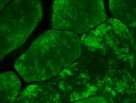
Immunocytochemistry/ Immunofluorescence: AlphaB Crystallin/CRYAB Antibody (OTI6A9) [NBP1-47708] -
Relationship between phosphorylation and subcellular localization of HSPB5. (a) Localization of overexpressed recombinant HSPB5 and endogenous PLK2. After 5 uM MG132 treatment (24 h), FLAG-tagged HSPB5 and endogenous PLK2 in undifferentiated L6 cells were immunofluorescently stained with anti-tag and anti-PLK2 antibodies. (b) Effect of PLK2 on localization of HSPB5 and desmin protein. Endogenous HSPB5 and desmin were immunofluorescently stained with anti-HSPB5 and anti-desmin antibodies. Undifferentiated L6 cells were transfected with siRNA to knockdown PLK2. After differentiation (48 h), L6 myotubes were treated with MG132 for 24 h. White arrowheads indicate cells with colocalization of HSPB5 and desmin at perinuclear ER region. Scale bar indicates 20 μm. Image collected and cropped by CiteAb from the following open publication (https://pubmed.ncbi.nlm.nih.gov/36232565), licensed under a CC-BY license. Not internally tested by Novus Biologicals.Applications for AlphaB Crystallin/CRYAB Antibody (OTI6A9)
Application
Recommended Usage
Flow Cytometry
1:100
Immunocytochemistry/ Immunofluorescence
1:50-100
Immunohistochemistry
1:50
Immunohistochemistry-Paraffin
1:50
Immunoprecipitation
2ug/500ul
Western Blot
1:500
Reviewed Applications
Read 1 review rated 5 using NBP1-47708 in the following applications:
Flow Cytometry Panel Builder
Bio-Techne Knows Flow Cytometry
Save time and reduce costly mistakes by quickly finding compatible reagents using the Panel Builder Tool.
Advanced Features
- Spectra Viewer - Custom analysis of spectra from multiple fluorochromes
- Spillover Popups - Visualize the spectra of individual fluorochromes
- Antigen Density Selector - Match fluorochrome brightness with antigen density

Formulation, Preparation, and Storage
Purification
Immunogen affinity purified
Formulation
PBS (pH 7.3), 1.0% BSA and 50% Glycerol
Preservative
0.02% Sodium Azide
Concentration
1 mg/ml
Shipping
The product is shipped with polar packs. Upon receipt, store it immediately at the temperature recommended below.
Stability & Storage
Store at -20C. Avoid freeze-thaw cycles.
Background: AlphaB Crystallin/CRYAB
Additional AlphaB Crystallin/CRYAB Products
Product Documents for AlphaB Crystallin/CRYAB Antibody (OTI6A9)
Certificate of Analysis
To download a Certificate of Analysis, please enter a lot or batch number in the search box below.
Product Specific Notices for AlphaB Crystallin/CRYAB Antibody (OTI6A9)
This product is for research use only and is not approved for use in humans or in clinical diagnosis. Primary Antibodies are guaranteed for 1 year from date of receipt.
Related Research Areas
Citations for AlphaB Crystallin/CRYAB Antibody (OTI6A9)
 Powered by Bioz
Customer Reviews for AlphaB Crystallin/CRYAB Antibody (OTI6A9) (1)
5 out of 5
1 Customer Rating
Have you used AlphaB Crystallin/CRYAB Antibody (OTI6A9)?
Submit a review and receive an Amazon gift card!
$25/€18/£15/$25CAN/¥2500 Yen for a review with an image
$10/€7/£6/$10CAN/¥1110 Yen for a review without an image
Submit a review
Customer Images
Showing
1
-
1 of
1 review
Showing All
Filter By:
-
Application: ImmunoflurescenceSample Tested: muscle tissueSpecies: MouseVerified Customer | Posted 08/17/2021AlphaB Crystallin/CRYAB Antibody in muscle

There are no reviews that match your criteria.
Protocols
Find general support by application which include: protocols, troubleshooting, illustrated assays, videos and webinars.
- 7-Amino Actinomycin D (7-AAD) Cell Viability Flow Cytometry Protocol
- Antigen Retrieval Protocol (PIER)
- Antigen Retrieval for Frozen Sections Protocol
- Appropriate Fixation of IHC/ICC Samples
- Cellular Response to Hypoxia Protocols
- Chromogenic IHC Staining of Formalin-Fixed Paraffin-Embedded (FFPE) Tissue Protocol
- Chromogenic Immunohistochemistry Staining of Frozen Tissue
- ClariTSA™ Fluorophore Kits
- Detection & Visualization of Antibody Binding
- Extracellular Membrane Flow Cytometry Protocol
- Flow Cytometry Protocol for Cell Surface Markers
- Flow Cytometry Protocol for Staining Membrane Associated Proteins
- Flow Cytometry Staining Protocols
- Flow Cytometry Troubleshooting Guide
- Fluorescent IHC Staining of Frozen Tissue Protocol
- Graphic Protocol for Heat-induced Epitope Retrieval
- Graphic Protocol for the Preparation and Fluorescent IHC Staining of Frozen Tissue Sections
- Graphic Protocol for the Preparation and Fluorescent IHC Staining of Paraffin-embedded Tissue Sections
- Graphic Protocol for the Preparation of Gelatin-coated Slides for Histological Tissue Sections
- ICC Cell Smear Protocol for Suspension Cells
- ICC Immunocytochemistry Protocol Videos
- ICC for Adherent Cells
- IHC Sample Preparation (Frozen sections vs Paraffin)
- Immunocytochemistry (ICC) Protocol
- Immunocytochemistry Troubleshooting
- Immunofluorescence of Organoids Embedded in Cultrex Basement Membrane Extract
- Immunofluorescent IHC Staining of Formalin-Fixed Paraffin-Embedded (FFPE) Tissue Protocol
- Immunohistochemistry (IHC) and Immunocytochemistry (ICC) Protocols
- Immunohistochemistry Frozen Troubleshooting
- Immunohistochemistry Paraffin Troubleshooting
- Immunoprecipitation Protocol
- Intracellular Flow Cytometry Protocol Using Alcohol (Methanol)
- Intracellular Flow Cytometry Protocol Using Detergents
- Intracellular Nuclear Staining Flow Cytometry Protocol Using Detergents
- Intracellular Staining Flow Cytometry Protocol Using Alcohol Permeabilization
- Intracellular Staining Flow Cytometry Protocol Using Detergents to Permeabilize Cells
- Preparing Samples for IHC/ICC Experiments
- Preventing Non-Specific Staining (Non-Specific Binding)
- Primary Antibody Selection & Optimization
- Propidium Iodide Cell Viability Flow Cytometry Protocol
- Protocol for Heat-Induced Epitope Retrieval (HIER)
- Protocol for Liperfluo
- Protocol for Making a 4% Formaldehyde Solution in PBS
- Protocol for VisUCyte™ HRP Polymer Detection Reagent
- Protocol for the Characterization of Human Th22 Cells
- Protocol for the Characterization of Human Th9 Cells
- Protocol for the Fluorescent ICC Staining of Cell Smears - Graphic
- Protocol for the Fluorescent ICC Staining of Cultured Cells on Coverslips - Graphic
- Protocol for the Preparation & Fixation of Cells on Coverslips
- Protocol for the Preparation and Chromogenic IHC Staining of Frozen Tissue Sections
- Protocol for the Preparation and Chromogenic IHC Staining of Frozen Tissue Sections - Graphic
- Protocol for the Preparation and Chromogenic IHC Staining of Paraffin-embedded Tissue Sections
- Protocol for the Preparation and Chromogenic IHC Staining of Paraffin-embedded Tissue Sections - Graphic
- Protocol for the Preparation and Fluorescent ICC Staining of Cells on Coverslips
- Protocol for the Preparation and Fluorescent ICC Staining of Non-adherent Cells
- Protocol for the Preparation and Fluorescent ICC Staining of Stem Cells on Coverslips
- Protocol for the Preparation and Fluorescent IHC Staining of Frozen Tissue Sections
- Protocol for the Preparation and Fluorescent IHC Staining of Paraffin-embedded Tissue Sections
- Protocol for the Preparation of Gelatin-coated Slides for Histological Tissue Sections
- Protocol for the Preparation of a Cell Smear for Non-adherent Cell ICC - Graphic
- Protocol: Annexin V and PI Staining by Flow Cytometry
- Protocol: Annexin V and PI Staining for Apoptosis by Flow Cytometry
- R&D Systems Quality Control Western Blot Protocol
- TUNEL and Active Caspase-3 Detection by IHC/ICC Protocol
- The Importance of IHC/ICC Controls
- Troubleshooting Guide: Fluorokine Flow Cytometry Kits
- Troubleshooting Guide: Immunohistochemistry
- Troubleshooting Guide: Western Blot Figures
- Western Blot Conditions
- Western Blot Protocol
- Western Blot Protocol for Cell Lysates
- Western Blot Troubleshooting
- Western Blot Troubleshooting Guide
- View all Protocols, Troubleshooting, Illustrated assays and Webinars
Loading...