
Angiopoietin-2 Antibody (MM0020-1F29) - Azide and BSA Free
Novus Biologicals | Catalog # NB110-85467

![Immunocytochemistry/ Immunofluorescence: Angiopoietin-2 Antibody (MM0020-1F29) - Azide and BSA Free [NB110-85467]](https://resources.rndsystems.com/images/products/Angiopoietin-2-Antibody-MM0020-1F29-Immunocytochemistry-Immunofluorescence-NB110-85467-img0004.jpg "Immunocytochemistry/ Immunofluorescence: Angiopoietin-2 Antibody (MM0020-1F29) - Azide and BSA Free [NB110-85467]")
Key Product Details
Species Reactivity
Validated:
Cited:
Applications
Validated:
Cited:
Label
Antibody Source
Format
Product Specifications
Immunogen
Reactivity Notes
Specificity
Clonality
Host
Isotype
Scientific Data Images for Angiopoietin-2 Antibody (MM0020-1F29) - Azide and BSA Free
Immunocytochemistry/ Immunofluorescence: Angiopoietin-2 Antibody (MM0020-1F29) - Azide and BSA Free [NB110-85467]
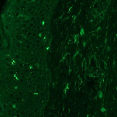
Immunocytochemistry/Immunofluorescence: Angiopoietin-2 Antibody (MM0020-1F29) [NB110-85467] - Staining of frozen human skin sections using Angiopoietin 2 antibody at 1:25 dilution. Image provided via product review by verified customer.![Immunohistochemistry: Angiopoietin-2 Antibody (MM0020-1F29) - Azide and BSA Free [NB110-85467]](https://resources.rndsystems.com/images/products/Angiopoietin-2-Antibody-MM0020-1F29-Immunohistochemistry-NB110-85467-img0007.jpg "Immunohistochemistry: Angiopoietin-2 Antibody (MM0020-1F29) - Azide and BSA Free [NB110-85467]")
Immunohistochemistry: Angiopoietin-2 Antibody (MM0020-1F29) - Azide and BSA Free [NB110-85467]
Immunohistochemistry: Angiopoietin-2 Antibody (MM0020-1F29) [NB110-85467] - Ang-2 (green) was detected in human skin (nevus) using Ang2-Alexa Fluor 488 antibody (1:40) in PBS for 1 hour. Nuclei were stained with DAPI (blue). Tissue was fixed with acetone. Image from a verified customer review. Image using the Alexa Fluor 488 format of this antibody.![Immunohistochemistry: Angiopoietin-2 Antibody (MM0020-1F29) - Azide and BSA Free [NB110-85467]](https://resources.rndsystems.com/images/products/Angiopoietin-2-Antibody-MM0020-1F29-Immunohistochemistry-NB110-85467-img0001.jpg "Immunohistochemistry: Angiopoietin-2 Antibody (MM0020-1F29) - Azide and BSA Free [NB110-85467]")
Immunohistochemistry: Angiopoietin-2 Antibody (MM0020-1F29) - Azide and BSA Free [NB110-85467]
Immunohistochemistry: Angiopoietin-2 Antibody (MM0020-1F29) [NB110-85467] - Normal Human placenta tissue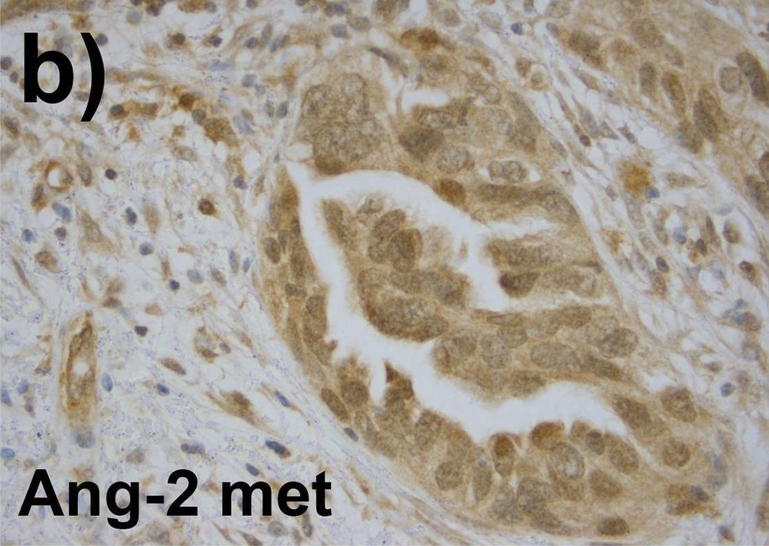
Immunohistochemistry: Angiopoietin-2 Antibody (MM0020-1F29) - Azide and BSA Free [NB110-85467] -
As compared to the primary tumors, related omental metastases showed stronger expression of Ang-2 (a and b) and Tie-2 (e and f). Primary tumors are shown on the left column and related metastases on the right. The vascular endothelial expression was strong in Ang-2 (black arrow in a). Tie-1 expression did not differ significantly between the primary and the distal metastatic tumors (c and d). Tie-1 was expressed in the tumor stroma (black arrow in d). Image collected and cropped by CiteAb from the following open publication (https://pubmed.ncbi.nlm.nih.gov/33151982), licensed under a CC-BY license. Not internally tested by Novus Biologicals.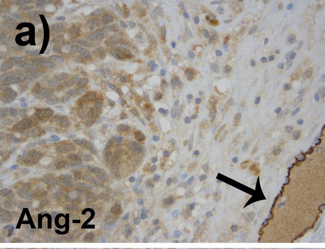
Immunohistochemistry: Angiopoietin-2 Antibody (MM0020-1F29) - Azide and BSA Free [NB110-85467] -
As compared to the primary tumors, related omental metastases showed stronger expression of Ang-2 (a and b) and Tie-2 (e and f). Primary tumors are shown on the left column and related metastases on the right. The vascular endothelial expression was strong in Ang-2 (black arrow in a). Tie-1 expression did not differ significantly between the primary and the distal metastatic tumors (c and d). Tie-1 was expressed in the tumor stroma (black arrow in d). Image collected and cropped by CiteAb from the following open publication (https://pubmed.ncbi.nlm.nih.gov/33151982), licensed under a CC-BY license. Not internally tested by Novus Biologicals.Applications for Angiopoietin-2 Antibody (MM0020-1F29) - Azide and BSA Free
Immunocytochemistry/ Immunofluorescence
Immunohistochemistry
Immunohistochemistry-Frozen
Immunohistochemistry-Paraffin
Western Blot
Reviewed Applications
Read 2 reviews rated 3 using NB110-85467 in the following applications:
Formulation, Preparation, and Storage
Purification
Reconstitution
Formulation
Format
Preservative
Concentration
Shipping
Stability & Storage
Calculators
Background: Angiopoietin-2
Additional Angiopoietin-2 Products
Product Documents for Angiopoietin-2 Antibody (MM0020-1F29) - Azide and BSA Free
Certificate of Analysis
To download a Certificate of Analysis, please enter a lot or batch number in the search box below.
Product Specific Notices for Angiopoietin-2 Antibody (MM0020-1F29) - Azide and BSA Free
This product is for research use only and is not approved for use in humans or in clinical diagnosis. Primary Antibodies are guaranteed for 1 year from date of receipt.
Citations for Angiopoietin-2 Antibody (MM0020-1F29) - Azide and BSA Free
 Powered by Bioz
Customer Reviews for Angiopoietin-2 Antibody (MM0020-1F29) - Azide and BSA Free (2)
Have you used Angiopoietin-2 Antibody (MM0020-1F29) - Azide and BSA Free?
Submit a review and receive an Amazon gift card!
$25/€18/£15/$25CAN/¥2500 for a review with an image
$10/€7/£6/$10CAN/¥1110 for a review without an image
Submit a review
Customer Images
-(01-mg)_NB110-85467_9571.jpg)
-
Application: ImmunofluorescenceSample Tested:Species: OtherVerified Customer | Posted 08/26/2014indirect immunofluorescence data with anti-Ang2 on chicken comb

-
Application: Immunohistochemistry-FrozenSample Tested: Frozen human skin sectionsSpecies: HumanVerified Customer | Posted 07/12/2013
There are no reviews that match your criteria.
Protocols
Find general support by application which include: protocols, troubleshooting, illustrated assays, videos and webinars.
- Antigen Retrieval Protocol (PIER)
- Antigen Retrieval for Frozen Sections Protocol
- Appropriate Fixation of IHC/ICC Samples
- Cellular Response to Hypoxia Protocols
- Chromogenic IHC Staining of Formalin-Fixed Paraffin-Embedded (FFPE) Tissue Protocol
- Chromogenic Immunohistochemistry Staining of Frozen Tissue
- ClariTSA™ Fluorophore Kits
- Detection & Visualization of Antibody Binding
- Fluorescent IHC Staining of Frozen Tissue Protocol
- Graphic Protocol for Heat-induced Epitope Retrieval
- Graphic Protocol for the Preparation and Fluorescent IHC Staining of Frozen Tissue Sections
- Graphic Protocol for the Preparation and Fluorescent IHC Staining of Paraffin-embedded Tissue Sections
- Graphic Protocol for the Preparation of Gelatin-coated Slides for Histological Tissue Sections
- ICC Cell Smear Protocol for Suspension Cells
- ICC Immunocytochemistry Protocol Videos
- ICC for Adherent Cells
- IHC Sample Preparation (Frozen sections vs Paraffin)
- Immunocytochemistry (ICC) Protocol
- Immunocytochemistry Troubleshooting
- Immunofluorescence of Organoids Embedded in Cultrex Basement Membrane Extract
- Immunofluorescent IHC Staining of Formalin-Fixed Paraffin-Embedded (FFPE) Tissue Protocol
- Immunohistochemistry (IHC) and Immunocytochemistry (ICC) Protocols
- Immunohistochemistry Frozen Troubleshooting
- Immunohistochemistry Paraffin Troubleshooting
- Preparing Samples for IHC/ICC Experiments
- Preventing Non-Specific Staining (Non-Specific Binding)
- Primary Antibody Selection & Optimization
- Protocol for Heat-Induced Epitope Retrieval (HIER)
- Protocol for Making a 4% Formaldehyde Solution in PBS
- Protocol for VisUCyte™ HRP Polymer Detection Reagent
- Protocol for the Fluorescent ICC Staining of Cell Smears - Graphic
- Protocol for the Fluorescent ICC Staining of Cultured Cells on Coverslips - Graphic
- Protocol for the Preparation & Fixation of Cells on Coverslips
- Protocol for the Preparation and Chromogenic IHC Staining of Frozen Tissue Sections
- Protocol for the Preparation and Chromogenic IHC Staining of Frozen Tissue Sections - Graphic
- Protocol for the Preparation and Chromogenic IHC Staining of Paraffin-embedded Tissue Sections
- Protocol for the Preparation and Chromogenic IHC Staining of Paraffin-embedded Tissue Sections - Graphic
- Protocol for the Preparation and Fluorescent ICC Staining of Cells on Coverslips
- Protocol for the Preparation and Fluorescent ICC Staining of Non-adherent Cells
- Protocol for the Preparation and Fluorescent ICC Staining of Stem Cells on Coverslips
- Protocol for the Preparation and Fluorescent IHC Staining of Frozen Tissue Sections
- Protocol for the Preparation and Fluorescent IHC Staining of Paraffin-embedded Tissue Sections
- Protocol for the Preparation of Gelatin-coated Slides for Histological Tissue Sections
- Protocol for the Preparation of a Cell Smear for Non-adherent Cell ICC - Graphic
- R&D Systems Quality Control Western Blot Protocol
- TUNEL and Active Caspase-3 Detection by IHC/ICC Protocol
- The Importance of IHC/ICC Controls
- Troubleshooting Guide: Immunohistochemistry
- Troubleshooting Guide: Western Blot Figures
- Western Blot Conditions
- Western Blot Protocol
- Western Blot Protocol for Cell Lysates
- Western Blot Troubleshooting
- Western Blot Troubleshooting Guide
- View all Protocols, Troubleshooting, Illustrated assays and Webinars
FAQs for Angiopoietin-2 Antibody (MM0020-1F29) - Azide and BSA Free
-
Q: How do you dissolve the lyophilized Ab? Water or PBS? And in what concentration?
A: The IgG fraction of culture supernatant was purified by Protein G affinity chromatography and lyophilized from a 0.2 um filtered solution in phosphate-buffered saline (PBS). 100 ug antibody is lyophilized from 40 ul of 1 x PBS. We suggest reconstituting the antibody with 500 ul sterile 1 x PBS (or to be perfect, with 40 ul H2O plus 460 ul PBS), to give a final concentration of 200 ug/ml. Alternatively, you may reconstitute the antibody in 40 ul H2O if a small volume is preferred.
-
Q: I have a question regarding your Angiopoietin 2 Antibody (MM0020-1F29). I am interested in differentiating Ang2 from mouse origin and Ang2 from human origin. Does this antibody cross react in any way with mouse Ang2?
A: This particular product has only been tested against human samples. We have not yet tested for cross reactivity with other species. Based on the homology of the full length protein, I would suspect that there may be some cross reaction. I located the amino acid range for this product. It is between aa70-aa496 of recombinant human Angiopoietin-2 (O15123). If you would be interested in testing this novel species, please take a look at our Innovator's Reward program.
-
Q: How do you dissolve the lyophilized Ab? Water or PBS? And in what concentration?
A: The IgG fraction of culture supernatant was purified by Protein G affinity chromatography and lyophilized from a 0.2 um filtered solution in phosphate-buffered saline (PBS). 100 ug antibody is lyophilized from 40 ul of 1 x PBS. We suggest reconstituting the antibody with 500 ul sterile 1 x PBS (or to be perfect, with 40 ul H2O plus 460 ul PBS), to give a final concentration of 200 ug/ml. Alternatively, you may reconstitute the antibody in 40 ul H2O if a small volume is preferred.
-
Q: I have a question regarding your Angiopoietin 2 Antibody (MM0020-1F29). I am interested in differentiating Ang2 from mouse origin and Ang2 from human origin. Does this antibody cross react in any way with mouse Ang2?
A: This particular product has only been tested against human samples. We have not yet tested for cross reactivity with other species. Based on the homology of the full length protein, I would suspect that there may be some cross reaction. I located the amino acid range for this product. It is between aa70-aa496 of recombinant human Angiopoietin-2 (O15123). If you would be interested in testing this novel species, please take a look at our Innovator's Reward program.