AP2 alpha Antibody (3B5)
Novus Biologicals | Catalog # NB100-74359
![Western Blot: AP2 alpha Antibody (3B5) [NB100-74359]](https://resources.rndsystems.com/images/products/AP2-alpha-Antibody-3B5-Western-Blot-NB100-74359-img0011.jpg "Western Blot: AP2 alpha Antibody (3B5) [NB100-74359]")
Loading...
Key Product Details
Species Reactivity
Validated:
Human, Mouse, Chicken
Cited:
Mouse, Avian - Chicken
Applications
Validated:
Immunohistochemistry, Immunohistochemistry-Paraffin, Immunohistochemistry-Frozen, Western Blot, Immunocytochemistry/ Immunofluorescence, Immunoprecipitation
Cited:
Immunohistochemistry-Frozen, Immunocytochemistry/ Immunofluorescence
Label
Unconjugated
Antibody Source
Monoclonal Mouse IgG2B Clone # 3B5
Loading...
Product Specifications
Immunogen
N-terminus of human AP2a.
Reactivity Notes
Please note that this antibody is reactive to Mouse and derived from the same host, Mouse. Additional Mouse on Mouse blocking steps may be required for IHC and ICC experiments. Please contact Technical Support for more information.
Specificity
AP-2 (3B5)
Clonality
Monoclonal
Host
Mouse
Isotype
IgG2B
Theoretical MW
48 kDa.
Disclaimer note: The observed molecular weight of the protein may vary from the listed predicted molecular weight due to post translational modifications, post translation cleavages, relative charges, and other experimental factors.
Disclaimer note: The observed molecular weight of the protein may vary from the listed predicted molecular weight due to post translational modifications, post translation cleavages, relative charges, and other experimental factors.
Scientific Data Images for AP2 alpha Antibody (3B5)
Western Blot: AP2 alpha Antibody (3B5) [NB100-74359]
Western Blot: AP2 alpha Antibody (3B5) [NB100-74359] - Analysis was performed on whole cell extracts (30 ug lysate) of PC-3 (Lane1), A-431 (Lane2), Mouse Placenta (Lane3) and RAW 264.7 (Lane5).![Immunocytochemistry/ Immunofluorescence: AP2 alpha Antibody (3B5) [NB100-74359]](https://resources.rndsystems.com/images/products/AP2-alpha-Antibody-3B5-Immunocytochemistry-Immunofluorescence-NB100-74359-img0008.jpg "Immunocytochemistry/ Immunofluorescence: AP2 alpha Antibody (3B5) [NB100-74359]")
Immunocytochemistry/ Immunofluorescence: AP2 alpha Antibody (3B5) [NB100-74359]
Immunocytochemistry/Immunofluorescence: AP2 alpha Antibody (3B5) [NB100-74359] - Analysis of AP2 using Anti-AP2 Monoclonal Antibody (3B5) shows staining in Hela Cells. AP2 staining (green), F-Actin staining with Phalloidin (red) and nuclei with DAPI (blue) is shown. Cells were grown on chamber slides and fixed with formaldehyde prior to staining. Cells were probed without (control) or with or an antibody recognizing AP2 at a dilution of 1:100 over night at 4C, washed with PBS and incubated with a DyLight-488 conjugated secondary antibody. Images were taken at 60X magnification.![Immunohistochemistry-Frozen: AP2 alpha Antibody (3B5) [NB100-74359]](https://resources.rndsystems.com/images/products/AP2-alpha-Antibody-3B5-Immunohistochemistry-Frozen-NB100-74359-img0012.jpg "Immunohistochemistry-Frozen: AP2 alpha Antibody (3B5) [NB100-74359]")
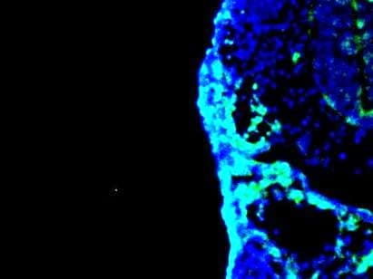
Immunohistochemistry-Frozen: AP2 alpha Antibody (3B5) [NB100-74359]
Immunohistochemistry-Frozen: AP2 alpha Antibody (3B5) [NB100-74359] - AP2alpha staining on an E9.5 mouse pharyngeal mesoderm region. IHC-Fr image submitted by a verified customer review.![Western Blot: AP2 alpha Antibody (3B5) [NB100-74359]](https://resources.rndsystems.com/images/products/AP2-alpha-Antibody-3B5-Western-Blot-NB100-74359-img0001.jpg "Western Blot: AP2 alpha Antibody (3B5) [NB100-74359]")
Western Blot: AP2 alpha Antibody (3B5) [NB100-74359]
Western Blot: AP2 alpha Antibody (3B5) [NB100-74359] - Figure 1 illustrates Western blot detection of AP-2 from MCF7 cell lysate.![Immunocytochemistry/ Immunofluorescence: AP2 alpha Antibody (3B5) [NB100-74359]](https://resources.rndsystems.com/images/products/AP2-alpha-Antibody-3B5-Immunocytochemistry-Immunofluorescence-NB100-74359-img0006.jpg "Immunocytochemistry/ Immunofluorescence: AP2 alpha Antibody (3B5) [NB100-74359]")
Immunocytochemistry/ Immunofluorescence: AP2 alpha Antibody (3B5) [NB100-74359]
Immunocytochemistry/Immunofluorescence: AP2 alpha Antibody (3B5) [NB100-74359] - Analysis of AP2 using Anti-AP2 Monoclonal Antibody (3B5) shows staining in U251 Cells. AP2 staining (green), F-Actin staining with Phalloidin (red) and nuclei with DAPI (blue) is shown.![Immunocytochemistry/ Immunofluorescence: AP2 alpha Antibody (3B5) [NB100-74359]](https://resources.rndsystems.com/images/products/AP2-alpha-Antibody-3B5-Immunocytochemistry-Immunofluorescence-NB100-74359-img0007.jpg "Immunocytochemistry/ Immunofluorescence: AP2 alpha Antibody (3B5) [NB100-74359]")
Immunocytochemistry/ Immunofluorescence: AP2 alpha Antibody (3B5) [NB100-74359]
Immunocytochemistry/Immunofluorescence: AP2 alpha Antibody (3B5) [NB100-74359] - Analysis of AP2 using Anti-AP2 Monoclonal Antibody (3B5) shows staining in MCF-7 Cells. AP2 staining (green), F-Actin staining with Phalloidin (red) and nuclei with DAPI (blue) is shown.![Immunohistochemistry-Paraffin: AP2 alpha Antibody (3B5) [NB100-74359]](https://resources.rndsystems.com/images/products/AP2-alpha-Antibody-3B5-Immunohistochemistry-Paraffin-NB100-74359-img0009.jpg "Immunohistochemistry-Paraffin: AP2 alpha Antibody (3B5) [NB100-74359]")
Immunohistochemistry-Paraffin: AP2 alpha Antibody (3B5) [NB100-74359]
Immunohistochemistry-Paraffin: AP2 alpha Antibody (3B5) [NB100-74359] - Immunohistochemistry was performed on cancer biopsies of deparaffinized Human breast carcinoma tissue.Applications for AP2 alpha Antibody (3B5)
Application
Recommended Usage
Immunocytochemistry/ Immunofluorescence
1:50 - 1:500
Immunohistochemistry
1:50
Immunohistochemistry-Frozen
1:50
Immunohistochemistry-Paraffin
1:50
Immunoprecipitation
1:100 - 1:250
Western Blot
1:50 - 1:200
Application Notes
In WB: Detects a 45 - 50 kDa protein representing AP-2. Block with BSA only (not milk). IHC-Fr usage was reported in scientific literature (PMID: 24996922).
Reviewed Applications
Read 1 review rated 5 using NB100-74359 in the following applications:
Formulation, Preparation, and Storage
Purification
Protein G purified
Formulation
PBS with 1 mg/ml BSA
Preservative
0.05% Sodium Azide
Concentration
Concentrations vary lot to lot. See vial label for concentration. If unlisted please contact technical services.
Shipping
The product is shipped with polar packs. Upon receipt, store it immediately at the temperature recommended below.
Stability & Storage
Store at -20C. Avoid freeze-thaw cycles.
Background: AP2 alpha
Alternate Names
Activating enhancer-binding protein 2-alpha, Activator protein 2, AP-2 transcription factor, AP2-alpha, AP2TFAP-2alpha, AP-2transcription factor AP-2 alpha (activating enhancer-binding protein 2 alpha), BOFS, FLJ51761, TFAP2, transcription factor AP-2 alpha (activating enhancer binding protein 2 alpha), transcription factor AP-2-alpha
Gene Symbol
TFAP2A
UniProt
Additional AP2 alpha Products
Product Documents for AP2 alpha Antibody (3B5)
Certificate of Analysis
To download a Certificate of Analysis, please enter a lot or batch number in the search box below.
Product Specific Notices for AP2 alpha Antibody (3B5)
This product is for research use only and is not approved for use in humans or in clinical diagnosis. Primary Antibodies are guaranteed for 1 year from date of receipt.
Citations for AP2 alpha Antibody (3B5)
 Powered by Bioz
Customer Reviews for AP2 alpha Antibody (3B5) (1)
5 out of 5
1 Customer Rating
Have you used AP2 alpha Antibody (3B5)?
Submit a review and receive an Amazon gift card!
$25/€18/£15/$25CAN/¥2500 Yen for a review with an image
$10/€7/£6/$10CAN/¥1110 Yen for a review without an image
Submit a review
Customer Images
Showing
1
-
1 of
1 review
Showing All
Filter By:
-
Application: Immunohistochemistry-FrozenSample Tested: E9.5 mouse embryo fixed in 4% PFASpecies: MouseVerified Customer | Posted 06/23/2021AP2alpha staining on an E9.5 mouse pharyngeal mesoderm region.Fixed 4% PFA overnight. Blocked with 1% BSA Primary antibody dilution - 20ug/mL Secondary antibody - Invitrogen Alexa Fluor 488 Secondary antibody dilution - 1:1000 Stained on a E9.5 mouse pharyngeal mesoderm region

There are no reviews that match your criteria.
Protocols
Find general support by application which include: protocols, troubleshooting, illustrated assays, videos and webinars.
- Antigen Retrieval Protocol (PIER)
- Antigen Retrieval for Frozen Sections Protocol
- Appropriate Fixation of IHC/ICC Samples
- Cellular Response to Hypoxia Protocols
- Chromogenic IHC Staining of Formalin-Fixed Paraffin-Embedded (FFPE) Tissue Protocol
- Chromogenic Immunohistochemistry Staining of Frozen Tissue
- ClariTSA™ Fluorophore Kits
- Detection & Visualization of Antibody Binding
- Fluorescent IHC Staining of Frozen Tissue Protocol
- Graphic Protocol for Heat-induced Epitope Retrieval
- Graphic Protocol for the Preparation and Fluorescent IHC Staining of Frozen Tissue Sections
- Graphic Protocol for the Preparation and Fluorescent IHC Staining of Paraffin-embedded Tissue Sections
- Graphic Protocol for the Preparation of Gelatin-coated Slides for Histological Tissue Sections
- ICC Cell Smear Protocol for Suspension Cells
- ICC Immunocytochemistry Protocol Videos
- ICC for Adherent Cells
- IHC Sample Preparation (Frozen sections vs Paraffin)
- Immunocytochemistry (ICC) Protocol
- Immunocytochemistry Troubleshooting
- Immunofluorescence of Organoids Embedded in Cultrex Basement Membrane Extract
- Immunofluorescent IHC Staining of Formalin-Fixed Paraffin-Embedded (FFPE) Tissue Protocol
- Immunohistochemistry (IHC) and Immunocytochemistry (ICC) Protocols
- Immunohistochemistry Frozen Troubleshooting
- Immunohistochemistry Paraffin Troubleshooting
- Immunoprecipitation Protocol
- Preparing Samples for IHC/ICC Experiments
- Preventing Non-Specific Staining (Non-Specific Binding)
- Primary Antibody Selection & Optimization
- Protocol for Heat-Induced Epitope Retrieval (HIER)
- Protocol for Making a 4% Formaldehyde Solution in PBS
- Protocol for VisUCyte™ HRP Polymer Detection Reagent
- Protocol for the Fluorescent ICC Staining of Cell Smears - Graphic
- Protocol for the Fluorescent ICC Staining of Cultured Cells on Coverslips - Graphic
- Protocol for the Preparation & Fixation of Cells on Coverslips
- Protocol for the Preparation and Chromogenic IHC Staining of Frozen Tissue Sections
- Protocol for the Preparation and Chromogenic IHC Staining of Frozen Tissue Sections - Graphic
- Protocol for the Preparation and Chromogenic IHC Staining of Paraffin-embedded Tissue Sections
- Protocol for the Preparation and Chromogenic IHC Staining of Paraffin-embedded Tissue Sections - Graphic
- Protocol for the Preparation and Fluorescent ICC Staining of Cells on Coverslips
- Protocol for the Preparation and Fluorescent ICC Staining of Non-adherent Cells
- Protocol for the Preparation and Fluorescent ICC Staining of Stem Cells on Coverslips
- Protocol for the Preparation and Fluorescent IHC Staining of Frozen Tissue Sections
- Protocol for the Preparation and Fluorescent IHC Staining of Paraffin-embedded Tissue Sections
- Protocol for the Preparation of Gelatin-coated Slides for Histological Tissue Sections
- Protocol for the Preparation of a Cell Smear for Non-adherent Cell ICC - Graphic
- R&D Systems Quality Control Western Blot Protocol
- TUNEL and Active Caspase-3 Detection by IHC/ICC Protocol
- The Importance of IHC/ICC Controls
- Troubleshooting Guide: Immunohistochemistry
- Troubleshooting Guide: Western Blot Figures
- Western Blot Conditions
- Western Blot Protocol
- Western Blot Protocol for Cell Lysates
- Western Blot Troubleshooting
- Western Blot Troubleshooting Guide
- View all Protocols, Troubleshooting, Illustrated assays and Webinars
Loading...