Apc11 Antibody - C-terminus
Novus Biologicals | Catalog # NBP1-78050
Loading...
Key Product Details
Species Reactivity
Validated:
Human
Cited:
Human
Applications
Validated:
Immunohistochemistry, Immunohistochemistry-Paraffin, Western Blot, ELISA, Immunoprecipitation
Cited:
Immunohistochemistry-Paraffin, Western Blot
Label
Unconjugated
Antibody Source
Polyclonal Rabbit Serum
Loading...
Product Specifications
Immunogen
This Apc11 Antibody was prepared from whole rabbit serum produced by repeated immunizations with a synthetic peptide corresponding to amino acids 76-84 of Human Apc11 (C-terminal) coupled to KLH. (Uniprot: Q9NYG5)
Reactivity Notes
Cross reactivity may also occur with Apc11 from other sources
Specificity
This product is monospecific antiserum processed by delipidation and defibrination followed by sterile filtration. This product reacts with human and mouse Apc11. Cross reactivity may also occur with Apc11 from other sources. Sufficient sequence differences exist to suggest that this antibody would not react with other RING box proteins such as ROC1 and ROC2.
Clonality
Polyclonal
Host
Rabbit
Isotype
Serum
Description
This product is monospecific antiserum processed by delipidation and defibrination followed by sterile filtration
Store vial at -20C prior to opening. Aliquot contents and freeze at -20C or below for extended storage. Avoid cycles of freezing and thawing. Centrifuge product if not completely clear after standing at room temperature. This product is stable for several weeks at 4C as an undiluted liquid. Dilute only prior to immediate use.
Store vial at -20C prior to opening. Aliquot contents and freeze at -20C or below for extended storage. Avoid cycles of freezing and thawing. Centrifuge product if not completely clear after standing at room temperature. This product is stable for several weeks at 4C as an undiluted liquid. Dilute only prior to immediate use.
Scientific Data Images for Apc11 Antibody - C-terminus
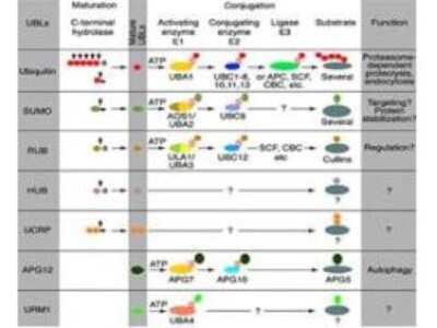
N/A: Apc11 Antibody [NBP1-78050] - Most modifiers mature by proteolytic processing from inactive precursors (a; amino acid). Arrowheads point to the cleavage sites. Ubiquitin is expressed either as polyubiquitin or as a fusion with ribosomal proteins. Conjugation requires activating (E1) & conjugating (E2) enzymes that form thiolesters (S) with the modifiers. Modification of cullins by RUB involves SCF(SKP1/cullin-1/F-box protein) /CBC(cullin-2/elongin B/elonginC) -like E3 enzymes that are also involved in ubiquitination. UBLs do not seem to form multi-UBL chains. UCRP(ISG15) resembles two ubiquitin moieties linked head-to-tail. Whether HUB1 functions as a modifier is currently unclear. APG12 and URM1 are distinct from the other modifiers because they are unrelated in sequence to ubiquitin.
Applications for Apc11 Antibody - C-terminus
Application
Recommended Usage
ELISA
1:2000-1:10000
Immunohistochemistry
1:10-1:500
Immunohistochemistry-Paraffin
1:10-1:500
Western Blot
1:500-1:1000
Application Notes
This antibody reacts with human APC11 by western blot and immunoprecipitation. The antibody immunoprecipitates in vitro translated protein and protein from overexpressing cell lysates (using HeLa and NIH-3T3, and others). Coimmunoprecipitation of related proteins (APC2) does occur. A 9.8 kDa band corresponding to human APC11 is detected. Most cell lines or tissues expressing APC11 can be used as a positive control. Researchers should determine optimal titers for other applications.
Formulation, Preparation, and Storage
Purification
Delipidation and Defibrination
Formulation
Antiserum
Preservative
0.01% Sodium Azide
Concentration
Please see the vial label for concentration. If unlisted please contact technical services.
Shipping
The product is shipped with polar packs. Upon receipt, store it immediately at the temperature recommended below.
Stability & Storage
Store at -20C short term. Aliquot and store at -80C long term. Avoid freeze-thaw cycles.
Background: Apc11
Alternate Names
anaphase promoting complex subunit 11, anaphase promoting complex subunit 11 (yeast APC11 homolog), anaphase-promoting complex subunit 11, APC11 anaphase promoting complex subunit 11 homolog, APC11Hepatocellular carcinoma-associated RING finger protein, Apc11p, Cyclosome subunit 11, HSPC214, MGC882
Gene Symbol
ANAPC11
UniProt
Additional Apc11 Products
Product Documents for Apc11 Antibody - C-terminus
Certificate of Analysis
To download a Certificate of Analysis, please enter a lot or batch number in the search box below.
Product Specific Notices for Apc11 Antibody - C-terminus
This product is for research use only and is not approved for use in humans or in clinical diagnosis. Primary Antibodies are guaranteed for 1 year from date of receipt.
Citations for Apc11 Antibody - C-terminus
 Powered by Bioz
Customer Reviews for Apc11 Antibody - C-terminus
There are currently no reviews for this product. Be the first to review Apc11 Antibody - C-terminus and earn rewards!
Have you used Apc11 Antibody - C-terminus?
Submit a review and receive an Amazon gift card!
$25/€18/£15/$25CAN/¥2500 Yen for a review with an image
$10/€7/£6/$10CAN/¥1110 Yen for a review without an image
Submit a review
Protocols
Find general support by application which include: protocols, troubleshooting, illustrated assays, videos and webinars.
- Antigen Retrieval Protocol (PIER)
- Antigen Retrieval for Frozen Sections Protocol
- Appropriate Fixation of IHC/ICC Samples
- Cellular Response to Hypoxia Protocols
- Chromogenic IHC Staining of Formalin-Fixed Paraffin-Embedded (FFPE) Tissue Protocol
- Chromogenic Immunohistochemistry Staining of Frozen Tissue
- ClariTSA™ Fluorophore Kits
- Detection & Visualization of Antibody Binding
- ELISA Sample Preparation & Collection Guide
- ELISA Troubleshooting Guide
- Fluorescent IHC Staining of Frozen Tissue Protocol
- Graphic Protocol for Heat-induced Epitope Retrieval
- Graphic Protocol for the Preparation and Fluorescent IHC Staining of Frozen Tissue Sections
- Graphic Protocol for the Preparation and Fluorescent IHC Staining of Paraffin-embedded Tissue Sections
- Graphic Protocol for the Preparation of Gelatin-coated Slides for Histological Tissue Sections
- How to Run an R&D Systems DuoSet ELISA
- How to Run an R&D Systems Quantikine ELISA
- How to Run an R&D Systems Quantikine™ QuicKit™ ELISA
- IHC Sample Preparation (Frozen sections vs Paraffin)
- Immunofluorescent IHC Staining of Formalin-Fixed Paraffin-Embedded (FFPE) Tissue Protocol
- Immunohistochemistry (IHC) and Immunocytochemistry (ICC) Protocols
- Immunohistochemistry Frozen Troubleshooting
- Immunohistochemistry Paraffin Troubleshooting
- Immunoprecipitation Protocol
- Preparing Samples for IHC/ICC Experiments
- Preventing Non-Specific Staining (Non-Specific Binding)
- Primary Antibody Selection & Optimization
- Protocol for Heat-Induced Epitope Retrieval (HIER)
- Protocol for Making a 4% Formaldehyde Solution in PBS
- Protocol for VisUCyte™ HRP Polymer Detection Reagent
- Protocol for the Preparation & Fixation of Cells on Coverslips
- Protocol for the Preparation and Chromogenic IHC Staining of Frozen Tissue Sections
- Protocol for the Preparation and Chromogenic IHC Staining of Frozen Tissue Sections - Graphic
- Protocol for the Preparation and Chromogenic IHC Staining of Paraffin-embedded Tissue Sections
- Protocol for the Preparation and Chromogenic IHC Staining of Paraffin-embedded Tissue Sections - Graphic
- Protocol for the Preparation and Fluorescent IHC Staining of Frozen Tissue Sections
- Protocol for the Preparation and Fluorescent IHC Staining of Paraffin-embedded Tissue Sections
- Protocol for the Preparation of Gelatin-coated Slides for Histological Tissue Sections
- Quantikine HS ELISA Kit Assay Principle, Alkaline Phosphatase
- Quantikine HS ELISA Kit Principle, Streptavidin-HRP Polymer
- R&D Systems Quality Control Western Blot Protocol
- Sandwich ELISA (Colorimetric) – Biotin/Streptavidin Detection Protocol
- Sandwich ELISA (Colorimetric) – Direct Detection Protocol
- TUNEL and Active Caspase-3 Detection by IHC/ICC Protocol
- The Importance of IHC/ICC Controls
- Troubleshooting Guide: ELISA
- Troubleshooting Guide: Immunohistochemistry
- Troubleshooting Guide: Western Blot Figures
- Western Blot Conditions
- Western Blot Protocol
- Western Blot Protocol for Cell Lysates
- Western Blot Troubleshooting
- Western Blot Troubleshooting Guide
- View all Protocols, Troubleshooting, Illustrated assays and Webinars
Loading...