![Western Blot: Apc4 Antibody [NB100-59824]](https://resources.rndsystems.com/images/products/Apc4-Antibody-Western-Blot-NB100-59824-img0002.jpg "Western Blot: Apc4 Antibody [NB100-59824]")
Loading...
Key Product Details
Validated by
Knockout/Knockdown, Independent Antibodies, Biological Validation
Species Reactivity
Validated:
Human, Mouse
Cited:
Mouse
Predicted:
Primate (100%). Backed by our 100% Guarantee.
Applications
Validated:
Knockout Validated, Western Blot, Immunocytochemistry/ Immunofluorescence, Immunoprecipitation
Cited:
Knockout Validated
Label
Unconjugated
Antibody Source
Polyclonal Rabbit IgG
Loading...
Product Specifications
Immunogen
The immunogen recognized by this antibody maps to a region between residue 758 and 808 of human anaphase-promoting complex subunit 4 using the numbering given in entry NP_037499.2 (GeneID 29945).
Reactivity Notes
Orangutan (100%).
Clonality
Polyclonal
Host
Rabbit
Isotype
IgG
Scientific Data Images for Apc4 Antibody

Immunocytochemistry/ Immunofluorescence: Rabbit Polyclonal Apc4 Antibody [NB100-59824]
Immunocytochemistry/ Immunofluorescence: Rabbit Polyclonal Apc4 Antibody [NB100-59824] - Imaging overexpressed HA-APC4. Scale bars: 10 μm. (A) HEK293 cells stained with DAPI (blue), GFP (green), HA (magenta), APC4 (red). (B) Wildtype primary mouse hippocampal cultures stained with DAPI (blue), MAP2 (magenta), HA (red), APC4 (green). 1:500 with 36-40hr primary incubation (24 or less does not work for both HEK293 and neurons). The signal that is there is absent in knockout cells. Image from a verified customer review.
Immunocytochemistry/ Immunofluorescence: Rabbit Polyclonal Apc4 Antibody [NB100-59824]
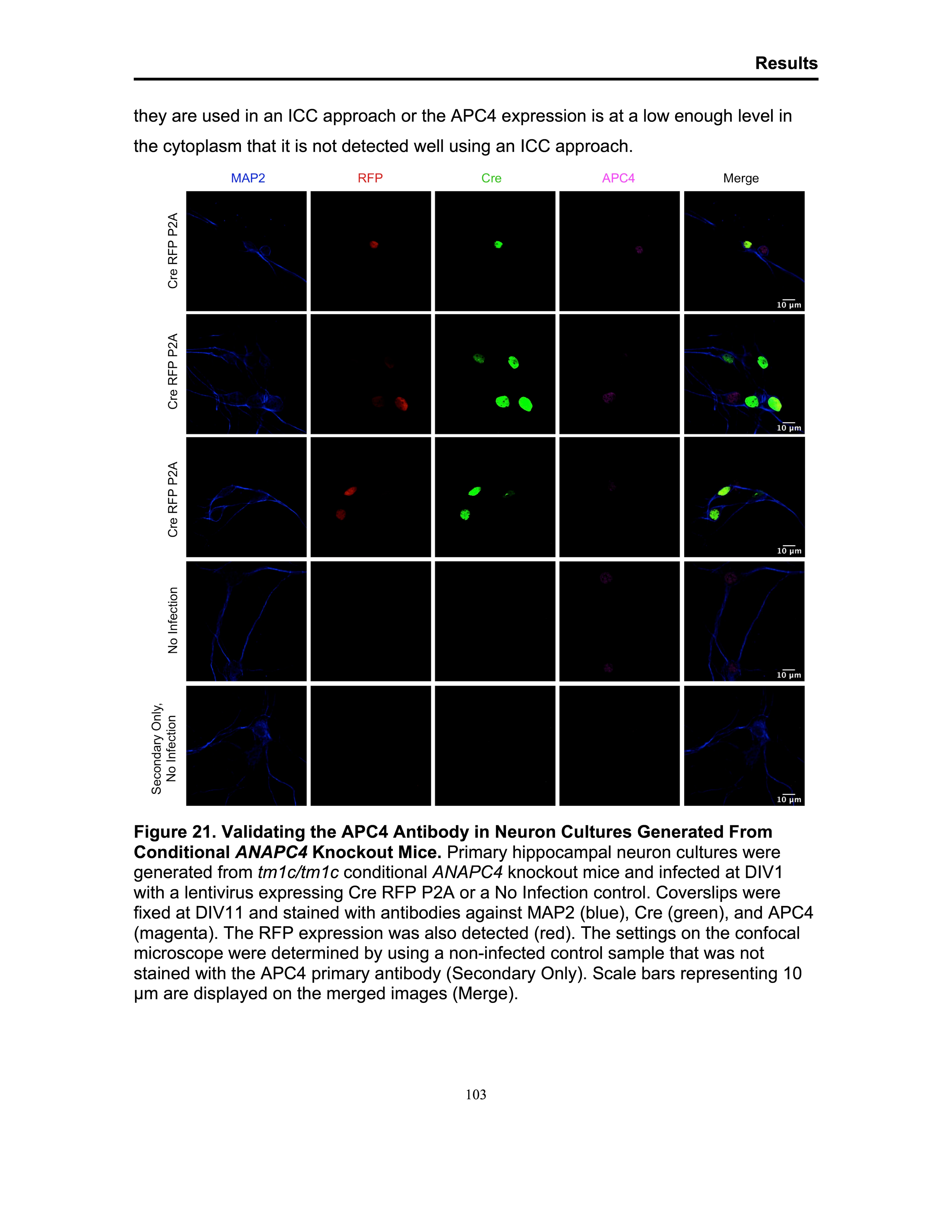
Immunocytochemistry/ Immunofluorescence: Rabbit Polyclonal Apc4 Antibody [NB100-59824] - Primary hippocampal neuron cultures were generated from tm1c/tm1c conditional ANAPC4 knockout mice and infected at DIV1 with a lentivirus expressing Cre RFP P2A or a No Infection control. Coverslips were fixed at DIV11 and stained with antibodies against MAP2 (blue), Cre (green), and APC4 (magenta). The RFP expression was also detected (red). The settings on the confocal microscope were determined by using a non-infected control sample that was not stained with the APC4 primary antibody (Secondary Only). Scale bars representing 10 um are displayed on the merged images (Merge). Image from a verified customer review.Applications for Apc4 Antibody
Application
Recommended Usage
Immunocytochemistry/ Immunofluorescence
Validated for Immunocytochemistry/Immunofluorescence from a verified customer review.
Immunoprecipitation
2-5 ug/mg lysate
Knockout Validated
Validated for Knockout from a verified customer review.
Western Blot
1:2000-1:10000
Reviewed Applications
Read 2 reviews rated 5 using NB100-59824 in the following applications:
Formulation, Preparation, and Storage
Purification
Immunogen affinity purified
Formulation
TBS and 0.1% BSA
Preservative
0.09% Sodium Azide
Concentration
0.2 mg/ml
Shipping
The product is shipped with polar packs. Upon receipt, store it immediately at the temperature recommended below.
Stability & Storage
Store at 4C. Do not freeze.
Background: Apc4
Alternate Names
anaphase promoting complex subunit 4, anaphase-promoting complex subunit 4, APC4Cyclosome subunit 4
Gene Symbol
ANAPC4
UniProt
Additional Apc4 Products
Product Documents for Apc4 Antibody
Certificate of Analysis
To download a Certificate of Analysis, please enter a lot or batch number in the search box below.
Product Specific Notices for Apc4 Antibody
This product is for research use only and is not approved for use in humans or in clinical diagnosis. Primary Antibodies are guaranteed for 1 year from date of receipt.
Citations for Apc4 Antibody
 Powered by Bioz
Customer Reviews for Apc4 Antibody (2)
5 out of 5
2 Customer Ratings
Have you used Apc4 Antibody?
Submit a review and receive an Amazon gift card!
$25/€18/£15/$25CAN/¥2500 Yen for a review with an image
$10/€7/£6/$10CAN/¥1110 Yen for a review without an image
Submit a review
Customer Images

Showing
1
-
2 of
2 reviews
Showing All
Filter By:
-
Application: ImmunocytochemistrySample Tested: primary neuron culturesSpecies: MouseVerified Customer | Posted 10/08/2024KO validation of APC4 antibody by ICC in neuron cultures
 Bio-Techne ResponseThis review reflects a new species or application tested on a primary antibody.
 -
Application: Western Blot and ImmunocytochemistrySample Tested: HEK293 human embryonic kidney cell line and primary cortical and hippocampal neuron culturesSpecies: Mouse and HumanVerified Customer | Posted 10/08/2024Figure 1. Imaging overexpressed HA-APC4. Scale bars: 10 μm. (A) HEK293 cells stained with DAPI (blue), GFP (green), HA (magenta), APC4 (red). (B) Wildtype primary mouse hippocampal cultures stained with DAPI (blue), MAP2 (magenta), HA (red), APC4 (green)WB: 1:5000 (1:2000 for lower expression) ICC: 1:500 with 36-40hr primary incubation (24 or less does not work for both HEK293 and neurons), and this is consistent with other antibodies. APC4 expression is very difficult to detect in general but the signal that is there is absent in knockout cells
Bio-Techne ResponseThis review reflects a new species or application tested on a primary antibody.
There are no reviews that match your criteria.
Protocols
Find general support by application which include: protocols, troubleshooting, illustrated assays, videos and webinars.
- Appropriate Fixation of IHC/ICC Samples
- Cellular Response to Hypoxia Protocols
- ClariTSA™ Fluorophore Kits
- Detection & Visualization of Antibody Binding
- ICC Cell Smear Protocol for Suspension Cells
- ICC Immunocytochemistry Protocol Videos
- ICC for Adherent Cells
- Immunocytochemistry (ICC) Protocol
- Immunocytochemistry Troubleshooting
- Immunofluorescence of Organoids Embedded in Cultrex Basement Membrane Extract
- Immunohistochemistry (IHC) and Immunocytochemistry (ICC) Protocols
- Immunoprecipitation Protocol
- Preparing Samples for IHC/ICC Experiments
- Preventing Non-Specific Staining (Non-Specific Binding)
- Primary Antibody Selection & Optimization
- Protocol for VisUCyte™ HRP Polymer Detection Reagent
- Protocol for the Fluorescent ICC Staining of Cell Smears - Graphic
- Protocol for the Fluorescent ICC Staining of Cultured Cells on Coverslips - Graphic
- Protocol for the Preparation and Fluorescent ICC Staining of Cells on Coverslips
- Protocol for the Preparation and Fluorescent ICC Staining of Non-adherent Cells
- Protocol for the Preparation and Fluorescent ICC Staining of Stem Cells on Coverslips
- Protocol for the Preparation of a Cell Smear for Non-adherent Cell ICC - Graphic
- R&D Systems Quality Control Western Blot Protocol
- TUNEL and Active Caspase-3 Detection by IHC/ICC Protocol
- The Importance of IHC/ICC Controls
- Troubleshooting Guide: Western Blot Figures
- Western Blot Conditions
- Western Blot Protocol
- Western Blot Protocol for Cell Lysates
- Western Blot Troubleshooting
- Western Blot Troubleshooting Guide
- View all Protocols, Troubleshooting, Illustrated assays and Webinars
Loading...