Aquaporin-4 Antibody - BSA Free
Novus Biologicals | Catalog # NBP1-87679
![Immunohistochemistry-Paraffin: Aquaporin-4 Antibody [NBP1-87679]](https://resources.rndsystems.com/images/products/Aquaporin-4-Antibody-Immunohistochemistry-Paraffin-NBP1-87679-img0027.jpg "Immunohistochemistry-Paraffin: Aquaporin-4 Antibody [NBP1-87679]")
Loading...
Key Product Details
Validated by
Orthogonal Validation
Species Reactivity
Validated:
Human, Mouse
Cited:
Human, Mouse, Rat, Porcine, Bovine
Predicted:
Rat (92%). Backed by our 100% Guarantee.
Applications
Validated:
Immunohistochemistry, Immunohistochemistry-Paraffin, Western Blot
Cited:
Immunohistochemistry, Immunohistochemistry-Paraffin, Immunohistochemistry-Frozen, Western Blot, Immunocytochemistry/ Immunofluorescence, Immunoprecipitation, IF/IHC
Label
Unconjugated
Antibody Source
Polyclonal Rabbit IgG
Format
BSA Free
Loading...
Product Specifications
Immunogen
This antibody was developed against Recombinant Protein corresponding to amino acids: CPDVEFKRRFKEAFSKAAQQTKGSYMEVEDNRSQVETDDLILKPGVVHVIDVDRGEEKKGKDQSGEVLSSV
Marker
Astrocytes Marker
Clonality
Polyclonal
Host
Rabbit
Isotype
IgG
Theoretical MW
35 kDa.
Disclaimer note: The observed molecular weight of the protein may vary from the listed predicted molecular weight due to post translational modifications, post translation cleavages, relative charges, and other experimental factors.
Disclaimer note: The observed molecular weight of the protein may vary from the listed predicted molecular weight due to post translational modifications, post translation cleavages, relative charges, and other experimental factors.
Scientific Data Images for Aquaporin-4 Antibody - BSA Free
![Western Blot: Aquaporin-4 Antibody [NBP1-87679]](https://resources.rndsystems.com/images/products/Aquaporin-4-Antibody-Western-Blot-NBP1-87679-img0017.jpg "Western Blot: Aquaporin-4 Antibody [NBP1-87679]")
Western Blot: Aquaporin-4 Antibody [NBP1-87679]
Western Blot: Aquaporin-4 Antibody [NBP1-87679] - Analysis in human brain tissue.![Immunohistochemistry: Aquaporin-4 Antibody [NBP1-87679]](https://resources.rndsystems.com/images/products/Aquaporin-4-Antibody-Immunohistochemistry-Paraffin-NBP1-87679-img0029.jpg "Immunohistochemistry: Aquaporin-4 Antibody [NBP1-87679]")
Immunohistochemistry: Aquaporin-4 Antibody [NBP1-87679]
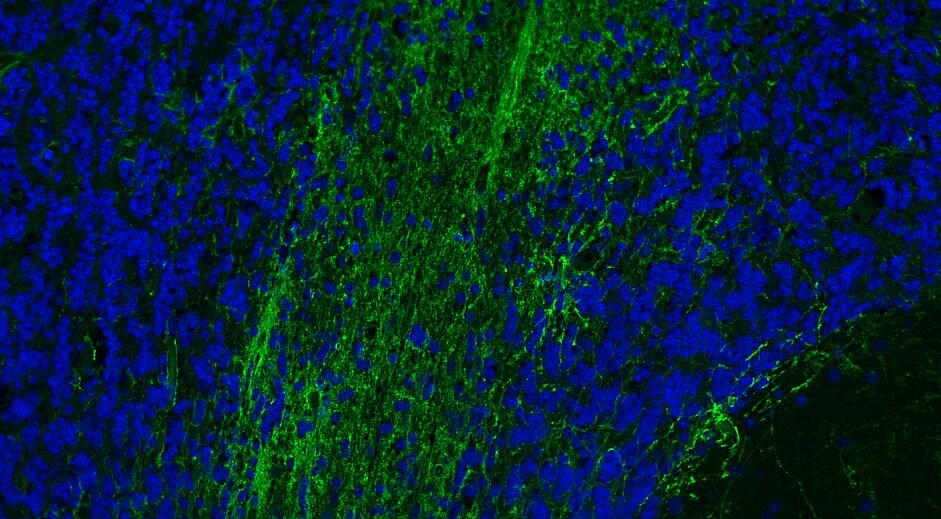
Immunohistochemistry: Aquaporin-4 Antibody [NBP1-87679] - Staining of mouse cerebellum shows positivity in astrocytes, mainly in the molecular layer.![Immunohistochemistry-Paraffin: Aquaporin-4 Antibody [NBP1-87679]](https://resources.rndsystems.com/images/products/Aquaporin-4-Antibody-Immunohistochemistry-Paraffin-NBP1-87679-img0030.jpg "Immunohistochemistry-Paraffin: Aquaporin-4 Antibody [NBP1-87679]")
Immunohistochemistry-Paraffin: Aquaporin-4 Antibody [NBP1-87679]
Immunohistochemistry-Paraffin: Aquaporin-4 Antibody [NBP1-87679] - Staining of human cerebellum shows moderate to strong membranous positivity in astrocytes, mainly in the molecular layer.![Immunohistochemistry-Paraffin: Aquaporin-4 Antibody [NBP1-87679]](https://resources.rndsystems.com/images/products/Aquaporin-4-Antibody-Immunohistochemistry-Paraffin-NBP1-87679-img0031.jpg "Immunohistochemistry-Paraffin: Aquaporin-4 Antibody [NBP1-87679]")
Immunohistochemistry-Paraffin: Aquaporin-4 Antibody [NBP1-87679]
Immunohistochemistry-Paraffin: Aquaporin-4 Antibody [NBP1-87679] - Staining of human cerebral cortex shows moderate to strong membranous positivity in astrocytes.![Immunohistochemistry-Paraffin: Aquaporin-4 Antibody [NBP1-87679]](https://resources.rndsystems.com/images/products/Aquaporin-4-Antibody-Immunohistochemistry-Paraffin-NBP1-87679-img0032.jpg "Immunohistochemistry-Paraffin: Aquaporin-4 Antibody [NBP1-87679]")
Immunohistochemistry-Paraffin: Aquaporin-4 Antibody [NBP1-87679]
Immunohistochemistry-Paraffin: Aquaporin-4 Antibody [NBP1-87679] - Staining of human liver shows no positivity in hepatocytes, as expected.![Immunohistochemistry-Paraffin: Aquaporin-4 Antibody [NBP1-87679]](https://resources.rndsystems.com/images/products/Aquaporin-4-Antibody-Immunohistochemistry-Paraffin-NBP1-87679-img0033.jpg "Immunohistochemistry-Paraffin: Aquaporin-4 Antibody [NBP1-87679]")
Immunohistochemistry-Paraffin: Aquaporin-4 Antibody [NBP1-87679]
Immunohistochemistry-Paraffin: Aquaporin-4 Antibody [NBP1-87679] - Staining of human stomach shows strong membranous positivity in glandular cells.
Western Blot: Aquaporin-4 Antibody - BSA Free [NBP1-87679] -
Induction of atrogin 1 mRNA and protein expression by rotator cuff tear (RCT). (a) Expression of transcripts encoding the E3 ubiquitin-protein ligases atrogin 1, MuRF1, and UBR2 in injured muscle after RCT (n = 12 per group). (b) Atrogin 1 protein expression in muscle isolated at different stages after RCT (n = 7 per group). Densitometric analyses of the western blots are shown on the right. Data represent means +/- standard errors of the means. *P < 0.05. (c) Immunofluorescence (IF) microscopy analyses of muscle isolated at different stages after RCT. The red signal represents atrogin 1 protein. The negative control (secondary antibody only) for the IF showed no specific signal (data not shown). (d) Loss of AQP4 protein by overexpression of atrogin 1. C2C12 cells were treated with an adenovirus expressing atrogin 1 [Ad-atrogin 1; multiplicity of infection (MOI) = 50, 100, 200] for 48 h, and western blot analysis was performed with each designated antibody. (e) Monomeric ubiquitin protein expression in muscle cells isolated at different stages after RCT. Image collected and cropped by CiteAb from the following open publication (https://pubmed.ncbi.nlm.nih.gov/32843684), licensed under a CC-BY license. Not internally tested by Novus Biologicals.
Western Blot: Aquaporin-4 Antibody - BSA Free [NBP1-87679] -
Aquaporin levels in the sulfatide deficient mouse brain. (A) Expression of AQP1 and AQP4 in Cre+ and Cre− cerebrum samples were measured by Western Blot and quantified (B,C). Multiple AQP4 bands with different levels of glycosylation were observed (bracket). (D) Representative immunofluorescence image for AQP4 (red) in the CST cKO mouse brain at 20 months of age. Scale bar: 250 μm. (E) AQP4 immunofluorescent area around the lateral ventricle was quantified using Image J. Unpaired two-tailed t-test (normality and equal variance were assumed/confirmed). ** p < 0.01. Image collected and cropped by CiteAb from the following open publication (https://pubmed.ncbi.nlm.nih.gov/36613677), licensed under a CC-BY license. Not internally tested by Novus Biologicals.
Western Blot: Aquaporin-4 Antibody - BSA Free [NBP1-87679] -
Increased levels of HMGB1 protein in muscle injured by rotator cuff tear (RCT). (a) Expression level of HMGB1 protein in muscle isolated at different stages after RCT (n = 7 per group). Densitometric analyses of the western blots are shown on the right. Data represent means +/- standard errors of the means. *P < 0.05. (b) Immunofluorescence (IF) microscopy analyses of muscle cells isolated at different stages after RCT. The green signal represents HMGB1 protein. The negative control (secondary antibody only) for the IF showed no specific signal (data not shown). (c) Expression of atrogin 1 mRNA in C2C12 myotubes treated with 200 ng/mL recombinant mouse HMGB1 for the indicated time periods. (d) Atrogin 1, ubiquitin, and AQP4 protein levels in C2C12 myotubes treated with 200 ng/mL HMGB1 and an adenovirus expressing atrogin 1 [Ad-atrogin 1; multiplicity of infection (MOI) = 100] for 48 h. (e) Ubiquitination of AQP4 caused by HMGB1 and atrogin 1. C2C12 myotubes were treated with 200 ng/mL HMGB1 and Ad-atrogin 1 (MOI = 100) with or without MG132. After 48 h of incubation, cell lysates were prepared for a ubiquitination assay using anti-AQP4 and anti-ubiquitin antibodies. (f) Expression of atrogin 1 mRNA in C2C12 myotubes exposed to 5 μM SB203580 (a p38 MAPK inhibitor), LY294002 (a PI3K inhibitor), BAY 11-7082 (an NF-kappa B inhibitor), PD98059 (an MEK inhibitor), or SP600125 (a JNK inhibitor). Cells were exposed to each inhibitor for 1 h prior to treatment with 200 ng/mL HMGB1 for 24 h. Image collected and cropped by CiteAb from the following open publication (https://pubmed.ncbi.nlm.nih.gov/32843684), licensed under a CC-BY license. Not internally tested by Novus Biologicals.
Western Blot: Aquaporin-4 Antibody - BSA Free [NBP1-87679] -
Reduced aquaporin 4 (AQP4) protein levels in atrophied muscle following rotator cuff tear (RCT). (a) Reduced myofiber size in muscle injured by RCT. Magnified view of the H&E-stained muscle. Magnification, × 400. Scale bar, 50 μm. The yellow dotted line indicates the muscle myofiber. (b) Myofiber size in control (CTL) and RCT muscles. The size of each dotted area was measured using ImageJ software. *P < 0.05. (c) Western blot analyses for myosin heavy chain (MYH) protein expression between control and the injured muscle after RCT (n = 7 per group). Densitometric analyses of the western blots are shown in the graphs. Data represents mean +/- SEM. *p < 0.05. (d) Reduced aquaporin 4 (AQP4) protein levels in atrophied muscle following RCT (n = 7 per group). Densitometric analyses of the western blots are shown on the right. Data represent means +/- standard errors of the means. *P < 0.05. (e) Immunofluorescence microscopy analyses of muscle cells isolated at different stages after RCT (n = 3 per group). The green signal in each panel represents membranous AQP4 protein and the blue signal represents cell nuclei. Magnification, × 200. Scale bar, 100 μm. Image collected and cropped by CiteAb from the following open publication (https://pubmed.ncbi.nlm.nih.gov/32843684), licensed under a CC-BY license. Not internally tested by Novus Biologicals.
Western Blot: Aquaporin-4 Antibody - BSA Free [NBP1-87679] -
Western blot results. (A) Western blot analysis of SNTA1 and AQP4 protein expression in Group C, Group PDN, and Group BHB. (B) Relative protein expression of SNTA1 and AQP4. Due to the reason that some blots in the original saved images were not mentioned in the present study, we spliced the blots and the typical ones were chosen to indicate the differences in the expression of proteins. Refer to Figure 1 for the meaning of the grouping abbreviation. Values were presented as mean +/- SEM. *p < 0.05 compared with values in Group PDN. #p < 0.05 compared with values in Group C. SNTA1 means alpha -syntrophin, and AQP4 means aquaporin-4 protein. Image collected and cropped by CiteAb from the following open publication (https://pubmed.ncbi.nlm.nih.gov/35898407), licensed under a CC-BY license. Not internally tested by Novus Biologicals.Applications for Aquaporin-4 Antibody - BSA Free
Application
Recommended Usage
Immunohistochemistry
1:2500 - 1:5000
Immunohistochemistry-Paraffin
1:2500 - 1:5000
Western Blot
0.04 - 0.4 ug/mL
Application Notes
IHC-Paraffin, HIER pH 6 retrieval is recommended.
Reviewed Applications
Read 2 reviews rated 4.5 using NBP1-87679 in the following applications:
Formulation, Preparation, and Storage
Purification
Affinity purified
Formulation
PBS (pH 7.2) and 40% Glycerol
Format
BSA Free
Preservative
0.02% Sodium Azide
Concentration
Concentrations vary lot to lot. See vial label for concentration. If unlisted please contact technical services.
Shipping
The product is shipped with polar packs. Upon receipt, store it immediately at the temperature recommended below.
Stability & Storage
Store at 4C short term. Aliquot and store at -20C long term. Avoid freeze-thaw cycles.
Background: Aquaporin 4/AQP4
Long Name
Aquaporin 4
Alternate Names
AQP-4, AQP4, MIWC, WCH4
Entrez Gene IDs
361 (Human)
Gene Symbol
AQP4
UniProt
Additional Aquaporin 4/AQP4 Products
Product Documents for Aquaporin-4 Antibody - BSA Free
Certificate of Analysis
To download a Certificate of Analysis, please enter a lot or batch number in the search box below.
Product Specific Notices for Aquaporin-4 Antibody - BSA Free
This product is for research use only and is not approved for use in humans or in clinical diagnosis. Primary Antibodies are guaranteed for 1 year from date of receipt.
Related Research Areas
Citations for Aquaporin-4 Antibody - BSA Free
 Powered by Bioz
Customer Reviews for Aquaporin-4 Antibody - BSA Free (2)
4.5 out of 5
2 Customer Ratings
Have you used Aquaporin-4 Antibody - BSA Free?
Submit a review and receive an Amazon gift card!
$25/€18/£15/$25CAN/¥2500 Yen for a review with an image
$10/€7/£6/$10CAN/¥1110 Yen for a review without an image
Submit a review
Customer Images
Showing
1
-
2 of
2 reviews
Showing All
Filter By:
-
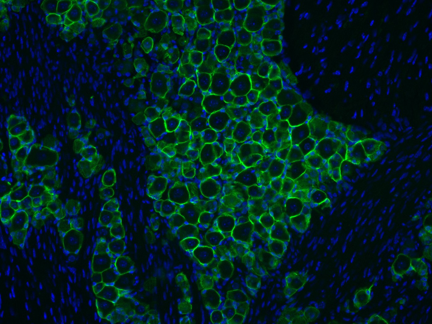
Application: Immunohistochemistry-ParaffinSample Tested: Brain (cerebral cortex)Species: HumanVerified Customer | Posted 12/24/2020AQP4 rabbit (1:2500) was incubated overnight at 4C on human cerebral cortex and visualised using AF Donkey anti-rabbit 488 (green).

-
Application: Immunohistochemistry-FrozenSample Tested: Trigeminal gangliaSpecies: MouseVerified Customer | Posted 11/22/2018Mouse trigeminal ganglion stained with aquaporin-4 antibody (1:5000 dilution).Mice were perfused with 0.9% saline and subsequently decapitated. The cerebrum was then dissected out of the cranium and the head was put in ~10mL of 4% PFA in 0.1M phosphate buffer overnight. The following morning the trigeminal ganglia were dissected and placed in 30% sucrose (sucrose dissolved in 0.1M phosphate buffer) for at least 48hr. The tissue was then blocked in OCT, frozen, cut at 10 microns (on a cryostat) and mounted on glass slides. Slides were washed three times with 1X PBS (diluted from a 10X stock (recipe available on thelabrat.com)) and blocked for 1hr in 1X PBS containing 0.2% triton X-100 and 10% goat serum. After the block, tissue was incubated overnight at room temperature in 1X PBS containing 0.2% triton X-100, 2% goat serum, 20mg/mL bovine serum albumin and the primary antibody diluted to a final concentration of 1:5000. The following morning the slides were washed twice with 1X PBS containing 0.2% tween-20 and once with 1X PBS. The slides were then incubated for 45 min (room temperature) in 1X PBS containing 0.2% triton X-100, 2% goat serum, 20mg/mL bovine serum albumin and 488-conjugated goat-anti rabbit secondary (ThermoFisher, 1:200). After the 45 min incubation period, slides were washed with washed twice with 1X PBS containing 0.2% tween 20 and once with 1X PBS. The slides were then coverslipped and mounted using vectashield mounting medium.
There are no reviews that match your criteria.
Protocols
Find general support by application which include: protocols, troubleshooting, illustrated assays, videos and webinars.
- Antigen Retrieval Protocol (PIER)
- Antigen Retrieval for Frozen Sections Protocol
- Appropriate Fixation of IHC/ICC Samples
- Cellular Response to Hypoxia Protocols
- Chromogenic IHC Staining of Formalin-Fixed Paraffin-Embedded (FFPE) Tissue Protocol
- Chromogenic Immunohistochemistry Staining of Frozen Tissue
- ClariTSA™ Fluorophore Kits
- Detection & Visualization of Antibody Binding
- Fluorescent IHC Staining of Frozen Tissue Protocol
- Graphic Protocol for Heat-induced Epitope Retrieval
- Graphic Protocol for the Preparation and Fluorescent IHC Staining of Frozen Tissue Sections
- Graphic Protocol for the Preparation and Fluorescent IHC Staining of Paraffin-embedded Tissue Sections
- Graphic Protocol for the Preparation of Gelatin-coated Slides for Histological Tissue Sections
- IHC Sample Preparation (Frozen sections vs Paraffin)
- Immunofluorescent IHC Staining of Formalin-Fixed Paraffin-Embedded (FFPE) Tissue Protocol
- Immunohistochemistry (IHC) and Immunocytochemistry (ICC) Protocols
- Immunohistochemistry Frozen Troubleshooting
- Immunohistochemistry Paraffin Troubleshooting
- Preparing Samples for IHC/ICC Experiments
- Preventing Non-Specific Staining (Non-Specific Binding)
- Primary Antibody Selection & Optimization
- Protocol for Heat-Induced Epitope Retrieval (HIER)
- Protocol for Making a 4% Formaldehyde Solution in PBS
- Protocol for VisUCyte™ HRP Polymer Detection Reagent
- Protocol for the Preparation & Fixation of Cells on Coverslips
- Protocol for the Preparation and Chromogenic IHC Staining of Frozen Tissue Sections
- Protocol for the Preparation and Chromogenic IHC Staining of Frozen Tissue Sections - Graphic
- Protocol for the Preparation and Chromogenic IHC Staining of Paraffin-embedded Tissue Sections
- Protocol for the Preparation and Chromogenic IHC Staining of Paraffin-embedded Tissue Sections - Graphic
- Protocol for the Preparation and Fluorescent IHC Staining of Frozen Tissue Sections
- Protocol for the Preparation and Fluorescent IHC Staining of Paraffin-embedded Tissue Sections
- Protocol for the Preparation of Gelatin-coated Slides for Histological Tissue Sections
- R&D Systems Quality Control Western Blot Protocol
- TUNEL and Active Caspase-3 Detection by IHC/ICC Protocol
- The Importance of IHC/ICC Controls
- Troubleshooting Guide: Immunohistochemistry
- Troubleshooting Guide: Western Blot Figures
- Western Blot Conditions
- Western Blot Protocol
- Western Blot Protocol for Cell Lysates
- Western Blot Troubleshooting
- Western Blot Troubleshooting Guide
- View all Protocols, Troubleshooting, Illustrated assays and Webinars
Loading...