ARHGAP22 [p Ser22] Antibody - Azide and BSA Free
Novus Biologicals | Catalog # NBP1-44072
![Western Blot: ARHGAP22 [p Ser22] Antibody [NBP1-44072]](https://resources.rndsystems.com/images/products/ARHGAP22-[p-Ser22]-Antibody-Western-Blot-NBP1-44072-img0004.jpg "Western Blot: ARHGAP22 [p Ser22] Antibody [NBP1-44072]")
Loading...
Key Product Details
Species Reactivity
Human, Mouse, Rat
Applications
Immunohistochemistry, Western Blot, ELISA, Immunocytochemistry/ Immunofluorescence
Label
Unconjugated
Antibody Source
Polyclonal Rabbit IgG
Format
Azide and BSA Free
Loading...
Product Specifications
Immunogen
ARHGAP22 [p Ser22] Antibody was prepared from whole rabbit serum produced by repeated immunizations with a synthetic phospho-peptide corresponding to the region surrounding mouse pS22 region of ARHGAP22. (Uniprot: Q8BL80)
Reactivity Notes
A BLAST analysis was used to suggest cross-reactivity with Mouse, Rat and Human based on 100% sequence homology. Cross-reactivity with ARHGAP22 pS22 from other sources has not been determined.
Modification
p Ser22
Specificity
This antibody is specific for phosphorylated ARHGAP22 at Serine 22. It also recognizes the S397->A mutation but not the S22->A mutation. A BLAST analysis was used to suggest cross-reactivity with Mouse, Rat and Human based on 100% sequence homology. Cross-reactivity with ARHGAP22 pS22 from other sources has not been determined.
Clonality
Polyclonal
Host
Rabbit
Isotype
IgG
Description
This antibody was affinity purified from monospecific antiserum by immunoaffinity chromatography
Store vial at -20C prior to opening. Aliquot contents and freeze at -20C or below for extended storage. Avoid cycles of freezing and thawing. Centrifuge product if not completely clear after standing at room temperature. This product is stable for several weeks at 4C as an undiluted liquid. Dilute only prior to immediate use.
Store vial at -20C prior to opening. Aliquot contents and freeze at -20C or below for extended storage. Avoid cycles of freezing and thawing. Centrifuge product if not completely clear after standing at room temperature. This product is stable for several weeks at 4C as an undiluted liquid. Dilute only prior to immediate use.
Scientific Data Images for ARHGAP22 [p Ser22] Antibody - Azide and BSA Free
Western Blot: ARHGAP22 [p Ser22] Antibody [NBP1-44072]
Western Blot: ARHGAP22 [p Ser22] Antibody [NBP1-44072] - Lane 1: NIH3T3 cells transfected with a null vector. Lane 2: NIH3T3 cells transfected with ARHGAP22. Lane 3: NIH3T3 cells transfected with ARHGAP22 S22 to alanine mutation. Lane 4: NIH3T3 cells transfected with ARHGAP22 S397 to alanine mutation. Primary antibody: Left: ARHGAP22 pS22, Right: ARHGAP22 pS397 antibody at 1ug/mL for overnight at 4C. Secondary antibody: IRDye800 rabbit secondary antibody at 1:10,000 for 45 min at RT. Block: 5% BLOTTO O/N at 4C. Predicted/Observed size: 68 kDa for ARHGAP22. Other band(s): Unmodified ARHGAP22. ARHGAP22-pS22 antibody recognizes the S397>A mutation, not the S22>mutation; ARHGAP22 pS397 recognizes the pS22>A mutation, not the pS397>A mutation; Confirms the specificity of each ARHGAP22 phospho specific antibody.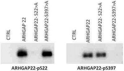
Western Blot: ARHGAP22 [p Ser22] Antibody [NBP1-44072] - ARHGAP22 [Phospo Ser22] Antibody [NBP1-44072] - Protein Molecular weight: 68 kDa Cell extracts from NIH 3T3cells that were transfected with either a null vector (CTRL), ARHGAP22, ARHGAP22-serine 22 to alanine mutation, ARHGAP22 serine 397 to alanine mutation were electroblotted. The left panel was probed with the NBP1-44072 antibody, while the right panel was probed with the NBP1-44073 antibody, each at 1ug/ml. Each antibody recognizes the unmodified ARHGAP22. NBP1-44072 antibody recognized the S397-A mutation but not the S22-A mutation, while NBP1-44073 antibody recognizes the pS22-A mutation and not the pS397-A mutation. This data confirms the specificity of each ARHGAP22 phospho specific antibody.
Applications for ARHGAP22 [p Ser22] Antibody - Azide and BSA Free
Application
Recommended Usage
ELISA
1:20000-1:60000
Immunocytochemistry/ Immunofluorescence
1:100-1:500
Immunohistochemistry
1:100-1:500
Western Blot
1 ug/ml
Application Notes
This product is tested in Western Blot and ELISA useful for Immunostaining. Specific conditions for reactivity should be optimized by the end user. Expect a band approximately ~77.8 kDa corresponding to the appropriate cell lysate or extract.
Formulation, Preparation, and Storage
Purification
Immunogen affinity purified
Formulation
0.02 M Potassium Phosphate, 0.15 M Sodium Chloride, pH 7.2, 50% (v/v) Glycerol
Format
Azide and BSA Free
Preservative
No Preservative
Concentration
Please see the vial label for concentration. If unlisted please contact technical services.
Shipping
The product is shipped with polar packs. Upon receipt, store it immediately at the temperature recommended below.
Stability & Storage
Store at -20C short term. Aliquot and store at -80C long term. Avoid freeze-thaw cycles.
Background: ARHGAP22
Alternate Names
Rho GTPase activating protein 22, rho GTPase-activating protein 22, RhoGAP2, Rho-type GTPase-activating protein 22
Entrez Gene IDs
58504 (Human)
Gene Symbol
ARHGAP22
UniProt
Additional ARHGAP22 Products
Product Documents for ARHGAP22 [p Ser22] Antibody - Azide and BSA Free
Certificate of Analysis
To download a Certificate of Analysis, please enter a lot or batch number in the search box below.
Product Specific Notices for ARHGAP22 [p Ser22] Antibody - Azide and BSA Free
This product is for research use only and is not approved for use in humans or in clinical diagnosis. Primary Antibodies are guaranteed for 1 year from date of receipt.
Citations for ARHGAP22 [p Ser22] Antibody - Azide and BSA Free
 Powered by Bioz
Customer Reviews for ARHGAP22 [p Ser22] Antibody - Azide and BSA Free
There are currently no reviews for this product. Be the first to review ARHGAP22 [p Ser22] Antibody - Azide and BSA Free and earn rewards!
Have you used ARHGAP22 [p Ser22] Antibody - Azide and BSA Free?
Submit a review and receive an Amazon gift card!
$25/€18/£15/$25CAN/¥2500 Yen for a review with an image
$10/€7/£6/$10CAN/¥1110 Yen for a review without an image
Submit a review
Protocols
Find general support by application which include: protocols, troubleshooting, illustrated assays, videos and webinars.
- Antigen Retrieval Protocol (PIER)
- Antigen Retrieval for Frozen Sections Protocol
- Appropriate Fixation of IHC/ICC Samples
- Cellular Response to Hypoxia Protocols
- Chromogenic IHC Staining of Formalin-Fixed Paraffin-Embedded (FFPE) Tissue Protocol
- Chromogenic Immunohistochemistry Staining of Frozen Tissue
- ClariTSA™ Fluorophore Kits
- Detection & Visualization of Antibody Binding
- ELISA Sample Preparation & Collection Guide
- ELISA Troubleshooting Guide
- Fluorescent IHC Staining of Frozen Tissue Protocol
- Graphic Protocol for Heat-induced Epitope Retrieval
- Graphic Protocol for the Preparation and Fluorescent IHC Staining of Frozen Tissue Sections
- Graphic Protocol for the Preparation and Fluorescent IHC Staining of Paraffin-embedded Tissue Sections
- Graphic Protocol for the Preparation of Gelatin-coated Slides for Histological Tissue Sections
- How to Run an R&D Systems DuoSet ELISA
- How to Run an R&D Systems Quantikine ELISA
- How to Run an R&D Systems Quantikine™ QuicKit™ ELISA
- ICC Cell Smear Protocol for Suspension Cells
- ICC Immunocytochemistry Protocol Videos
- ICC for Adherent Cells
- IHC Sample Preparation (Frozen sections vs Paraffin)
- Immunocytochemistry (ICC) Protocol
- Immunocytochemistry Troubleshooting
- Immunofluorescence of Organoids Embedded in Cultrex Basement Membrane Extract
- Immunofluorescent IHC Staining of Formalin-Fixed Paraffin-Embedded (FFPE) Tissue Protocol
- Immunohistochemistry (IHC) and Immunocytochemistry (ICC) Protocols
- Immunohistochemistry Frozen Troubleshooting
- Immunohistochemistry Paraffin Troubleshooting
- Preparing Samples for IHC/ICC Experiments
- Preventing Non-Specific Staining (Non-Specific Binding)
- Primary Antibody Selection & Optimization
- Protocol for Heat-Induced Epitope Retrieval (HIER)
- Protocol for Making a 4% Formaldehyde Solution in PBS
- Protocol for VisUCyte™ HRP Polymer Detection Reagent
- Protocol for the Fluorescent ICC Staining of Cell Smears - Graphic
- Protocol for the Fluorescent ICC Staining of Cultured Cells on Coverslips - Graphic
- Protocol for the Preparation & Fixation of Cells on Coverslips
- Protocol for the Preparation and Chromogenic IHC Staining of Frozen Tissue Sections
- Protocol for the Preparation and Chromogenic IHC Staining of Frozen Tissue Sections - Graphic
- Protocol for the Preparation and Chromogenic IHC Staining of Paraffin-embedded Tissue Sections
- Protocol for the Preparation and Chromogenic IHC Staining of Paraffin-embedded Tissue Sections - Graphic
- Protocol for the Preparation and Fluorescent ICC Staining of Cells on Coverslips
- Protocol for the Preparation and Fluorescent ICC Staining of Non-adherent Cells
- Protocol for the Preparation and Fluorescent ICC Staining of Stem Cells on Coverslips
- Protocol for the Preparation and Fluorescent IHC Staining of Frozen Tissue Sections
- Protocol for the Preparation and Fluorescent IHC Staining of Paraffin-embedded Tissue Sections
- Protocol for the Preparation of Gelatin-coated Slides for Histological Tissue Sections
- Protocol for the Preparation of a Cell Smear for Non-adherent Cell ICC - Graphic
- Quantikine HS ELISA Kit Assay Principle, Alkaline Phosphatase
- Quantikine HS ELISA Kit Principle, Streptavidin-HRP Polymer
- R&D Systems Quality Control Western Blot Protocol
- Sandwich ELISA (Colorimetric) – Biotin/Streptavidin Detection Protocol
- Sandwich ELISA (Colorimetric) – Direct Detection Protocol
- TUNEL and Active Caspase-3 Detection by IHC/ICC Protocol
- The Importance of IHC/ICC Controls
- Troubleshooting Guide: ELISA
- Troubleshooting Guide: Immunohistochemistry
- Troubleshooting Guide: Western Blot Figures
- Western Blot Conditions
- Western Blot Protocol
- Western Blot Protocol for Cell Lysates
- Western Blot Troubleshooting
- Western Blot Troubleshooting Guide
- View all Protocols, Troubleshooting, Illustrated assays and Webinars
Loading...