Asialoglycoprotein Receptor 2 Antibody - BSA Free
Novus Biologicals | Catalog # NBP1-85578
![Immunohistochemistry-Paraffin: Asialoglycoprotein Receptor 2 Antibody [NBP1-85578]](https://resources.rndsystems.com/images/products/Asialoglycoprotein-Receptor-2-Antibody-Immunohistochemistry-Paraffin-NBP1-85578-img0011.jpg "Immunohistochemistry-Paraffin: Asialoglycoprotein Receptor 2 Antibody [NBP1-85578]")
Loading...
Key Product Details
Validated by
Orthogonal Validation, Independent Antibodies
Species Reactivity
Human
Applications
Immunohistochemistry, Immunohistochemistry-Paraffin, Western Blot, Simple Western
Label
Unconjugated
Antibody Source
Polyclonal Rabbit IgG
Format
BSA Free
Loading...
Product Specifications
Immunogen
This antibody was developed against Recombinant Protein corresponding to amino acids: QSAQLQAELRSLKEAFSNFSSSTLTEVQAISTHGGSVGDKITSLGAKLEKQQQDLKADHDALLFHLKHFPVDLRFVACQMELLHSNGSQRTCC
Clonality
Polyclonal
Host
Rabbit
Isotype
IgG
Scientific Data Images for Asialoglycoprotein Receptor 2 Antibody - BSA Free
Immunohistochemistry-Paraffin: Asialoglycoprotein Receptor 2 Antibody [NBP1-85578]
Immunohistochemistry-Paraffin: Asialoglycoprotein Receptor 2 Antibody [NBP1-85578] - Staining in human liver and pancreas tissues using anti-ASGR2 antibody. Corresponding ASGR2 RNA-seq data are presented for the same tissues.![Immunohistochemistry-Paraffin: Asialoglycoprotein Receptor 2 Antibody [NBP1-85578]](https://resources.rndsystems.com/images/products/Asialoglycoprotein-Receptor-2-Antibody-Immunohistochemistry-Paraffin-NBP1-85578-img0014.jpg "Immunohistochemistry-Paraffin: Asialoglycoprotein Receptor 2 Antibody [NBP1-85578]")
Immunohistochemistry-Paraffin: Asialoglycoprotein Receptor 2 Antibody [NBP1-85578]
Immunohistochemistry-Paraffin: Asialoglycoprotein Receptor 2 Antibody [NBP1-85578] - Staining of human colon, liver, lymph node and pancreas using Anti-ASGR2 antibody NBP1-85578 (A) shows similar protein distribution across tissues to independent antibody NBP1-85579 (B).![Immunohistochemistry-Paraffin: Asialoglycoprotein Receptor 2 Antibody [NBP1-85578]](https://resources.rndsystems.com/images/products/Asialoglycoprotein-Receptor-2-Antibody-Immunohistochemistry-Paraffin-NBP1-85578-img0018.jpg "Immunohistochemistry-Paraffin: Asialoglycoprotein Receptor 2 Antibody [NBP1-85578]")
Immunohistochemistry-Paraffin: Asialoglycoprotein Receptor 2 Antibody [NBP1-85578]
Immunohistochemistry-Paraffin: Asialoglycoprotein Receptor 2 Antibody [NBP1-85578] - Staining of human pancreas using Anti-ASGR2 antibody NBP1-85578.![Immunohistochemistry-Paraffin: Asialoglycoprotein Receptor 2 Antibody [NBP1-85578]](https://resources.rndsystems.com/images/products/Asialoglycoprotein-Receptor-2-Antibody-Immunohistochemistry-Paraffin-NBP1-85578-img0009.jpg "Immunohistochemistry-Paraffin: Asialoglycoprotein Receptor 2 Antibody [NBP1-85578]")
Immunohistochemistry-Paraffin: Asialoglycoprotein Receptor 2 Antibody [NBP1-85578]
Immunohistochemistry-Paraffin: Asialoglycoprotein Receptor 2 Antibody [NBP1-85578] - Staining of human liver shows high expression.![Immunohistochemistry-Paraffin: Asialoglycoprotein Receptor 2 Antibody [NBP1-85578]](https://resources.rndsystems.com/images/products/Asialoglycoprotein-Receptor-2-Antibody-Immunohistochemistry-Paraffin-NBP1-85578-img0010.jpg "Immunohistochemistry-Paraffin: Asialoglycoprotein Receptor 2 Antibody [NBP1-85578]")
Immunohistochemistry-Paraffin: Asialoglycoprotein Receptor 2 Antibody [NBP1-85578]
Immunohistochemistry-Paraffin: Asialoglycoprotein Receptor 2 Antibody [NBP1-85578] - Staining of human pancreas shows low expression as expected.![Immunohistochemistry-Paraffin: Asialoglycoprotein Receptor 2 Antibody [NBP1-85578]](https://resources.rndsystems.com/images/products/Asialoglycoprotein-Receptor-2-Antibody-Immunohistochemistry-Paraffin-NBP1-85578-img0015.jpg "Immunohistochemistry-Paraffin: Asialoglycoprotein Receptor 2 Antibody [NBP1-85578]")
Immunohistochemistry-Paraffin: Asialoglycoprotein Receptor 2 Antibody [NBP1-85578]
Immunohistochemistry-Paraffin: Asialoglycoprotein Receptor 2 Antibody [NBP1-85578] - Staining of human lymph node.![Immunohistochemistry-Paraffin: Asialoglycoprotein Receptor 2 Antibody [NBP1-85578]](https://resources.rndsystems.com/images/products/Asialoglycoprotein-Receptor-2-Antibody-Immunohistochemistry-Paraffin-NBP1-85578-img0016.jpg "Immunohistochemistry-Paraffin: Asialoglycoprotein Receptor 2 Antibody [NBP1-85578]")
Immunohistochemistry-Paraffin: Asialoglycoprotein Receptor 2 Antibody [NBP1-85578]
Immunohistochemistry-Paraffin: Asialoglycoprotein Receptor 2 Antibody [NBP1-85578] - Staining of human colon.![Immunohistochemistry-Paraffin: Asialoglycoprotein Receptor 2 Antibody [NBP1-85578]](https://resources.rndsystems.com/images/products/Asialoglycoprotein-Receptor-2-Antibody-Immunohistochemistry-Paraffin-NBP1-85578-img0017.jpg "Immunohistochemistry-Paraffin: Asialoglycoprotein Receptor 2 Antibody [NBP1-85578]")
Immunohistochemistry-Paraffin: Asialoglycoprotein Receptor 2 Antibody [NBP1-85578]
Immunohistochemistry-Paraffin: Asialoglycoprotein Receptor 2 Antibody [NBP1-85578] - Staining of human liver using Anti-ASGR2 antibody NBP1-85578.![Simple Western: Asialoglycoprotein Receptor 2 Antibody [NBP1-85578]](https://resources.rndsystems.com/images/products/Asialoglycoprotein-Receptor-2-Antibody-Simple-Western-NBP1-85578-img0006.jpg "Simple Western: Asialoglycoprotein Receptor 2 Antibody [NBP1-85578]")
Simple Western: Asialoglycoprotein Receptor 2 Antibody [NBP1-85578]
Simple Western: Asialoglycoprotein Receptor 2 Antibody [NBP1-85578] - Simple Western lane view shows a specific band for ASGR2 in 0.2 mg/ml of HepG2 lysate. This experiment was performed under reducing conditions using the 12-230 kDa separation system.![Asialoglycoprotein Receptor 2 Antibody - BSA Free Western Blot: Asialoglycoprotein Receptor 2 Antibody - BSA Free [NBP1-85578]](https://resources.rndsystems.com/images/products/nbp1-85578_rabbit-polyclonal-asialoglycoprotein-receptor-2-antibody-24420259402423.jpg "Western Blot: Asialoglycoprotein Receptor 2 Antibody - BSA Free [NBP1-85578]")
Western Blot: Asialoglycoprotein Receptor 2 Antibody - BSA Free [NBP1-85578]
Analysis in human cell line HepG2.Applications for Asialoglycoprotein Receptor 2 Antibody - BSA Free
Application
Recommended Usage
Immunohistochemistry
1:5000 - 1:10000
Immunohistochemistry-Paraffin
1:5000 - 1:10000
Simple Western
1:20
Western Blot
0.04 - 0.4 ug/ml
Application Notes
IHC-Paraffin, HIER pH 6 retrieval is recommended.
In Simple Western only 10 - 15 uL of the recommended dilution is used per data point.
See Simple Western Antibody Database for Simple Western validation: Tested in HepG2, separated by Size, antibody dilution of 1:20, apparent MW was 66 kDa. Separated by Size-Wes, Sally Sue/Peggy Sue.
In Simple Western only 10 - 15 uL of the recommended dilution is used per data point.
See Simple Western Antibody Database for Simple Western validation: Tested in HepG2, separated by Size, antibody dilution of 1:20, apparent MW was 66 kDa. Separated by Size-Wes, Sally Sue/Peggy Sue.
Reviewed Applications
Read 1 review rated 5 using NBP1-85578 in the following applications:
Formulation, Preparation, and Storage
Purification
Affinity purified
Formulation
PBS (pH 7.2) and 40% Glycerol
Format
BSA Free
Preservative
0.02% Sodium Azide
Concentration
Concentrations vary lot to lot. See vial label for concentration. If unlisted please contact technical services.
Shipping
The product is shipped with polar packs. Upon receipt, store it immediately at the temperature recommended below.
Stability & Storage
Store at 4C short term. Aliquot and store at -20C long term. Avoid freeze-thaw cycles.
Background: ASGR2
Long Name
Asialoglycoprotein Receptor 2
Alternate Names
ASGPR2, CLEC4H2, HBXBP
Gene Symbol
ASGR2
Additional ASGR2 Products
Product Documents for Asialoglycoprotein Receptor 2 Antibody - BSA Free
Certificate of Analysis
To download a Certificate of Analysis, please enter a lot or batch number in the search box below.
Product Specific Notices for Asialoglycoprotein Receptor 2 Antibody - BSA Free
This product is for research use only and is not approved for use in humans or in clinical diagnosis. Primary Antibodies are guaranteed for 1 year from date of receipt.
Related Research Areas
Customer Reviews for Asialoglycoprotein Receptor 2 Antibody - BSA Free (1)
5 out of 5
1 Customer Rating
Have you used Asialoglycoprotein Receptor 2 Antibody - BSA Free?
Submit a review and receive an Amazon gift card!
$25/€18/£15/$25CAN/¥2500 Yen for a review with an image
$10/€7/£6/$10CAN/¥1110 Yen for a review without an image
Submit a review
Customer Images
Showing
1
-
1 of
1 review
Showing All
Filter By:
-
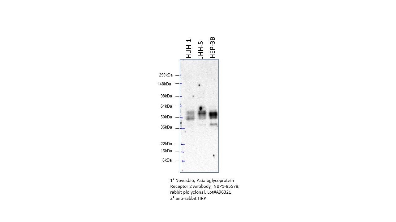
Application: Western BlotSample Tested: 3 human cancer cell linesSpecies: HumanVerified Customer | Posted 10/02/2020Clean antibody with no other non-specific bands. Bands showing at higher MW than calculated MW due to glycosylation of ASGR2.

There are no reviews that match your criteria.
Protocols
Find general support by application which include: protocols, troubleshooting, illustrated assays, videos and webinars.
- Antigen Retrieval Protocol (PIER)
- Antigen Retrieval for Frozen Sections Protocol
- Appropriate Fixation of IHC/ICC Samples
- Cellular Response to Hypoxia Protocols
- Chromogenic IHC Staining of Formalin-Fixed Paraffin-Embedded (FFPE) Tissue Protocol
- Chromogenic Immunohistochemistry Staining of Frozen Tissue
- ClariTSA™ Fluorophore Kits
- Detection & Visualization of Antibody Binding
- Fluorescent IHC Staining of Frozen Tissue Protocol
- Graphic Protocol for Heat-induced Epitope Retrieval
- Graphic Protocol for the Preparation and Fluorescent IHC Staining of Frozen Tissue Sections
- Graphic Protocol for the Preparation and Fluorescent IHC Staining of Paraffin-embedded Tissue Sections
- Graphic Protocol for the Preparation of Gelatin-coated Slides for Histological Tissue Sections
- IHC Sample Preparation (Frozen sections vs Paraffin)
- Immunofluorescent IHC Staining of Formalin-Fixed Paraffin-Embedded (FFPE) Tissue Protocol
- Immunohistochemistry (IHC) and Immunocytochemistry (ICC) Protocols
- Immunohistochemistry Frozen Troubleshooting
- Immunohistochemistry Paraffin Troubleshooting
- Preparing Samples for IHC/ICC Experiments
- Preventing Non-Specific Staining (Non-Specific Binding)
- Primary Antibody Selection & Optimization
- Protocol for Heat-Induced Epitope Retrieval (HIER)
- Protocol for Making a 4% Formaldehyde Solution in PBS
- Protocol for VisUCyte™ HRP Polymer Detection Reagent
- Protocol for the Preparation & Fixation of Cells on Coverslips
- Protocol for the Preparation and Chromogenic IHC Staining of Frozen Tissue Sections
- Protocol for the Preparation and Chromogenic IHC Staining of Frozen Tissue Sections - Graphic
- Protocol for the Preparation and Chromogenic IHC Staining of Paraffin-embedded Tissue Sections
- Protocol for the Preparation and Chromogenic IHC Staining of Paraffin-embedded Tissue Sections - Graphic
- Protocol for the Preparation and Fluorescent IHC Staining of Frozen Tissue Sections
- Protocol for the Preparation and Fluorescent IHC Staining of Paraffin-embedded Tissue Sections
- Protocol for the Preparation of Gelatin-coated Slides for Histological Tissue Sections
- R&D Systems Quality Control Western Blot Protocol
- TUNEL and Active Caspase-3 Detection by IHC/ICC Protocol
- The Importance of IHC/ICC Controls
- Troubleshooting Guide: Immunohistochemistry
- Troubleshooting Guide: Western Blot Figures
- Western Blot Conditions
- Western Blot Protocol
- Western Blot Protocol for Cell Lysates
- Western Blot Troubleshooting
- Western Blot Troubleshooting Guide
- View all Protocols, Troubleshooting, Illustrated assays and Webinars
Loading...