ATF6 Antibody - BSA Free
Novus Biologicals | Catalog # NBP1-75478
![Western Blot: ATF6 AntibodyBSA Free [NBP1-75478]](https://resources.rndsystems.com/images/products/ATF6-Antibody-Western-Blot-NBP1-75478-img0007.jpg "Western Blot: ATF6 AntibodyBSA Free [NBP1-75478]")
![Western Blot: ATF6 AntibodyBSA Free [NBP1-75478]](https://resources.rndsystems.com/images/products/ATF6-Antibody-Western-Blot-NBP1-75478-img0006.jpg "Western Blot: ATF6 AntibodyBSA Free [NBP1-75478]")
Key Product Details
Validated by
Species Reactivity
Validated:
Cited:
Applications
Validated:
Cited:
Label
Antibody Source
Format
Product Specifications
Immunogen
Reactivity Notes
Localization
Clonality
Host
Isotype
Scientific Data Images for ATF6 Antibody - BSA Free
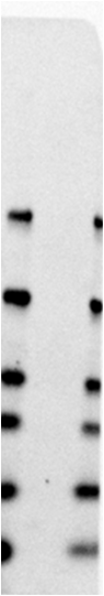
Western Blot: ATF6 AntibodyBSA Free [NBP1-75478]
Western Blot: ATF6 Antibody [NBP1-75478] - Total protein from HeLa cells treated with and without 1 mM DTT for 24 hours was separated on a 7.5% gel by SDS-PAGE, transferred to PVDF membrane and blocked in 1% BSA. The membrane was probed with 2.0 ug/ml anti-ATF6 in block buffer and detected with an anti-rabbit HRP secondary antibody using chemiluminescence. Note the decrease in full length ATF6 (90 kDa, arrow) and increase in cleaved ATF6 (55 kDa, arrowhead) upon DTT treatment and the activation of the unfolded protein response pathway.![Simple Western: ATF6 AntibodyBSA Free [NBP1-75478]](https://resources.rndsystems.com/images/products/ATF6-Antibody-Simple-Western-NBP1-75478-img0004.jpg "Simple Western: ATF6 AntibodyBSA Free [NBP1-75478]")
Simple Western: ATF6 AntibodyBSA Free [NBP1-75478]
Simple Western: ATF6 Antibody [NBP1-75478] - Simple Western lane view shows a specific band for ATF6 in 1.0 mg/ml of HeLa lysate. This experiment was performed under reducing conditions using the 12-230kDa separation system.![Immunocytochemistry/ Immunofluorescence: ATF6 Antibody - BSA Free [NBP1-75478]](https://resources.rndsystems.com/images/products/ATF6-Antibody-Immunocytochemistry-Immunofluorescence-NBP1-75478-img0005.jpg "Immunocytochemistry/ Immunofluorescence: ATF6 Antibody - BSA Free [NBP1-75478]")
Immunocytochemistry/ Immunofluorescence: ATF6 Antibody - BSA Free [NBP1-75478]
Immunocytochemistry/Immunofluorescence: ATF6 Antibody [NBP1-75478] - HeLa cells were fixed and permeabilized for 10 minutes using -20C MeOH. The cells were incubated with anti-ATF6 at 10 ug/ml overnight at 4C and detected with an anti-rabbit Dylight 488 (Green) at a 1:500 dilution. Nuclei were counterstained with DAPI (Blue). Cells were imaged using a 40X objective.![Immunocytochemistry/ Immunofluorescence: ATF6 Antibody - BSA Free [NBP1-75478]](https://resources.rndsystems.com/images/products/ATF6-Antibody-Immunocytochemistry-Immunofluorescence-NBP1-75478-img0002.jpg "Immunocytochemistry/ Immunofluorescence: ATF6 Antibody - BSA Free [NBP1-75478]")
Immunocytochemistry/ Immunofluorescence: ATF6 Antibody - BSA Free [NBP1-75478]
Immunocytochemistry/Immunofluorescence: ATF6 Antibody [NBP1-75478] - Antibody was tested in HeLa cells with FITC (green). Nuclei were counterstained with Dapi (blue).Applications for ATF6 Antibody - BSA Free
Immunocytochemistry/ Immunofluorescence
Simple Western
Western Blot
In Simple Western only 10 - 15 uL of the recommended dilution is used per data point.
See Simple Western Antibody Database for Simple Western validation: Tested in HeLa lysate 1.0 mg/mL, separated by Size, antibody dilution of 1:25, apparent MW was 101 kDa. Separated by Size-Wes, Sally Sue/Peggy Sue.
Reviewed Applications
Read 1 review rated 1 using NBP1-75478 in the following applications:
Formulation, Preparation, and Storage
Purification
Formulation
Format
Preservative
Concentration
Shipping
Stability & Storage
Background: ATF6
Long Name
Alternate Names
Entrez Gene IDs
Gene Symbol
UniProt
Additional ATF6 Products
Product Documents for ATF6 Antibody - BSA Free
Certificate of Analysis
To download a Certificate of Analysis, please enter a lot or batch number in the search box below.
Product Specific Notices for ATF6 Antibody - BSA Free
Manufactured by Genomic Antibody Technology™. GAT FAQs
This product is for research use only and is not approved for use in humans or in clinical diagnosis. Primary Antibodies are guaranteed for 1 year from date of receipt.
Citations for ATF6 Antibody - BSA Free
 Powered by Bioz
Customer Reviews for ATF6 Antibody - BSA Free (1)
Have you used ATF6 Antibody - BSA Free?
Submit a review and receive an Amazon gift card!
$25/€18/£15/$25CAN/¥2500 Yen for a review with an image
$10/€7/£6/$10CAN/¥1110 Yen for a review without an image
Submit a review
Customer Images
-
Application: Western BlotSample Tested: Human primary fibroblastSpecies: HumanVerified Customer | Posted 10/31/2020ATF6 in human primary fibroblasts. No band detected. Primary antibody incubation O/N 4ºC 1:1000 in BSA 3% PBST (0.1% Tween). Secondary ab: Donkey anti-rabit Alexa Fluor 647 1:5000 1h RT in BSA 3% PBST (0.1% Tween).TGX 12% gel.10 ug of protein loaded reduced with DTT. Primary antibody incubation O/N 4ºC 1:1000 in BSA 3% PBST (0.1% Tween). Secondary ab: Donkey anti-rabit Alexa Fluor 647 1:5000 1h RT in BSA 3% PBST (0.1% Tween).
 Bio-Techne ResponseThank you for reviewing our product. We are sorry to that that this antibody did not perform as expected. We have been in touch with the customer to resolve this issue according to our Product Guarantee and to the customer’s satisfaction.
There are no reviews that match your criteria.
Protocols
View specific protocols for ATF6 Antibody - BSA Free (NBP1-75478):
Immunocytochemistry Protocol
Culture cells to appropriate density in 35 mm culture dishes or 6-well plates.
1. Remove culture medium and add 10% formalin to the dish. Fix at room temperature for 30 minutes.
2. Remove the formalin and add ice cold methanol. Incubate for 5-10 minutes.
3. Remove methanol and add washing solution (i.e. PBS). Be sure to not let the specimen dry out. Wash three times for 10 minutes.
4. To block nonspecific antibody binding incubate in 10% normal goat serum from 1 hour to overnight at room temperature.
5. Add primary antibody at appropriate dilution and incubate at room temperature from 2 hours to overnight at room temperature.
6. Remove primary antibody and replace with washing solution. Wash three times for 10 minutes.
7. Add secondary antibody at appropriate dilution. Incubate for 1 hour at room temperature.
8. Remove antibody and replace with wash solution, then wash for 10 minutes. Add Hoechst 33258 to wash solution at 1:25,0000 and incubate for 10 minutes. Wash a third time for 10 minutes.
9. Cells can be viewed directly after washing. The plates can also be stored in PBS containing Azide covered in Parafilm (TM). Cells can also be cover-slipped using Fluoromount, with appropriate sealing.
*The above information is only intended as a guide. The researcher should determine what protocol best meets their needs. Please follow safe laboratory procedures.
Find general support by application which include: protocols, troubleshooting, illustrated assays, videos and webinars.
- Appropriate Fixation of IHC/ICC Samples
- Cellular Response to Hypoxia Protocols
- ClariTSA™ Fluorophore Kits
- Detection & Visualization of Antibody Binding
- ICC Cell Smear Protocol for Suspension Cells
- ICC Immunocytochemistry Protocol Videos
- ICC for Adherent Cells
- Immunocytochemistry (ICC) Protocol
- Immunocytochemistry Troubleshooting
- Immunofluorescence of Organoids Embedded in Cultrex Basement Membrane Extract
- Immunohistochemistry (IHC) and Immunocytochemistry (ICC) Protocols
- Preparing Samples for IHC/ICC Experiments
- Preventing Non-Specific Staining (Non-Specific Binding)
- Primary Antibody Selection & Optimization
- Protocol for VisUCyte™ HRP Polymer Detection Reagent
- Protocol for the Fluorescent ICC Staining of Cell Smears - Graphic
- Protocol for the Fluorescent ICC Staining of Cultured Cells on Coverslips - Graphic
- Protocol for the Preparation and Fluorescent ICC Staining of Cells on Coverslips
- Protocol for the Preparation and Fluorescent ICC Staining of Non-adherent Cells
- Protocol for the Preparation and Fluorescent ICC Staining of Stem Cells on Coverslips
- Protocol for the Preparation of a Cell Smear for Non-adherent Cell ICC - Graphic
- R&D Systems Quality Control Western Blot Protocol
- TUNEL and Active Caspase-3 Detection by IHC/ICC Protocol
- The Importance of IHC/ICC Controls
- Troubleshooting Guide: Western Blot Figures
- Western Blot Conditions
- Western Blot Protocol
- Western Blot Protocol for Cell Lysates
- Western Blot Troubleshooting
- Western Blot Troubleshooting Guide
- View all Protocols, Troubleshooting, Illustrated assays and Webinars
FAQs for ATF6 Antibody - BSA Free
-
Q: We are looking for an antibody specific to ATF6 alpha that will not cross-react with ATF6 beta. Would you please help confirm if any of the following are ATF6 alpha specific antibodies: NBP1-40256, NBP1-77251, NBP1-76675 & NBP1-41439?
A:
The antibodies NBP1-77251 and NBP1-76675 are specific to ATF6 alpha and should not cross-react with ATF6 beta.
-
Q: Will this antibody detect the cleaved portion of ATF6 (60-80kDa), as well as the non-cleaved portion (110 kDa)? We are using human lymphocyte cell lysates (samples of patients with leukemia) on a proteinsimple Wes machine.
A: Thank you for contacting Novus regarding the ATF6 Antibody (NBP1-75478). We do see the full length in Simple Western but not the cleavage product. Nevertheless we assume you should see the 46kD cleavage product since the immunogen is located there but only under ER stress. The actual epitope is not mapped but the immunogen is with in the region of a.a. 200-250. Since the cleavage site is at 419-420 then we assume it is possible to detect this portion of the cleavge product but not the 27KD product without the immunogen sequence. 74 kD full length aa1-420=46kD aa420-670=27kD
-
Q: We are looking for an antibody specific to ATF6 alpha that will not cross-react with ATF6 beta. Would you please help confirm if any of the following are ATF6 alpha specific antibodies: NBP1-40256, NBP1-77251, NBP1-76675 & NBP1-41439?
A:
The antibodies NBP1-77251 and NBP1-76675 are specific to ATF6 alpha and should not cross-react with ATF6 beta.
-
Q: Will this antibody detect the cleaved portion of ATF6 (60-80kDa), as well as the non-cleaved portion (110 kDa)? We are using human lymphocyte cell lysates (samples of patients with leukemia) on a proteinsimple Wes machine.
A: Thank you for contacting Novus regarding the ATF6 Antibody (NBP1-75478). We do see the full length in Simple Western but not the cleavage product. Nevertheless we assume you should see the 46kD cleavage product since the immunogen is located there but only under ER stress. The actual epitope is not mapped but the immunogen is with in the region of a.a. 200-250. Since the cleavage site is at 419-420 then we assume it is possible to detect this portion of the cleavge product but not the 27KD product without the immunogen sequence. 74 kD full length aa1-420=46kD aa420-670=27kD