ATP7b Antibody - BSA Free
Novus Biologicals | Catalog # NB100-360
![Western Blot: ATP7b AntibodyBSA Free [NB100-360]](https://resources.rndsystems.com/images/products/ATP7b-Antibody-Western-Blot-NB100-360-img0003.jpg "Western Blot: ATP7b AntibodyBSA Free [NB100-360]")
Key Product Details
Species Reactivity
Validated:
Cited:
Applications
Validated:
Cited:
Label
Antibody Source
Format
Product Specifications
Immunogen
Localization
Clonality
Host
Isotype
Theoretical MW
Disclaimer note: The observed molecular weight of the protein may vary from the listed predicted molecular weight due to post translational modifications, post translation cleavages, relative charges, and other experimental factors.
Scientific Data Images for ATP7b Antibody - BSA Free
Western Blot: ATP7b AntibodyBSA Free [NB100-360]
Western Blot: ATP7b Antibody [NB100-360] - ATP7b detected in 20 ug of mouse brain membrane fraction.![Immunocytochemistry/ Immunofluorescence: ATP7b Antibody - BSA Free [NB100-360]](https://resources.rndsystems.com/images/products/ATP7b-Antibody-Immunocytochemistry-Immunofluorescence-NB100-360-img0005.jpg "Immunocytochemistry/ Immunofluorescence: ATP7b Antibody - BSA Free [NB100-360]")
Immunocytochemistry/ Immunofluorescence: ATP7b Antibody - BSA Free [NB100-360]
Immunocytochemistry/Immunofluorescence: ATP7b Antibody [NB100-360] - HeLa cells were fixed for 10 minutes using 10% formalin and then permeabilized for 5 minutes using 1X TBS + 0.5% Triton X-100. The cells were incubated with anti-ATP7b [NB100-360] at a 1:100 dilution overnight at 4C and detected with an anti-rabbit DyLight 488 (Green) at a 1:500 dilution. Alpha tubulin (DM1A) NB100-690 was used as a co-stain at a 1:1000 dilution and detected with an anti-mouse DyLight 550 (Red) at 1:500. Nuclei were counterstained with DAPI (Blue). Cells were imaged using a 40X objective.![Immunohistochemistry: ATP7b Antibody - BSA Free [NB100-360]](https://resources.rndsystems.com/images/products/ATP7b-Antibody-Immunohistochemistry-NB100-360-img0004.jpg "Immunohistochemistry: ATP7b Antibody - BSA Free [NB100-360]")
Immunohistochemistry: ATP7b Antibody - BSA Free [NB100-360]
Immunohistochemistry: ATP7b Antibody [NB100-360] - Analysis of ATP7b in mouse liver using DAB with hematoxylin counterstain.
Western Blot: ATP7b Antibody - BSA Free [NB100-360] -
Copper contributes to the production of MB in Wilson disease through inhibiting autophagy. (A) Differential detergent fractionation was performed on WT HepG2 cells or GFP-Ctr1 HepG2 after treatment with 1 mM of copper ions for 6 h. Western blotting was carried out to detect the protein levels. (B) Colocalization analysis of the markers of MB such p62, K8/K18, and Ub before and after treatment of copper ions. The arrows mean colocalization. (C) Differential detergent fractionation was performed on WT HepaRG cells or ATP7B knockdown HepRG cells treated with 1 mM of copper ions for 6 h and western blotting was carried out to detect the protein levels. (D) Differential detergent fractionation followed by western blotting was performed on HepaRG cells treated with 1 mM of copper ions in the presence or absence of 40 μM of CQ for 6 h. (E) Differential detergent fractionation was performed on ATP7B knockdown HepRG cells treated with 1 mM of copper ions for 6 h and western blotting was carried out to detect the protein levels. (F) Densitometry analysis of protein bands of (E) was performed. The ratio of K8/K18 and LC3B in soluble fraction, p62 and ubiquitin in insoluble fraction was calculated. Image collected and cropped by CiteAb from the following open publication (https://pubmed.ncbi.nlm.nih.gov/35349929), licensed under a CC-BY license. Not internally tested by Novus Biologicals.Applications for ATP7b Antibody - BSA Free
Immunoblotting
Immunocytochemistry/ Immunofluorescence
Immunohistochemistry
Immunohistochemistry-Paraffin
Western Blot
Reviewed Applications
Read 1 review rated 4 using NB100-360 in the following applications:
Formulation, Preparation, and Storage
Purification
Formulation
Format
Preservative
Concentration
Shipping
Stability & Storage
Background: ATP7b
Alternate Names
Gene Symbol
Additional ATP7b Products
Product Documents for ATP7b Antibody - BSA Free
Certificate of Analysis
To download a Certificate of Analysis, please enter a lot or batch number in the search box below.
Product Specific Notices for ATP7b Antibody - BSA Free
This product is for research use only and is not approved for use in humans or in clinical diagnosis. Primary Antibodies are guaranteed for 1 year from date of receipt.
Citations for ATP7b Antibody - BSA Free
 Powered by Bioz
Customer Reviews for ATP7b Antibody - BSA Free (1)
Have you used ATP7b Antibody - BSA Free?
Submit a review and receive an Amazon gift card!
$25/€18/£15/$25CAN/¥2500 Yen for a review with an image
$10/€7/£6/$10CAN/¥1110 Yen for a review without an image
Submit a review
Customer Images
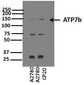
-
Application: Western BlotSample Tested: Whole cell lysates for ovarian cancer cell lines A2780 and its cisplatin resistant variant A2780-CP20.Species: HumanVerified Customer | Posted 09/07/2015Western Blot for ATP7b

There are no reviews that match your criteria.
Protocols
View specific protocols for ATP7b Antibody - BSA Free (NB100-360):
Immunocytochemistry Protocol
Culture cells to appropriate density in 35 mm culture dishes or 6-well plates.
1. Remove culture medium and add 10% formalin to the dish. Fix at room temperature for 30 minutes.
2. Remove the formalin and add ice cold methanol. Incubate for 5-10 minutes.
3. Remove methanol and add washing solution (i.e. PBS). Be sure to not let the specimen dry out. Wash three times for 10 minutes.
4. To block nonspecific antibody binding incubate in 10% normal goat serum from 1 hour to overnight at room temperature.
5. Add primary antibody at appropriate dilution and incubate at room temperature from 2 hours to overnight at room temperature.
6. Remove primary antibody and replace with washing solution. Wash three times for 10 minutes.
7. Add secondary antibody at appropriate dilution. Incubate for 1 hour at room temperature.
8. Remove antibody and replace with wash solution, then wash for 10 minutes. Add Hoechst 33258 to wash solution at 1:25,0000 and incubate for 10 minutes. Wash a third time for 10 minutes.
9. Cells can be viewed directly after washing. The plates can also be stored in PBS containing Azide covered in Parafilm (TM). Cells can also be cover-slipped using Fluoromount, with appropriate sealing.
*The above information is only intended as a guide. The researcher should determine what protocol best meets their needs. Please follow safe laboratory procedures.
Immunohistochemistry Procedure
Cell Preparation (At least 108 cells were used per block)
1. Harvesting cells:
A. Trypsinization
B. 15 minute centrifugation at 2,500 RPM
C. PBS rinse
D. 15 minute centrifugation at 2,500 RPM
2. Suspend cells in 10 ml of 10% formaldehyde in PBS, overnight @ RT.
3. Centrifuge cells at 2,500 RPM for 10 minutes.
4. Resuspend cells in 10 ml of 70% ethanol.
5. Centrifuge cells at 2,500 RPM and taken into 70% ethanol.
Cell Staining
1. Ribbon Thickness: 5 um
2. Deparaffination Agent: Xylin
3. Hydration: Ethanol in PBS
4. Antigen Retrieval: 10 minute microwave retrieval in citrate buffer; 20 minute cooling
5. Blocking:
A. endogeneous peroxidase: 0.3% H2O2 in PBS for 10 minutes
B. endogeneous protein: 1% BSA for 20 minutes
6. Primary antibody, polyclonal anti-ATP7b (NB 100-360): 1:500, overnight @ 4 degrees Celcius
7. Secondary antibody, anti-rabbit (HRP): (dilute per manufacturer recommendation), 30 minutes @ RT
8. Wash 3x 15 minutes
9. Chromogen: AEC
10. Counterstain: Mayers hematoxylin
*The above information is only intended as a guide. The researcher should determine what protocol best meets their needs. Please follow safe laboratory procedures.
Western Blot Protocol
1. Perform SDS-PAGE on samples to be analyzed, loading 40 ug of total protein per lane.
2. Transfer proteins to membrane according to the instructions provided by the manufacturer of the membrane and transfer apparatus.
3. Stain according to standard Ponceau S procedure (or similar product) to assess transfer success, and mark molecular weight standards where appropriate.
4. Rinse the blot.
5. Block the membrane using standard blocking buffer for at least 1 hour.
6. Wash the membrane in wash buffer three times for 10 minutes each.
7. Dilute primary antibody in blocking buffer and incubate 1 hour at room temperature.
8. Wash the membrane in wash buffer three times for 10 minutes each.
9. Apply the diluted HRP conjugated secondary antibody in blocking buffer (as per manufacturers instructions) and incubate 1 hour at room temperature.
10. Wash the blot in wash buffer three times for 10 minutes each (this step can be repeated as required to reduce background).
11. Apply the detection reagent of choice in accordance with the manufacturers instructions.
Note: Tween-20 can be added to the blocking or antibody dilution buffer at a final concentration of 0.05-0.2%.
*The above information is only intended as a guide. The researcher should determine what protocol best meets their needs. Please follow safe laboratory procedures.
Find general support by application which include: protocols, troubleshooting, illustrated assays, videos and webinars.
- Antigen Retrieval Protocol (PIER)
- Antigen Retrieval for Frozen Sections Protocol
- Appropriate Fixation of IHC/ICC Samples
- Cellular Response to Hypoxia Protocols
- Chromogenic IHC Staining of Formalin-Fixed Paraffin-Embedded (FFPE) Tissue Protocol
- Chromogenic Immunohistochemistry Staining of Frozen Tissue
- ClariTSA™ Fluorophore Kits
- Detection & Visualization of Antibody Binding
- Fluorescent IHC Staining of Frozen Tissue Protocol
- Graphic Protocol for Heat-induced Epitope Retrieval
- Graphic Protocol for the Preparation and Fluorescent IHC Staining of Frozen Tissue Sections
- Graphic Protocol for the Preparation and Fluorescent IHC Staining of Paraffin-embedded Tissue Sections
- Graphic Protocol for the Preparation of Gelatin-coated Slides for Histological Tissue Sections
- ICC Cell Smear Protocol for Suspension Cells
- ICC Immunocytochemistry Protocol Videos
- ICC for Adherent Cells
- IHC Sample Preparation (Frozen sections vs Paraffin)
- Immunocytochemistry (ICC) Protocol
- Immunocytochemistry Troubleshooting
- Immunofluorescence of Organoids Embedded in Cultrex Basement Membrane Extract
- Immunofluorescent IHC Staining of Formalin-Fixed Paraffin-Embedded (FFPE) Tissue Protocol
- Immunohistochemistry (IHC) and Immunocytochemistry (ICC) Protocols
- Immunohistochemistry Frozen Troubleshooting
- Immunohistochemistry Paraffin Troubleshooting
- Immunoprecipitation Protocol
- Preparing Samples for IHC/ICC Experiments
- Preventing Non-Specific Staining (Non-Specific Binding)
- Primary Antibody Selection & Optimization
- Protocol for Heat-Induced Epitope Retrieval (HIER)
- Protocol for Making a 4% Formaldehyde Solution in PBS
- Protocol for VisUCyte™ HRP Polymer Detection Reagent
- Protocol for the Fluorescent ICC Staining of Cell Smears - Graphic
- Protocol for the Fluorescent ICC Staining of Cultured Cells on Coverslips - Graphic
- Protocol for the Preparation & Fixation of Cells on Coverslips
- Protocol for the Preparation and Chromogenic IHC Staining of Frozen Tissue Sections
- Protocol for the Preparation and Chromogenic IHC Staining of Frozen Tissue Sections - Graphic
- Protocol for the Preparation and Chromogenic IHC Staining of Paraffin-embedded Tissue Sections
- Protocol for the Preparation and Chromogenic IHC Staining of Paraffin-embedded Tissue Sections - Graphic
- Protocol for the Preparation and Fluorescent ICC Staining of Cells on Coverslips
- Protocol for the Preparation and Fluorescent ICC Staining of Non-adherent Cells
- Protocol for the Preparation and Fluorescent ICC Staining of Stem Cells on Coverslips
- Protocol for the Preparation and Fluorescent IHC Staining of Frozen Tissue Sections
- Protocol for the Preparation and Fluorescent IHC Staining of Paraffin-embedded Tissue Sections
- Protocol for the Preparation of Gelatin-coated Slides for Histological Tissue Sections
- Protocol for the Preparation of a Cell Smear for Non-adherent Cell ICC - Graphic
- R&D Systems Quality Control Western Blot Protocol
- TUNEL and Active Caspase-3 Detection by IHC/ICC Protocol
- The Importance of IHC/ICC Controls
- Troubleshooting Guide: Immunohistochemistry
- Troubleshooting Guide: Western Blot Figures
- Western Blot Conditions
- Western Blot Protocol
- Western Blot Protocol for Cell Lysates
- Western Blot Troubleshooting
- Western Blot Troubleshooting Guide
- View all Protocols, Troubleshooting, Illustrated assays and Webinars
FAQs for ATP7b Antibody - BSA Free
-
Q: Can you disclose any further detail related to the immunogen sequence used to generate NB100-360?
A: A more specific range for the immunogen of our NB100-360 is aa300-350 of the human ATP7b protein
-
Q: Which isoform of the protein does NB100-360 recognize?
A: The immunogen for this product is to the N-terminal, so it will recognize all 4 known isoforms of ATP7b.
-
Q: Would you be able to provide any additional information about the sequence or location of the immunogenic peptide?
A: I cannot give the exact immunogen sequence as it is considered to be propriety information; however, I am able to provide a range. The immunogen of NB100-360 lies within aa 300-350 of the human ATP7b protein.
-
Q: Can you disclose any further detail related to the immunogen sequence used to generate NB100-360?
A: A more specific range for the immunogen of our NB100-360 is aa300-350 of the human ATP7b protein
-
Q: Which isoform of the protein does NB100-360 recognize?
A: The immunogen for this product is to the N-terminal, so it will recognize all 4 known isoforms of ATP7b.
-
Q: Would you be able to provide any additional information about the sequence or location of the immunogenic peptide?
A: I cannot give the exact immunogen sequence as it is considered to be propriety information; however, I am able to provide a range. The immunogen of NB100-360 lies within aa 300-350 of the human ATP7b protein.