Aurora B Antibody - BSA Free
Novus Biologicals | Catalog # NB100-294
![Western Blot: Aurora B AntibodyBSA Free [NB100-294]](https://resources.rndsystems.com/images/products/Aurora-B-Antibody---BSA-Free-Western-Blot-NB100-294-img0002.jpg "Western Blot: Aurora B AntibodyBSA Free [NB100-294]")
Key Product Details
Species Reactivity
Validated:
Human, Mouse, Rat
Cited:
Human
Applications
Validated:
Immunohistochemistry, Immunohistochemistry-Paraffin, Western Blot, Immunocytochemistry/ Immunofluorescence, Immunoprecipitation, Microarray
Cited:
Western Blot, Immunocytochemistry/ Immunofluorescence, IF/IHC
Label
Unconjugated
Antibody Source
Polyclonal Rabbit IgG
Format
BSA Free
Loading...
Product Specifications
Immunogen
Synthetic peptide corresponding to amino acids 1-19 of human Aurora B with C-terminal added cysteine conjugated to KLH. Uniprot accession no. Q96GD4
Marker
Mitosis Marker
Specificity
Aurora B
Clonality
Polyclonal
Host
Rabbit
Isotype
IgG
Theoretical MW
41 kDa.
Disclaimer note: The observed molecular weight of the protein may vary from the listed predicted molecular weight due to post translational modifications, post translation cleavages, relative charges, and other experimental factors.
Disclaimer note: The observed molecular weight of the protein may vary from the listed predicted molecular weight due to post translational modifications, post translation cleavages, relative charges, and other experimental factors.
Scientific Data Images for Aurora B Antibody - BSA Free
Western Blot: Aurora B AntibodyBSA Free [NB100-294]
Western Blot: Aurora B Antibody - BSA Free [NB100-294] - Whole extract of PC-12 cells were separated on SDS-PAGE and probed with Anti-Aurora B antibody produced in Rabbit. The antibody was developed using 1:10,000 Anti-Rabbit IgG (whole molecule)-Peroxidase antibody produced in Goat.Lanes:1. 1:1000 antibody2. 1:2000 antibody3. Negative Control![Immunocytochemistry/ Immunofluorescence: Aurora B Antibody - BSA Free [NB100-294]](https://resources.rndsystems.com/images/products/Aurora-B-Antibody---BSA-Free-Immunocytochemistry-Immunofluorescence-NB100-294-img0001.jpg "Immunocytochemistry/ Immunofluorescence: Aurora B Antibody - BSA Free [NB100-294]")
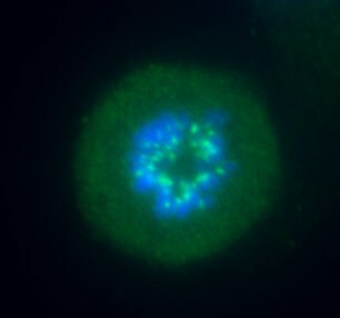
Immunocytochemistry/ Immunofluorescence: Aurora B Antibody - BSA Free [NB100-294]
Immunocytochemistry/Immunofluorescence: Aurora B Antibody - BSA Free [NB100-294] - HeLa human cervix epitheloid carcinoma cells were fixed and permeabilized with 4% paraformaldehyde followed by cold methanol. Fixed cells were stained with 1:50 Anti-Aurora B antibody produced in rabbit. The antibody was developed using Anti-Rabbit IgG (whole molecule)-FITC antibody produced in goat.
Western Blot: Aurora B Antibody - BSA Free [NB100-294] -
AURKB mediates EGF associated BRAF/MEK/ERKs and PI3-K/AKT pathways. (A, B) Knocking down of AURKB suppressed the level of EGF in A375 and A375R tumor from xenograft mice compared with shcontrol. The asterisks indicate a significant difference compared with the shcontrol group (*, p < 0.05; ** p < 0.01). (C) Knocking down of AURKB increased levels of c-PARP, c-caspase 3, and decreased of Bcl-2 level of A375 and A375R cell lines. (D, E) Western blot analysis of activation of BRAF/MEK/ERKs and PI3-K/AKT by knocking down of AURKB in A375 and A375R cell lines. Image collected and cropped by CiteAb from the following open publication (https://pubmed.ncbi.nlm.nih.gov/32863956), licensed under a CC-BY license. Not internally tested by Novus Biologicals.
Western Blot: Aurora B Antibody - BSA Free [NB100-294] -
Database analysis and AURKB expression level in melanoma. (A) Volcano plot of gene expression analysis in GSE 4587. The X-axis indicates the fold change between normal skin or nevi and melanoma samples, and the Y-axis indicates on a log10 scale the p-values obtained from a supervised logistic regression analysis testing the association of gene expression between normal skin or nevi/melanoma. The horizontal dotted lines mark the significance cutoffs (i.e., fold change more than 4 times or less than 0.25, and p-value less than 0.001). (B) Heat map of analysis of the expression of these selected genes in GSE 4587. AURKB has a higher expression level in melanoma compared with non-melanoma (normal skin and nevi). (C) Pathway enrichment analysis of differential gene analysis. The top 2 pathways were selected, fold change more than 4 times, p-value less than 0.0001. (D) The Venn diagram lists the common genes in both pathways. (E) Melanoma patients with high expression of AURKB show a significantly lower overall survival rate. (F)AURKB is highly expressed in melanoma compared with normal skin as determined by GEPIA. (G) AURKB is significantly overexpressed in melanoma tissue compared with normal skin as shown in a tissue array analysis; the scale bar = 100 um. (H) AURKB is overexpressed in drug-resistant melanoma cell lines (A375R, M238R, and M249R) compared with drug-sensitive melanoma cell lines (A375, M238 and M249). Statistical significance was determined by Student's t-test and the asterisks indicate a significant change compared with the control group (*, p < 0.05; ***, p < 0.001). Image collected and cropped by CiteAb from the following open publication (https://pubmed.ncbi.nlm.nih.gov/32863956), licensed under a CC-BY license. Not internally tested by Novus Biologicals.
Western Blot: Aurora B Antibody - BSA Free [NB100-294] -
AURKB mediates EGF associated BRAF/MEK/ERKs and PI3-K/AKT pathways. (A, B) Knocking down of AURKB suppressed the level of EGF in A375 and A375R tumor from xenograft mice compared with shcontrol. The asterisks indicate a significant difference compared with the shcontrol group (*, p < 0.05; ** p < 0.01). (C) Knocking down of AURKB increased levels of c-PARP, c-caspase 3, and decreased of Bcl-2 level of A375 and A375R cell lines. (D, E) Western blot analysis of activation of BRAF/MEK/ERKs and PI3-K/AKT by knocking down of AURKB in A375 and A375R cell lines. Image collected and cropped by CiteAb from the following open publication (https://pubmed.ncbi.nlm.nih.gov/32863956), licensed under a CC-BY license. Not internally tested by Novus Biologicals.
Western Blot: Aurora B Antibody - BSA Free [NB100-294] -
HI-511 binds to and effectively suppresses the activation of AURKB and BRAF V600E. (A) The structure of HI-511. (B) Computer docking model of HI-511 binding with AURKB. (C) Computer docking model of HI-511 binding with BRAF V600E. (D) HI-511 inhibits AURKB in vitro. Active kinase AURKB and HI-511 (0, 1, 5 and10 uM) or APIO-EE-9 (5 uM) were mixed with the substrate histone H3. The relative amounts of phosphorylated substrate were visualized by Western blot. (E) HI-511 inhibits BRAF V600E in vitro. Active kinase BRAF V600E and HI-511 (0, 1, 5, 10 uM) or vemurafenib (5 uM) were mixed with the substrate phosphatidylinositol. The relative amounts of phosphorylated substrate were visualized by Western blot. Image collected and cropped by CiteAb from the following open publication (https://pubmed.ncbi.nlm.nih.gov/32863956), licensed under a CC-BY license. Not internally tested by Novus Biologicals.Applications for Aurora B Antibody - BSA Free
Application
Recommended Usage
Immunocytochemistry/ Immunofluorescence
1:50
Immunohistochemistry
1:10-1:500
Immunohistochemistry-Paraffin
1:10-1:500
Immunoprecipitation
50-60 ug
Western Blot
1:1000-1:2000
Application Notes
By Western blot, it detects a band of approximately 41 kDa. Detection of the Aurora B band by immunoblotting is specifically inhibited with the immunizing peptide.
Reviewed Applications
Read 5 reviews rated 4.8 using NB100-294 in the following applications:
Formulation, Preparation, and Storage
Purification
IgG purified
Formulation
10mM PBS (pH 7.4)
Format
BSA Free
Preservative
0.09% Sodium Azide
Concentration
12.5 mg/ml
Shipping
The product is shipped with polar packs. Upon receipt, store it immediately at the temperature recommended below.
Stability & Storage
Store at 4C short term. Aliquot and store at -20C long term. Avoid freeze-thaw cycles.
Background: Aurora B
Alternate Names
Aik2, AIM-1, AIM1, ARK2, AURKB, IPL1, STK12, STK5
Gene Symbol
AURKB
Additional Aurora B Products
Product Documents for Aurora B Antibody - BSA Free
Certificate of Analysis
To download a Certificate of Analysis, please enter a lot or batch number in the search box below.
Product Specific Notices for Aurora B Antibody - BSA Free
This product is for research use only and is not approved for use in humans or in clinical diagnosis. Primary Antibodies are guaranteed for 1 year from date of receipt.
Related Research Areas
Citations for Aurora B Antibody - BSA Free
 Powered by Bioz
Customer Reviews for Aurora B Antibody - BSA Free (5)
4.8 out of 5
5 Customer Ratings
Have you used Aurora B Antibody - BSA Free?
Submit a review and receive an Amazon gift card!
$25/€18/£15/$25CAN/¥2500 Yen for a review with an image
$10/€7/£6/$10CAN/¥1110 Yen for a review without an image
Submit a review
Customer Images
Showing
1
-
5 of
5 reviews
Showing All
Filter By:
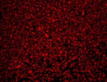
-
Application: ImmunofluorescenceSample Tested: RPE1 cellsSpecies: HumanVerified Customer | Posted 04/21/2016Prometaphase Aurora B

-
Application: Immunohistochemistry-ParaffinSample Tested: 293T cell lineSpecies: HumanVerified Customer | Posted 10/02/2015Aurora B on 293T
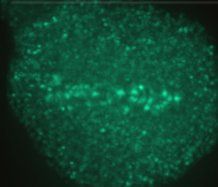
-
Application: ImmunocytochemistrySample Tested: RPE-1 cellsSpecies: HumanVerified Customer | Posted 11/06/2013
-
Application: ImmunofluorescenceSample Tested:Species: HumanVerified Customer | Posted 10/04/2013
-
Application: ImmunofluorescenceSample Tested: RPE-1Species: HumanVerified Customer | Posted 05/02/2012
There are no reviews that match your criteria.
Protocols
Find general support by application which include: protocols, troubleshooting, illustrated assays, videos and webinars.
- Antigen Retrieval Protocol (PIER)
- Antigen Retrieval for Frozen Sections Protocol
- Appropriate Fixation of IHC/ICC Samples
- Cellular Response to Hypoxia Protocols
- Chromogenic IHC Staining of Formalin-Fixed Paraffin-Embedded (FFPE) Tissue Protocol
- Chromogenic Immunohistochemistry Staining of Frozen Tissue
- ClariTSA™ Fluorophore Kits
- Detection & Visualization of Antibody Binding
- Fluorescent IHC Staining of Frozen Tissue Protocol
- Graphic Protocol for Heat-induced Epitope Retrieval
- Graphic Protocol for the Preparation and Fluorescent IHC Staining of Frozen Tissue Sections
- Graphic Protocol for the Preparation and Fluorescent IHC Staining of Paraffin-embedded Tissue Sections
- Graphic Protocol for the Preparation of Gelatin-coated Slides for Histological Tissue Sections
- ICC Cell Smear Protocol for Suspension Cells
- ICC Immunocytochemistry Protocol Videos
- ICC for Adherent Cells
- IHC Sample Preparation (Frozen sections vs Paraffin)
- Immunocytochemistry (ICC) Protocol
- Immunocytochemistry Troubleshooting
- Immunofluorescence of Organoids Embedded in Cultrex Basement Membrane Extract
- Immunofluorescent IHC Staining of Formalin-Fixed Paraffin-Embedded (FFPE) Tissue Protocol
- Immunohistochemistry (IHC) and Immunocytochemistry (ICC) Protocols
- Immunohistochemistry Frozen Troubleshooting
- Immunohistochemistry Paraffin Troubleshooting
- Immunoprecipitation Protocol
- Preparing Samples for IHC/ICC Experiments
- Preventing Non-Specific Staining (Non-Specific Binding)
- Primary Antibody Selection & Optimization
- Protocol for Heat-Induced Epitope Retrieval (HIER)
- Protocol for Making a 4% Formaldehyde Solution in PBS
- Protocol for VisUCyte™ HRP Polymer Detection Reagent
- Protocol for the Fluorescent ICC Staining of Cell Smears - Graphic
- Protocol for the Fluorescent ICC Staining of Cultured Cells on Coverslips - Graphic
- Protocol for the Preparation & Fixation of Cells on Coverslips
- Protocol for the Preparation and Chromogenic IHC Staining of Frozen Tissue Sections
- Protocol for the Preparation and Chromogenic IHC Staining of Frozen Tissue Sections - Graphic
- Protocol for the Preparation and Chromogenic IHC Staining of Paraffin-embedded Tissue Sections
- Protocol for the Preparation and Chromogenic IHC Staining of Paraffin-embedded Tissue Sections - Graphic
- Protocol for the Preparation and Fluorescent ICC Staining of Cells on Coverslips
- Protocol for the Preparation and Fluorescent ICC Staining of Non-adherent Cells
- Protocol for the Preparation and Fluorescent ICC Staining of Stem Cells on Coverslips
- Protocol for the Preparation and Fluorescent IHC Staining of Frozen Tissue Sections
- Protocol for the Preparation and Fluorescent IHC Staining of Paraffin-embedded Tissue Sections
- Protocol for the Preparation of Gelatin-coated Slides for Histological Tissue Sections
- Protocol for the Preparation of a Cell Smear for Non-adherent Cell ICC - Graphic
- R&D Systems Quality Control Western Blot Protocol
- TUNEL and Active Caspase-3 Detection by IHC/ICC Protocol
- The Importance of IHC/ICC Controls
- Troubleshooting Guide: Immunohistochemistry
- Troubleshooting Guide: Western Blot Figures
- Western Blot Conditions
- Western Blot Protocol
- Western Blot Protocol for Cell Lysates
- Western Blot Troubleshooting
- Western Blot Troubleshooting Guide
- View all Protocols, Troubleshooting, Illustrated assays and Webinars
Loading...